
Detailed answer for Bethesda system for thyroid cytology
"Bethesda system" AND "thyroid cytology"
Bethesda system thyroid cytology FNA categories
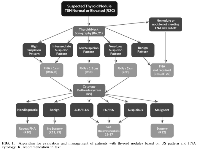
Summary : This figure presents an algorithmic flowchart for the evaluation and management of patients with suspected thyroid nodules, based on ultrasound (US) pattern and fine-needle aspiration (FNA) cytology, referencing the Bethesda system and specific recommendations. flowchart: # Nodes : • Start (rectangle): "Suspected Thyroid Nodule TSH Normal or Elevated (R2C)" • Decision (rectangle): "Thyroid/Neck Sonography (R6, 21)" • Branch (rectangle): "High Suspicion Pattern" • Branch (rectangle): "Intermediate Suspicion Pattern" • Branch (rectangle): "Low Suspicion Pattern" • Branch (rectangle): "Very Low Suspicion Pattern" • Branch (rectangle): "Benign Pattern" • Terminal (rectangle): "No nodule or nodule not meeting FNA size cutoff" • Action (rectangle): "FNA ≥ 1 cm (R8A, B)" • Action (rectangle): "FNA ≥ 1.5 cm (R8C)" • Action (rectangle): "FNA ≥ 2 cm (R8D)" • Terminal (rectangle): "FNA not required (R8E, 8F, 23)" • Action (rectangle): "Cytology Bethesda system (R9)" • Outcome (rectangle): "Nondiagnostic" • Outcome (rectangle): "Benign" • Outcome (rectangle): "AUS/FLUS" • Outcome (rectangle): "FN/FSN" • Outcome (rectangle): "Suspicious" • Outcome (rectangle): "Malignant" • Action (rectangle): "Repeat FNA (R10)" • Action (rectangle): "No Surgery (R11, 23)" • Action (rectangle): "See Recommendations 13–17" • Action (rectangle): "Surgery (R12)" # Connectors : • Arrows direct flow from "Suspected Thyroid Nodule" to "Thyroid/Neck Sonography" and to "No nodule or nodule not meeting FNA size cutoff". • From "Thyroid/Neck Sonography", arrows branch to "High Suspicion Pattern", "Intermediate Suspicion Pattern", "Low Suspicion Pattern", "Very Low Suspicion Pattern", and "Benign Pattern". • Each suspicion pattern node leads to a corresponding FNA size threshold or "FNA not required". • All FNA nodes converge to "Cytology Bethesda system (R9)". • "Cytology Bethesda system (R9)" branches to six outcomes: "Nondiagnostic", "Benign", "AUS/FLUS", "FN/FSN", "Suspicious", "Malignant". • Each outcome leads to a specific management action (e.g., "Repeat FNA", "No Surgery", "See Recommendations", "Surgery"). # Layout : • Top-down hierarchical structure. • Initial decision splits into five main US pattern branches. • FNA thresholds filter which nodules proceed to cytology. • Cytology results further stratify management. • Terminal actions at the bottom row. # Analysis : • The flowchart provides a stepwise, pattern-based approach to thyroid nodule management, integrating US risk stratification and cytology. • FNA is only performed if nodules meet specific size and suspicion criteria. • Cytology results (Bethesda system) dictate subsequent management, ranging from repeat FNA, observation, further recommendations, or surgery. • The algorithm emphasizes minimizing unnecessary procedures for benign or low-risk nodules and prioritizing intervention for suspicious or malignant findings.
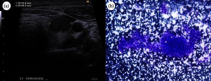
This dual-panel image displays diagnostic findings for thyroid pathology, combining ultrasonography and cytology. (a) A transverse thyroid ultrasound image reveals a localized, solid, hypoechoic nodule in the left lobe. The lesion is characterized by a wider-than-tall orientation and well-defined, smooth margins without internal calcifications. Caliper measurements indicate the nodule size is approximately 11.8 mm x 10.2 mm. (b) A corresponding cytological smear from fine-needle aspiration (FNA) shows a highly cellular specimen with densely packed clusters. The predominant cellular morphology includes spindle-shaped and tall cells with elongated nuclei and an increased nucleus-to-cytoplasm (N:C) ratio, characteristic of papillary growth patterns. Together, these images illustrate the diagnostic workup for thyroid cancer, specifically highlighting features that led to a Bethesda category VI classification and the eventual diagnosis of cribriform-morular thyroid carcinoma. This content is intended for intermediate to advanced medical learners studying endocrinology, radiology, and pathology.
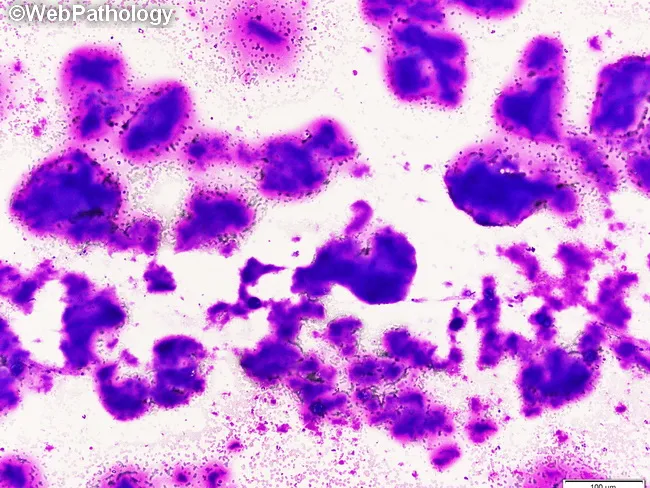
Imaging modality and technique: Light microscopy of a stained cytology smear from thyroid fine-needle aspiration (FNA). The specimen shows thyroid follicular cells set in a predominantly colloid-rich background, with occasional microfollicular clusters and scattered single cells. Staining: Papanicolaou stain, bright-field optics, high contrast between dense nuclear detail and translucent colloid. Anatomical context: endocrine organ pathology involving the thyroid gland in the anterior neck. The histologic plane is cytology; intact tissue architecture is limited, but overall cellularity is low-to-moderate with abundant colloid. Visual features: widespread, translucent, watery colloid covering large areas; occasional thick, opaque colloid with well-defined outlines; background may contain bubbles and folds; some circular or pseudopapillary arrangements can form; follicular cells display round to oval nuclei with even chromatin and inconspicuous nucleoli; lack of significant nuclear atypia is noted. Pathology and diagnostic relevance: colloid-rich aspirates with a high colloid-to-cell ratio are characteristic of benign thyroid nodules (colloid goiter) and hyperplastic nodules; differential considerations include follicular adenoma or carcinoma, but heavy colloid typically argues against malignancy. Clinical correlation: integrate with ultrasound nodule features, serum thyroid function tests, and repeat sampling if needed; educational value: reinforces FNAC interpretation and colloid morphology, including descriptors such as 'bubble-gum' colloid and translucent sliding film.
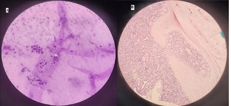
This comparative diagnostic image presents two different pathological preparations of a follicular thyroid nodule. Panel A displays a Fine-Needle Aspiration Cytology (FNAC) smear, characterized by dispersed individual follicular cells with small, dark purple-stained nuclei. The background contains amorphous light purple extracellular material and thin fibrous strands, representing a benign cytological appearance (Bethesda Category II). Panel B shows a postoperative histopathological section stained with Hematoxylin and Eosin (H&E) at 40x magnification. This section demonstrates a structured follicular architecture consisting of varied-sized thyroid follicles lined by follicular epithelium. Key features include dense follicular crowding and a prominent pink-stained fibrous capsule with clear evidence of neoplastic tissue invasion. The visual comparison highlights the diagnostic progression from benign cytological features to a definitive histological diagnosis of follicular thyroid carcinoma based on capsular invasion, a finding not visible on cytology alone. The image is intended for medical education regarding the limitations of FNAC and the surgical pathology of thyroid malignancies.
The Bethesda System for Reporting Thyroid Cytopathology (TBSRTC)
Background and History
The Six Bethesda Categories
Bethesda I - Nondiagnostic / Unsatisfactory
- Definition: The specimen does not meet adequacy criteria. This occurs when there are fewer than 6 groups of well-visualized follicular cells with at least 10 cells each, or when the smear is obscured by blood, air-drying artifact, clotting, or inflammation.
- Risk of malignancy: 5-20% (NIFTP = cancer); 5-18% (NIFTP ≠ cancer)
- Why high ROM?: The actual operative risk (20%, range 9-32%) is high because clinicians often proceed to surgery when repeat FNA also fails - this introduces selection bias.
- Management: Repeat FNA with ultrasound guidance. If repeat FNA is again nondiagnostic, diagnostic lobectomy is appropriate.
- Note: Ultrasound guidance reduces the nondiagnostic rate from 15-20% down to approximately 7%.
Bethesda II - Benign
- Definition: The most common category. Includes colloid nodules, benign follicular nodules, thyroiditis (Hashimoto's, subacute, lymphocytic), cysts, and hyperplastic/adenomatous nodules.
- Cytological features: Bland-appearing follicular cells arranged in macrofollicular patterns; abundant colloid in background; no significant nuclear atypia.
- Risk of malignancy: 0-4% (NIFTP ≠ cancer); 2-7% (NIFTP = cancer). Actual operative risk: 2.5% (range 1-10%).
- Management: Clinical and sonographic follow-up. Surgery is not routinely indicated.
- Caveats:
- Routine repeat FNA during surveillance is NOT indicated; reserve only for significant changes in nodule characteristics.
- A higher index of suspicion is warranted for nodules >3-4 cm, as sampling error and false-negative rates increase with size.
- Retrospective surgical series found malignancy in 11-13% of patients with initially benign cytology and nodules >3-4 cm (though likely overestimated due to selection bias). - Mulholland & Greenfield's Surgery, p. 4044
Bethesda III - Atypia of Undetermined Significance (AUS) / Follicular Lesion of Undetermined Significance (FLUS)
- Definition: Cytological findings that are neither clearly benign nor clearly neoplastic. This is the most heterogeneous and subjective category - interpathologist variability is most pronounced here.
- Subcategories (2023 update): The 3rd edition introduced cytological subcategories within AUS:
- Nuclear atypia (most common) - mild nuclear enlargement, grooves, occasional intranuclear pseudoinclusions
- Architectural atypia - microfollicular arrangements without sufficient atypia for FN
- Both nuclear and architectural atypia
- Oncocytic/Hurthle cell atypia
- Other (lymphoid, inflammatory)
- Risk of malignancy: 13-30% (NIFTP = cancer); 6-24% (NIFTP ≠ cancer)
- Management: Options include observation, repeat FNA (may yield specific diagnosis in >50% of cases), molecular testing, or diagnostic lobectomy. Preparation quality and cellular adequacy heavily influence interpretation.
- Key point: The recommended rate of AUS diagnoses in a practice should not exceed 10% of all FNA interpretations, as over-use of this category undermines clinical utility. - Sabiston Textbook of Surgery, p. 1507
Bethesda IV - Follicular Neoplasm (FN) / Suspicious for Follicular Neoplasm (SFN)
- Definition: A cellular specimen composed predominantly of follicular cells with architectural features concerning for a follicular neoplasm. Includes Hurthle cell (oncocytic) variant.
- Cytological features: Highly cellular aspirate; uniform follicular cells arranged in microfollicles (small rings of follicular cells around a central lumen); scant colloid; uniform nuclei with inconspicuous nucleoli.
- Why FNA cannot distinguish benign from malignant here: Follicular carcinoma is diagnosed by capsular invasion or vascular invasion - architectural features that can only be evaluated on full histological sections of the entire nodule, not on individual aspirated cells. - Sabiston Textbook of Surgery, p. 1507
- Risk of malignancy: 23-34% (NIFTP = cancer); 17-28% (NIFTP ≠ cancer). Actual operative risk: 25% (range 14-34%).
- Management: Molecular testing or diagnostic lobectomy remains standard of care. A majority are benign on final pathology, hence efforts to use molecular testing to avoid unnecessary surgery.
Bethesda V - Suspicious for Malignancy (SFM)
- Definition: Features highly suggestive but not conclusive for malignancy. Most commonly represents suspicious for papillary thyroid carcinoma (PTC); can also be suspicious for medullary carcinoma, lymphoma, or metastatic tumor.
- Cytological features: Nuclear enlargement with pale chromatin ("ground glass" nuclei), nuclear grooves, intranuclear pseudoinclusions (rare but highly specific), microfollicular groups, psammoma bodies may be present.
- Risk of malignancy: 67-83% (NIFTP = cancer); 58-74% (NIFTP ≠ cancer). Actual operative risk: 70% (range 53-97%).
- Management: Near-total thyroidectomy or lobectomy. Ultrasound assessment of cervical lymph nodes is mandatory before surgery. Though technically "indeterminate," the high malignancy risk means these are managed similarly to Bethesda VI in most situations. - Harrison's Principles of Internal Medicine 22E, p. 3092
Bethesda VI - Malignant
- Definition: Cytological findings conclusive for malignancy.
- Most common diagnoses:
- Papillary thyroid carcinoma (PTC) - by far the most common (~98% of Bethesda VI)
- Medullary thyroid carcinoma
- Poorly differentiated / anaplastic thyroid carcinoma
- Metastatic carcinoma
- Primary lymphoma (though core biopsy is often preferred for lymphoma)
- Cytological features of PTC: Large sheets of neoplastic cells; enlarged oval "Orphan Annie eye" nuclei (optically clear chromatin); multiple intranuclear pseudoinclusions; nuclear grooves; psammoma bodies.
- Risk of malignancy: 97-100% (NIFTP = cancer); 94-96% (NIFTP ≠ cancer). Actual operative risk: 99% (range 94-100%).
- Management: Near-total thyroidectomy or lobectomy depending on tumor characteristics, size, and need for postoperative radioiodine therapy.
- Note: The overall false-positive rate for Bethesda VI should be 0-0.5%, and false-negative rate for Bethesda II should be 1-6% at high-volume centers. - Mulholland & Greenfield's Surgery, p. 4043
Summary Table: 2023 Bethesda System (Version 3)
| Category | Name | ROM (NIFTP = cancer) | ROM (NIFTP ≠ cancer) | Usual Management |
|---|---|---|---|---|
| I | Nondiagnostic / Unsatisfactory | 5-20% | 5-18% | Repeat FNA with US guidance |
| II | Benign | 2-7% | 0-4% | Clinical + sonographic follow-up |
| III | AUS / FLUS | 13-30% | 6-24% | Repeat FNA, molecular testing, or lobectomy |
| IV | Follicular Neoplasm / SFN | 23-34% | 17-28% | Molecular testing or lobectomy |
| V | Suspicious for Malignancy | 67-83% | 58-74% | Lobectomy or near-total thyroidectomy |
| VI | Malignant | 97-100% | 94-96% | Lobectomy or near-total thyroidectomy |
- Source: The 2023 Bethesda System (Ali SZ et al., Thyroid 2023;33:1039-1044), as cited in Sabiston Textbook of Surgery, p. 1507
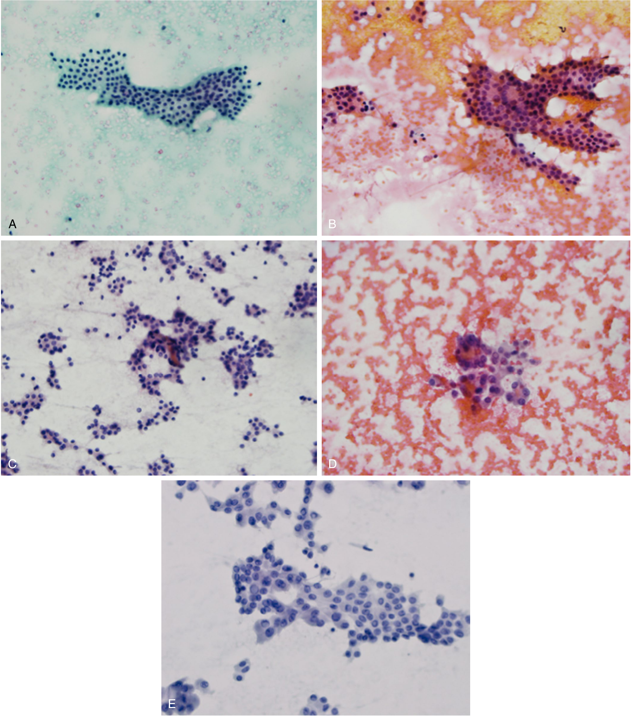
Cytological Images by Category
NIFTP and Its Impact on Risk Categories
Indeterminate Categories and Molecular Testing
- Afirma GSC (Genomic Sequence Classifier): 10,196-gene mRNA microarray. A "benign" result has negative predictive value (NPV) >95%, enabling surveillance instead of surgery.
- ThyroSeq v3: Next-generation DNA/RNA sequencing. Detects point mutations (BRAF, RAS, TERT), gene fusions (RET/PTC, PAX8-PPARγ), and copy number alterations. A "negative" result has NPV >95% for Bethesda III/IV.
- Key mutations by cancer type:
- PTC: BRAF V600E (45%), RAS (10%), RET/PTC rearrangements (20%), TERT promoter (10-12%)
- FTC: RAS (45%), PAX8-PPARγ rearrangement (35%), TERT promoter (17%)
Management Algorithm
Practical Points and Limitations
- Bethesda I has a paradoxically high operative malignancy rate (~20%) not because the nodule is more dangerous, but because only high-risk patients proceed to surgery after repeated nondiagnostic FNA.
- Bethesda II is highly accurate (<3% malignancy), but false-negatives increase with nodule size >3-4 cm.
- Bethesda III has the greatest interobserver variability and each institution should validate its own ROM for this category by correlating cytology with surgical histopathology.
- Bethesda IV cannot be definitively diagnosed as benign or malignant by FNA because the diagnosis of follicular carcinoma requires demonstration of capsular or vascular invasion, which demands histological evaluation of the entire capsule.
- Core needle biopsy is preferred over FNA for suspected thyroid lymphoma, as architectural assessment is required.
- The combined false-positive (0-0.5%) and false-negative (1-6%) rates for Bethesda VI and II respectively reflect the high accuracy of the system at the diagnostic extremes.