
Neurogenic bladder dysfunction diagnosis nursing
neurogenic bladder anatomy diagram spinal cord levels
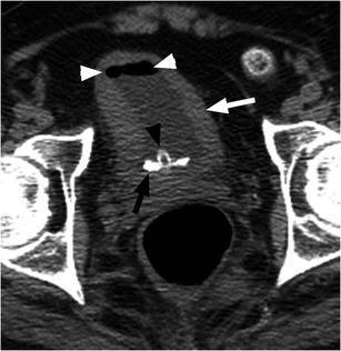
This axial non-contrast computerized tomography (CT) image of the pelvis demonstrates a neurogenic bladder in a patient with a spinal cord injury. Key visual findings include significant circumferential thickening of the urinary bladder wall (white arrow), characteristic of chronic bladder outlet obstruction or neurogenic dysfunction. Within the bladder lumen, a high-attenuation, calcified bladder stone is visible layering dependently (black arrow). An indwelling urinary catheter is present, identified by the catheter tubing and balloon (black arrowhead). Small pockets of gas are also noted within the superior aspect of the bladder lumen (white arrowheads), which can be secondary to instrumentation from the catheter or indicative of infection by gas-forming organisms. The image serves as a clinical example of the secondary complications of neurogenic bladder, including urolithiasis and chronic cystitis, frequently encountered in patients requiring long-term urinary catheterization.
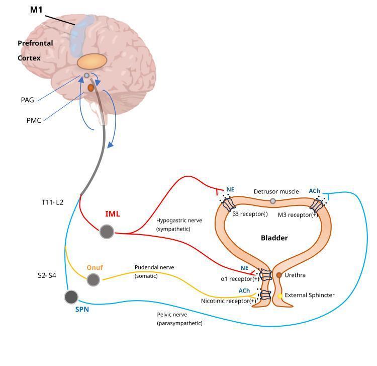
This medical illustration depicts the neural control of the lower urinary tract, specifically the micturition reflex and voluntary bladder control. It shows the hierarchical integration between the brain (primary motor cortex M1, prefrontal cortex, periaqueductal gray PAG, and pontine micturition center PMC) and the spinal cord. The diagram illustrates three distinct pathways: 1) The sympathetic pathway (red), originating from the T11-L2 intermediolateral cell column (IML) via the hypogastric nerve, releasing norepinephrine (NE) to inhibit the detrusor muscle (beta-3 receptors) and contract the bladder neck (alpha-1 receptors). 2) The parasympathetic pathway (blue), originating from the S2-S4 sacral parasympathetic nucleus (SPN) via the pelvic nerve, releasing acetylcholine (ACh) to contract the detrusor muscle (M3 receptors). 3) The somatic motor pathway (yellow), originating from Onuf's nucleus (S2-S4) via the pudendal nerve, releasing ACh to contract the external urethral sphincter (nicotinic receptors). This diagram serves as a pathophysiology and anatomical guide to neurogenic bladder and the physiological mechanisms of urination.
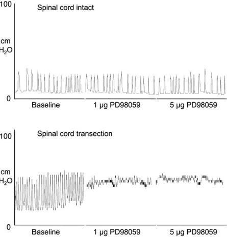
This diagnostic image consists of two cystometrogram (bladder pressure) tracings comparing 'Spinal cord intact' and 'Spinal cord transection' conditions in an experimental model. The vertical axis measures bladder pressure in cm H2O, and the horizontal axis represents time under three conditions: Baseline, 1 μg PD98059, and 5 μg PD98059. In the 'Spinal cord intact' panel, regular, low-amplitude bladder contractions are visible at baseline, and these remain unchanged following the intrathecal administration of increasing doses of PD98059 (a MEK inhibitor). In the 'Spinal cord transection' panel, the baseline shows high-frequency, high-amplitude bladder reflex activity, characteristic of neurogenic detrusor overactivity. Following the administration of PD98059, there is a dose-dependent decrease in both the frequency and amplitude of these contractions, with the 5 μg dose resulting in a significant stabilization of bladder pressure. The content illustrates the role of MAP kinase signaling in neurogenic bladder dysfunction and the potential therapeutic effect of pathway inhibition on reflex micturition in spinal cord injury scenarios.
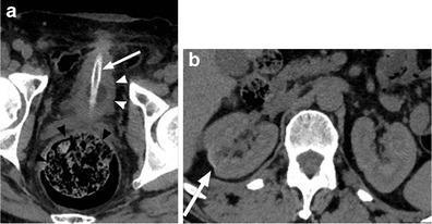
This composite of axial non-contrast computed tomography (CT) images illustrates secondary complications of chronic neurogenic bladder in a patient with a long-term spinal cord injury (SCI). Image (a) focuses on the pelvic region, demonstrating a small-capacity, thick-walled urinary bladder (white arrowheads), consistent with detrusor hyperactivity. A high-attenuation suprapubic indwelling catheter (white arrow) is positioned within the bladder lumen. Posterior to the bladder, the rectum is significantly distended with mottled fecal material (black arrowheads), indicating fecal impaction, a common manifestation of neurogenic bowel. Image (b) presents an axial slice of the upper abdomen showing the kidneys. The right kidney exhibits focal renal cortical scarring (white arrow), visualized as a peripheral indentation and loss of parenchymal volume. These findings highlight the chronic genitourinary and gastrointestinal sequelae of SCI, specifically the impact of long-term catheterization and dysfunctional voiding/elimination patterns on the urinary tract and bowel.
"neurogenic bladder" nursing assessment management
Neurogenic Bladder Dysfunction: Diagnosis and Nursing
What Is Neurogenic Bladder?
- Bailey and Love's Short Practice of Surgery, 28th Edition, p. 1526
Classification by Lesion Level
| Lesion Level | Causes | Bladder/Sphincter Effect | Key Risk |
|---|---|---|---|
| Suprapontine | CVA, Parkinson's, TBI, brain tumor | Neurogenic detrusor overactivity (NDO); voiding phase intact (sacral reflexes preserved) | Incontinence, urgency |
| Spinal (infrapontine - suprasacral) | SCI, myelitis, MS, disc herniation | Detrusor overactivity + sphincter overactivity (DSD); high-pressure voiding | Upper tract deterioration, autonomic dysreflexia |
| Sacral/Infrasacral | Spina bifida, cauda equina, pelvic surgery | Detrusor areflexia/underactivity; low sphincter tone | Retention, overflow incontinence |
- Bailey and Love's Short Practice of Surgery, 28th Edition, p. 1526
- Smith and Tanagho's General Urology, 19th Edition, p. 509
Neural Control Pathway (Pathophysiology)
- Sympathetic (T11-L2, via hypogastric nerve): Norepinephrine inhibits detrusor (β3) and contracts bladder neck (α1) - promotes storage
- Parasympathetic (S2-S4, via pelvic nerve): Acetylcholine contracts detrusor (M3) - promotes voiding
- Somatic (S2-S4, Onuf's nucleus, via pudendal nerve): Acetylcholine contracts external urethral sphincter - voluntary continence
Nursing Assessment
1. History
- Urinary symptoms: frequency, urgency, urge incontinence, hesitancy, incomplete emptying, nocturia, urinary retention
- Neurological diagnosis and course: type, duration, progression of neurologic disease
- Bowel symptoms: neurogenic bowel often coexists
- Sexual function
- Medications (especially those affecting bladder function)
- History of UTIs, decubitus ulcers, prior urologic procedures
- In men: prostate enlargement; in women: pelvic floor prolapse or urethral hypermobility
- Comorbidities: diabetes, cardiovascular disease
-
Mobility and hand function - determines feasibility of self-catheterization
-
Cognitive function - affects compliance and behavioral strategies
-
Social support - essential for caregiver-assisted bladder programs
-
Smith and Tanagho's General Urology, 19th Edition, p. 491
2. Physical Examination
-
Blood pressure measurement (baseline, and to monitor for autonomic dysreflexia)
-
Abdominal examination (bladder distension, suprapubic tenderness)
-
External genitalia examination in males; vaginal/pelvic floor exam in females as indicated
-
Rectal examination: anal tone, fecal loading
-
Neurological examination:
- Cognitive function assessment
- Ambulation and mobility
- Hand function (for CIC feasibility)
- Lumbar and spinal segment testing
- Perianal sensation, bulbocavernosus reflex (S2-S4 integrity)
- Sensation and reflexes in the urogenital area
-
Smith and Tanagho's General Urology, 19th Edition, p. 491
3. Nursing-Specific Assessment Tools
- Bladder diary/voiding diary: Document voiding frequency, volumes, incontinence episodes, urgency severity
- Post-void residual (PVR) via bladder scan or catheterization: essential to determine emptying efficiency. An elevated PVR indicates incomplete emptying and infection risk
- Symptom questionnaires: IPSS (International Prostate Symptom Score), OAB-q, or NBSS (Neurogenic Bladder Symptom Score)
- Pain and quality of life assessment
Diagnostic Investigations
A. Urine Testing
- Urinalysis and urine culture: detect blood, glucose, protein, leukocytosis, nitrites
- Important: collect clean specimens - never sample from leg drainage bags
- Many patients with neurogenic bladder have asymptomatic bacteriuria (ASB) - this does NOT require antibiotic treatment unless the patient has clinical symptoms (consensus guidelines). Treating ASB promotes bacterial resistance.
B. Renal Function
- Serum creatinine and GFR: note that GFR calculations overestimate renal function in patients with low muscle mass (e.g., SCI patients) since most formulas are muscle-mass based
- Serial monitoring for signs of renal deterioration
C. Upper Tract Evaluation (Imaging)
- Upper tract evaluation every 6 months to 1 year in high-risk patients (SCI, spina bifida)
- Renal ultrasound: first-line for hydronephrosis surveillance
- CT or MRI urography for detailed evaluation
- Look for: hydronephrosis, cortical scarring, vesicoureteral reflux (VUR), bladder wall thickening, calculi
D. Urodynamic Studies (UDS) - The Gold Standard
- Post-void residual (PVR) assessment
- Complex cystometrogram (CMG): assess bladder sensation, detrusor compliance, capacity, and overactivity during filling
- Pressure-flow study: assess voiding efficiency
- Videourodynamics (fluoroscopy + UDS): when available - detects silent upper tract changes
- Electromyography (EMG): sphincter activity, detrusor-sphincter dyssynergia (DSD/DESD)
- Document the effect of neurologic disease on the lower urinary tract
- Correlate patient symptoms with urodynamic signs
- Assess urologic risk factors for complications: DESD, poor bladder compliance, sustained high-pressure detrusor contractions, vesicoureteral reflux
-
Bladder compliance: low compliance = high storage pressures = upper tract risk
-
Detrusor leak point pressure (DLPP): >40 cmH₂O is associated with upper tract damage
-
Cystometric capacity
-
Detrusor-sphincter dyssynergia (DSD)
-
Smith and Tanagho's General Urology, 19th Edition, p. 509-510
- Do NOT offer UDS routinely to low-risk patients (e.g., MS without red flags)
- Offer videourodynamics to high-risk patients (spina bifida, SCI, anorectal anomalies)
- Always perform UDS before surgical treatment of neurogenic LUT dysfunction
Nursing Diagnoses (NANDA-based)
- Impaired urinary elimination r/t neurological impairment
- Urinary retention r/t detrusor underactivity or DSD
- Urge urinary incontinence r/t neurogenic detrusor overactivity
- Risk for infection (UTI/CAUTI) r/t catheter use, incomplete bladder emptying
- Risk for autonomic dysreflexia r/t bladder distension (in SCI above T6)
- Deficient knowledge r/t self-catheterization technique, bladder management
- Disturbed body image / impaired social interaction r/t incontinence
- Risk for impaired skin integrity r/t urinary incontinence
Management (Nursing Interventions)
Behavioral/Conservative (Nursing-Led)
- Timed voiding: fixed-interval toilet schedules
- Habit retraining: identify natural voiding patterns, prompt voiding before involuntary leakage
- Verbal prompts and positive reinforcement: for patients with cognitive deficits
- Pelvic floor exercises (PFPT): enhances inhibitory feedback, effective in women with MS
- Fluid management: adequate hydration (avoid excessive restriction which concentrates urine and increases infection risk); restrict caffeine and alcohol
Clean Intermittent Catheterization (CIC) - First-Line for Retention
- Frequency: typically every 4-6 hours; tailor to prevent volumes >400-500 mL
- Risk of ASB: 40-80% of CIC patients develop bacteriuria; do not treat unless symptomatic
- Catheter type: sterile vs. non-sterile, single-use vs. reusable - no proven difference in UTI rates
- Risk factors for UTI: high catheterization volumes (women), low catheterization frequency (men)
- Prophylactic antibiotics: not recommended long-term - increase bacterial resistance without reducing clinical UTIs
Pharmacological (Nursing Monitoring)
| Drug Class | Drug Examples | Purpose | Nursing Monitoring |
|---|---|---|---|
| Anticholinergics | Oxybutynin, tolterodine, solifenacin, trospium | Reduce NDO, increase capacity | Dry mouth, constipation, blurred vision, tachycardia, confusion, urinary retention |
| β3-agonist | Mirabegron | Detrusor relaxation | Blood pressure (mean rise ~2.4 mmHg), heart rate |
| Desmopressin | DDAVP | Reduce nocturnal polyuria | Serum Na⁺ (hyponatremia risk), use caution in >65 years, edema |
| OnabotulinumtoxinA (Botox) | Intravesical | Reduce NDO/compliance | Post-injection urinary retention (up to 39% in neurogenic pts); need for CIC |
Monitoring for Complications
| Complication | Signs/Symptoms | Nursing Action |
|---|---|---|
| Autonomic Dysreflexia (SCI ≥T6) | Severe HTN, pounding headache, flushing above lesion, sweating, bradycardia | IMMEDIATE: sit patient upright, check/drain bladder, remove catheter obstruction, check for fecal impaction; call physician |
| UTI | Fever, increased spasticity, worsening incontinence, cloudy/malodorous urine, change in mental status | Clean-catch UA and culture; avoid treating ASB |
| Upper tract deterioration | Flank pain, rising creatinine, hydronephrosis on US | Monitor renal function; arrange urological review |
| Bladder stones | Recurrent UTI, hematuria, catheter obstruction | Imaging (KUB, CT) |
Patient and Carer Education
- CIC technique (sterile/clean; step-by-step)
- Signs of UTI vs. expected ASB
- Recognition of autonomic dysreflexia and immediate management
- Voiding diary completion
- Medication side effects and compliance
- Skin care (incontinence-associated dermatitis prevention)
- Bowel management program (neurogenic bowel commonly coexists)
- Sexuality and fertility resources
Summary Table: Assessment Goals
| Goal | Rationale |
|---|---|
| Preserve upper urinary tract | High bladder pressures → hydronephrosis → renal failure |
| Minimize lower tract complications | UTI, stones, DSD |
| Maintain quality of life | Compatible bladder management with patient's lifestyle |
| Prevent autonomic dysreflexia | Life-threatening if bladder distension occurs in SCI ≥T6 |
- Smith and Tanagho's General Urology, 19th Edition (Chapter 27, pp. 491-493, 509-510)
- Bailey and Love's Short Practice of Surgery, 28th Edition (p. 1525-1526)
- Campbell-Walsh-Wein Urology, 4-Volume Set (Part XIII: Neurogenic Bladder)
- Goldman-Cecil Medicine, International Edition (Focal CNS disorders)
- Cao W et al. (2025). Clamping Catheter Versus Free Drainage for Neurogenic Bladder. Biol Res Nurs [PMID: 39809550]