
Histology of Loose areolar tissue, Compare and contrast the microscopic structures of various types of muscles With suitable examples.
Reading File
Now I'll run parallel searches for both topics.
Finding Sources
Finding Sources
Finding Sources
Finding Sources
Finding Sources
Finding Sources
Finding Sources
Finding Sources
Now let me read the key sections from both histology textbooks in parallel.
Reading File
Reading File
Excellent data retrieved. Now let me read more of the muscle tissue section and get areolar tissue cell details in parallel.
Reading File
Reading File
Now let me search for medical images to accompany the response, and read more about cardiac/smooth muscle.
Searching Images
loose areolar connective tissue histology microscopy
Searching Images
skeletal muscle histology cross section longitudinal striations
Searching Images
cardiac muscle smooth muscle histology comparison
Reading File
Reading File
I now have comprehensive content from the textbooks. Let me also get the Junqueira's content on areolar tissue cells and fetch the key histology images.
Reading File
Loading Image
Loading Image
Loading Image
I now have comprehensive textbook content and images. Let me compile the full detailed answer.
Histology of Loose Areolar Tissue & Comparison of Muscle Types
PART 1: HISTOLOGY OF LOOSE AREOLAR TISSUE
Loose connective tissue (also called areolar tissue) is a cellular connective tissue with thin and relatively sparse collagen fibers. The ground substance occupies more volume than the fibers do - it has a viscous to gel-like consistency and plays an important role in the diffusion of oxygen, nutrients, CO₂, and metabolic wastes between small vessels and cells.
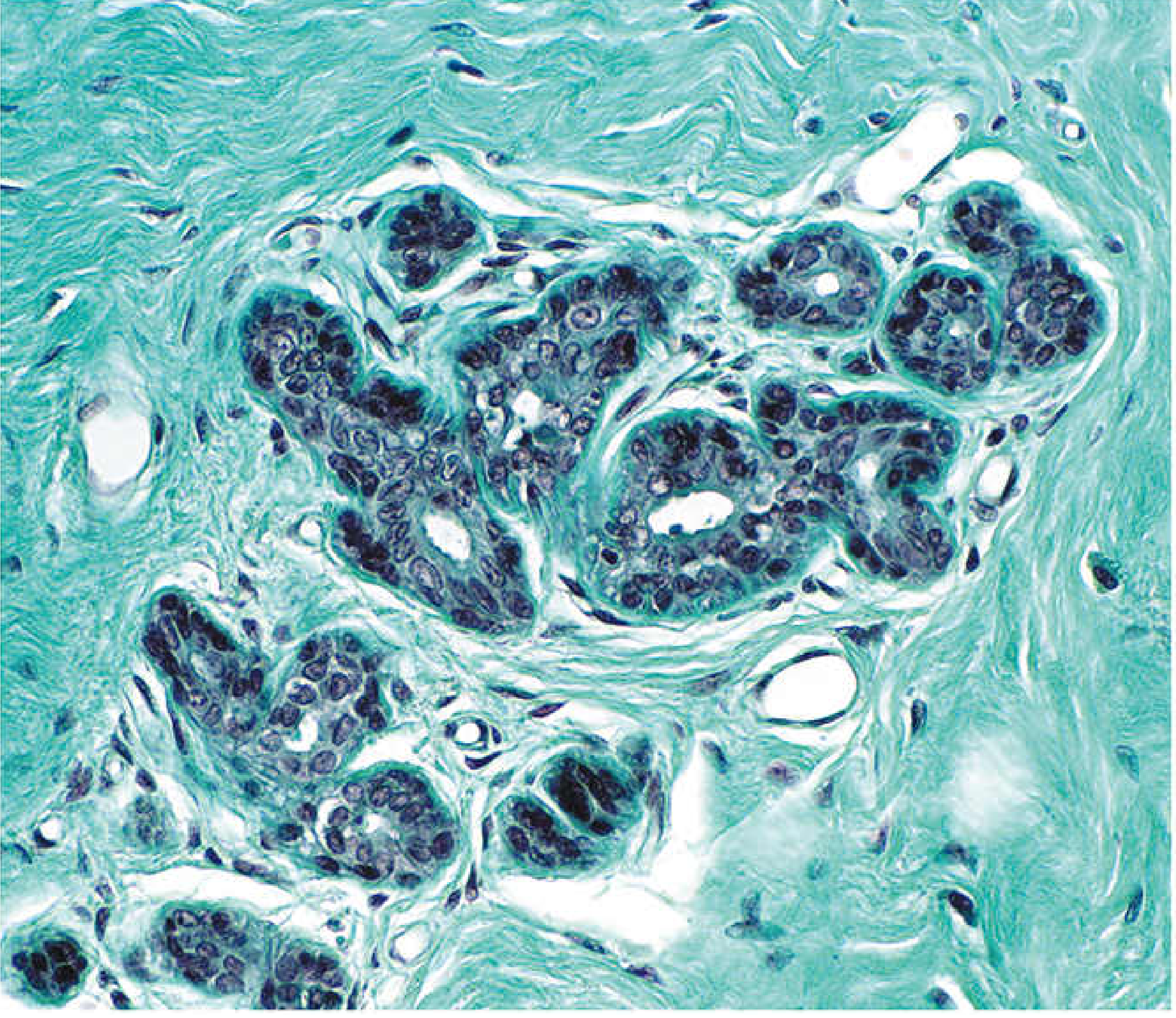
Location
- Beneath epithelia covering body surfaces and lining internal surfaces
- Associated with the epithelium of glands
- Surrounds the smallest blood vessels (capillaries)
- Forms the lamina propria of mucous membranes (respiratory and alimentary tracts)
- Fills spaces between muscle fibers and nerves
Microscopic Components
Loose areolar tissue contains cells, fibers, and ground substance in roughly equal proportions.
A. Cells
| Cell Type | Appearance under Microscope | Function |
|---|---|---|
| Fibroblast (predominant) | Flat, spindle-shaped; elongated pale nucleus; inconspicuous cytoplasm | Produces collagen, reticular, and elastic fibers; synthesizes ground substance |
| Macrophage (Histiocyte) | Irregular shape; kidney-shaped or indented nucleus; lysosomes visible on EM | Phagocytosis; antigen presentation; immune defense |
| Mast cell | Large, round to oval; nucleus hidden by dark metachromatic granules (toluidine blue) | Granules contain heparin, histamine, tryptase; role in allergy and inflammation |
| Plasma cell | Round; eccentric "clock-face" or "cartwheel" nucleus; basophilic cytoplasm; prominent Golgi ("hof") | Secretes immunoglobulins (antibodies) |
| Adipocyte | Large; signet-ring appearance - nucleus pushed to periphery by large lipid droplet | Fat storage; insulation |
| Lymphocytes | Small, dark round nucleus, scant cytoplasm | Immune surveillance; transient |
| Eosinophils | Bilobed nucleus; bright eosinophilic (red) granules | Anti-parasitic; allergy |
| Neutrophils | Multilobed nucleus | Acute inflammation; transient |
Most cells in loose connective tissue are transient wandering cells that migrate from local blood vessels in response to stimuli - making this tissue the primary site of inflammatory and immune reactions.
B. Fibers
Three types of fibers are present, all produced by fibroblasts:
-
Collagen fibers (most abundant) - Wavy bundles; stain pink with eosin; stain green/blue with Masson/Mallory trichrome. High tensile strength but flexible. Composed of fibrils with a 68 nm repeat banding pattern on EM. In loose tissue, collagen bundles are thin and dispersed.
-
Reticular fibers - Thin (type III collagen); form a delicate meshwork. Stained by silver impregnation (argyrophilic - appear black). Not visible on routine H&E. Support soft organs (lymph nodes, liver, spleen).
-
Elastic fibers - Thin, branching fibers that can stretch and recoil. Stain with orcein or Verhoeff's stain. Contain elastin core with fibrillin scaffold.
C. Ground Substance (Extracellular Matrix)
- Abundant and occupies more space than fibers in loose tissue
- Composed of: proteoglycans (aggrecan, versican), glycosaminoglycans (hyaluronic acid, chondroitin sulfate, heparan sulfate), and glycoproteins (fibronectin, laminin)
- Gel-like consistency; acts as a diffusion medium for nutrients and gases
- Creates a selective barrier to microorganisms
PART 2: COMPARISON OF MUSCLE TYPES
The three major types of muscle tissue - skeletal, cardiac, and smooth - differ in their microscopic structure, nuclear position, striations, control, and regenerative capacity.
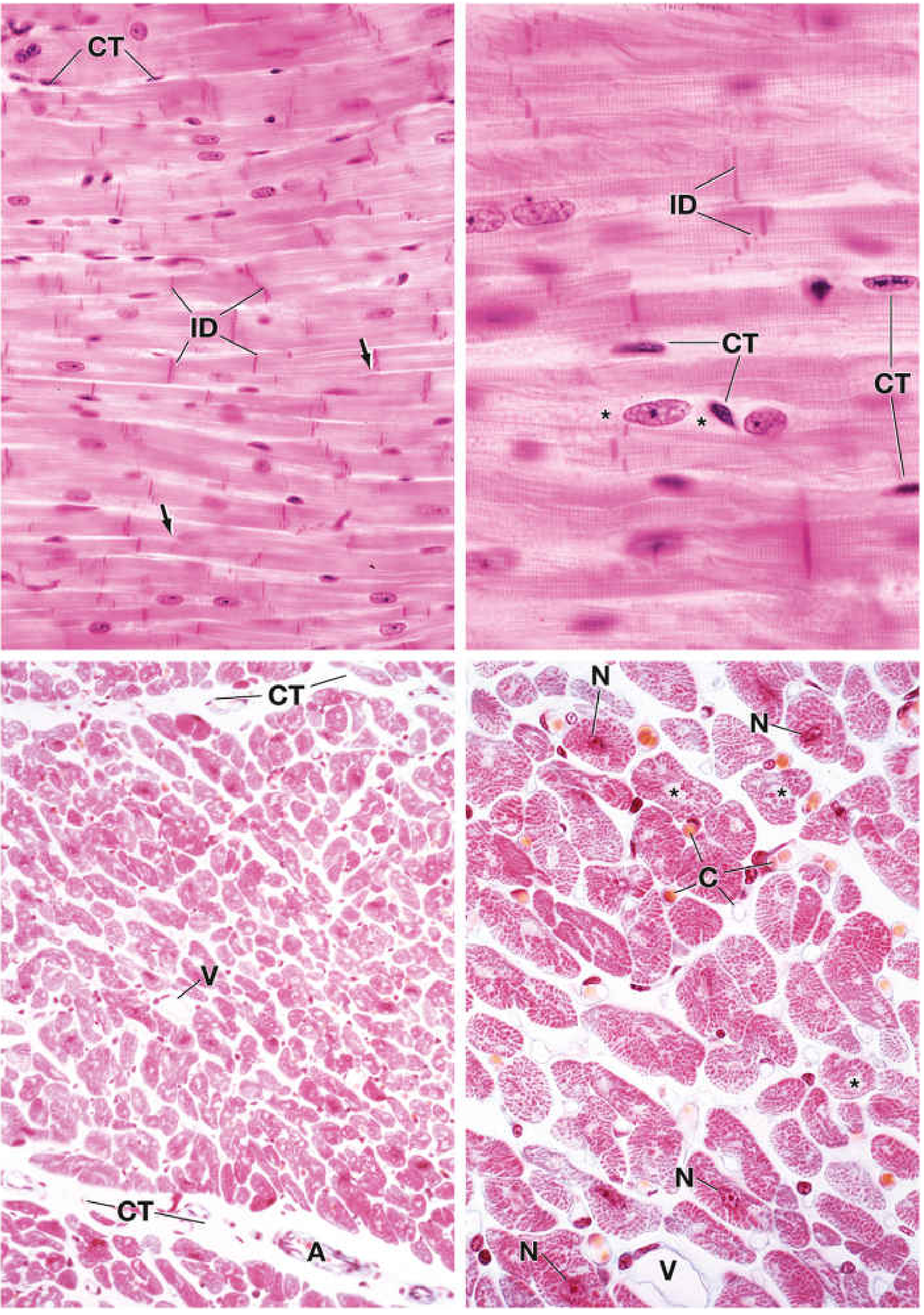
Detailed Comparison Table
| Feature | Skeletal Muscle | Cardiac Muscle | Smooth Muscle |
|---|---|---|---|
| Location (example) | Biceps, quadriceps, diaphragm | Heart wall (myocardium) | Wall of gut, uterus, blood vessels, iris |
| Cell shape | Long, cylindrical fibers (10-100 μm diameter) | Short, branching, cylindrical cells | Fusiform (spindle-shaped), small, finely tapered ends |
| Cell length | Very long (up to several cm) | 50-100 μm | 20-200 μm |
| Nuclei - number | Multiple (multinucleated syncytium) | Usually 1, occasionally 2 | Single |
| Nuclei - position | Peripheral (subsarcolemmal) | Central | Central (cigar-shaped; corrugated/folded when contracted) |
| Striations | Yes - prominent cross-striations | Yes - cross-striations (less distinct than skeletal) | No - no striations |
| Branching | No | Yes - cells branch and anastomose | No |
| Intercalated discs | Absent | Present (unique to cardiac) - appear as dark transverse lines | Absent |
| Connective tissue | Endomysium (around each fiber), Perimysium (around fascicles), Epimysium (around muscle) | Endomysium between cells, rich in capillaries | Thin endomysium |
| Sarcomere / Myofibrils | Highly organized; prominent sarcomeres. I band (light, actin), A band (dark, myosin), Z line, H zone, M line | Organized sarcomeres similar to skeletal; myofibrils less tightly packed | NO sarcomeres; actin and myosin filaments arranged obliquely; attached to dense bodies (analogous to Z lines) |
| T-tubule system | T-tubules at A-I junction; form TRIADS (T-tubule + 2 terminal cisternae) | T-tubules at Z line level; form DIADS (T-tubule + 1 terminal cisterna) | No T-tubules; Ca²⁺ enters via caveolae |
| Sarcoplasmic reticulum | Well developed; large terminal cisternae | Less developed; smaller cisternae | Poorly developed; sparse SR |
| Ca²⁺ trigger | Ca²⁺ from SR (RyR1 receptors); T-tubule voltage sensor = DHSR | Ca²⁺ from SR + Ca²⁺ entry from extracellular space (Ca²⁺-induced Ca²⁺ release) | Ca²⁺-calmodulin → activates MLCK (myosin light-chain kinase) |
| Contraction regulation | Troponin-tropomyosin complex (Ca²⁺ binds troponin C) | Same as skeletal | Via caldesmon/calponin; Ca²⁺-calmodulin-MLCK pathway; no troponin |
| Contraction type | Voluntary, rapid, forceful, fatigable | Involuntary, rhythmic, continuous, resistant to fatigue | Involuntary, slow, prolonged, sustained |
| Innervation | Somatic motor neuron; neuromuscular junction (motor end plate) | Autonomic (modulates rate only - SA node generates intrinsic rhythm) | Autonomic + hormones + local stimuli (mechanical stretch, chemical) |
| Regeneration | Via satellite cells (Pax7⁺ myogenic stem cells) | Very limited; repaired by fibrous connective tissue (scar) | Yes - smooth muscle cells can divide (hyperplasia) |
| Fiber types | Type I (slow oxidative/red), Type IIa (fast oxidative-glycolytic/intermediate), Type IIb (fast glycolytic/white) | Single functional type; Purkinje fibers are specialized conducting cells | Single type |
| Staining (H&E) | Eosinophilic; striations and peripheral nuclei clearly visible | Eosinophilic; central nuclei, intercalated discs (dark lines), branching | Pale eosinophilic; central oval nuclei; no striations |
Individual Descriptions
1. Skeletal Muscle (Voluntary / Striated)
- Longitudinal section: Long parallel fibers with alternating light (I band) and dark (A band) cross-striations. Multiple flattened nuclei lie at the periphery under the sarcolemma. Striations are the most prominent of all three types.
- Cross-section: Polygonal fibers of varying size. Peripheral nuclei visible as small dense profiles at the edge. Myofibrils appear as punctate (dot-like) structures. Fascicles are separated by perimysium (CT); individual fibers by endomysium.
- Key identifier: Multiple peripheral nuclei + prominent striations + no branching + no intercalated discs.
2. Cardiac Muscle (Involuntary / Striated)
- Longitudinal section: Short branching cylindrical fibers. Single central oval nucleus (occasionally two) surrounded by a clear zone of perinuclear cytoplasm (glycogen-rich). Cross-striations are present but less regular than skeletal. Intercalated discs appear as dark, transverse or staircase-like lines - these contain fascia adherens, gap junctions (electrical coupling), and desmosomes (mechanical coupling).
- Cross-section: Round cells of variable size with large central nuclei. Rich endomysial connective tissue with numerous capillaries (one per cell).
- Key identifier: Central nucleus + branching + intercalated discs + striations.
3. Smooth Muscle (Involuntary / Non-striated)
- Longitudinal section: Long, fusiform cells with a single central elongated (cigar-shaped) nucleus - when contracted, the nucleus becomes corkscrew/wavy in shape. No striations at all. Cells arranged in sheets or bundles. Cytoplasm appears homogeneously pale pink.
- Cross-section: Round to oval profiles of varying diameter (due to tapered ends). Central nucleus visible only in the widest cells (those sectioned through the mid-region). Others show no nucleus in cross-section.
- Key identifier: Fusiform shape + central nucleus + NO striations + no intercalated discs.
Summary Mnemonic
| Feature | Skeletal | Cardiac | Smooth |
|---|---|---|---|
| Nuclei position | Peripheral | Central | Central |
| Number of nuclei | Many | 1 (rarely 2) | 1 |
| Striations | Yes | Yes | No |
| Branching | No | Yes | No |
| Intercalated discs | No | Yes | No |
| Voluntary | Yes | No | No |
Sources
- Histology: A Text and Atlas with Correlated Cell and Molecular Biology (Ross & Pawlina), Chapters 6 & 11
- Junqueira's Basic Histology: Text and Atlas, 17e, Chapter 5 & 10
This is a shared conversation. Sign in to Orris to start your own chat.