
Lower left side back pain
lower left back pain causes diagnosis treatment 2025
lower back pain anatomy lumbar spine causes diagram
"low back pain"[MeSH Terms] AND diagnosis AND management
lumbar disc herniation nerve root compression MRI
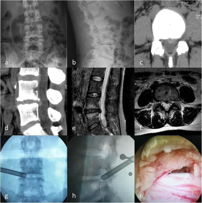
A multi-modal imaging series and clinical photograph detailing a lumbar disc herniation and its surgical management. (a-b) Preoperative lumbar X-rays in anteroposterior and lateral views showing spinal alignment and vertebral anatomy. (c-d) CT scans in axial and sagittal planes demonstrate a focal disc protrusion in the posterior left portion of the L4/5 intervertebral disc. (e-f) MRI scans (T2-weighted) confirm the L4/5 left-sided disc herniation, showing significant compression of the left L5 nerve root within the spinal canal. (g-h) Intraoperative fluoroscopic X-rays (anteroposterior and lateral) visualize the precise placement of a percutaneous endoscopic lumbar discectomy (PELD) working channel at the L4/5 level. (i) An endoscopic clinical photograph provides a direct visualization of the exposed left L5 nerve root following decompression. The collection serves as an educational sequence from diagnostic imaging through surgical intervention for lumbar radiculopathy.
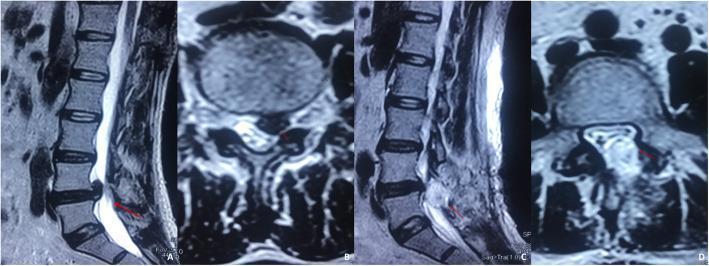
This composite diagnostic image features four MRI views (T2-weighted sequence) of the lumbar spine, specifically focusing on the L4/L5 segment in a 37-year-old male. Panels (A) and (B) demonstrate preoperative lumbar disc herniation. Panel (A), a sagittal view, shows a large posterior herniation of the L4/L5 intervertebral disc significantly encroaching upon the spinal canal. Panel (B), a transverse view, displays the herniated nucleus pulposus lateralized to the left, causing direct compression of the left exiting nerve root. Panels (C) and (D) illustrate postoperative complications following discectomy. Panel (C), a sagittal view, reveals an accumulation of altered signal intensity posterior to the L4/L5 disc space, consistent with a symptomatic postoperative epidural hematoma. Panel (D), an axial view, demonstrates this hematoma occupying the epidural space, resulting in mass effect with marked compression and displacement of the dural sac and the left nerve root. Red arrows highlight the specific sites of herniation and subsequent hematoma-induced compression, serving as a clinical example of postoperative spinal epidural hematoma (SEH).
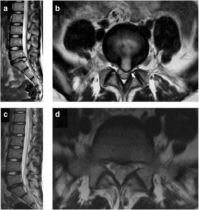
This diagnostic image set consists of four T2-weighted magnetic resonance imaging (MRI) scans of the lumbar spine, providing a pre- and post-operative comparison of a lumbar disc herniation. Images (a) and (b) represent the preoperative state. The sagittal view (a) shows a posterior disc protrusion at the L5-S1 level, narrowing the spinal canal. The corresponding axial view (b) demonstrates a significant left-sided paramedian disc herniation (indicated by a white arrow) causing clear compression of the exiting nerve root and displacement of the thecal sac. Images (c) and (d) show the same patient six months following surgical decompression. The postoperative sagittal view (c) illustrates restored spinal canal patency at L5-S1. The postoperative axial view (d) confirms the complete removal of the herniated disc material, showing a decompressed nerve root and restored epidural space. This comparison is an educational example of degenerative disc disease and the efficacy of surgical intervention in relieving mechanical neural compression.
Lower Left Side Back Pain
Common Causes
1. Musculoskeletal (Most Common)
- Involves muscles, ligaments, and fascia (semispinalis, erector spinae, multifidus, latissimus dorsi)
- Active trigger points show elevated substance P, inflammatory cytokines, and higher myoelectric tone
- Pain is focal to diffuse with tenderness, limited range of motion, and a normal neurologic exam
- Source: Harrison's Principles of Internal Medicine 22E, p. 1907
- Accounts for 26-42% of axial low back pain
- Pain is typically worsened by sitting or bending forward
- A left-sided disc herniation (most common at L4/L5 or L5/S1) will cause pain radiating down the left leg (sciatica), often in a dermatomal pattern
- As disc degenerates, nerve fibers grow into the inner annulus/nucleus pulposus, sensitizing them to mechanical and chemical stimuli
- Source: Harrison's, p. 1911
- Affects ~10-15% of patients with axial low back pain; prevalence increases with age
- Pain is typically unilateral and paraspinal - left facet pathology = left-sided pain
- Unlike disc pain, sitting may relieve facet pain; morning stiffness is common
- Source: Harrison's, p. 1914
- The left SI joint can refer pain to the lower left back, buttock, and posterior thigh
- Common in pregnancy, after trauma, or with inflammatory arthritis (ankylosing spondylitis)
- A stress fracture of the pars interarticularis - common in athletes, especially those with repetitive hyperextension (gymnastics, football)
- Source: Textbook of Family Medicine 9e, p. 794
2. Radiculopathy ("Pinched Nerve")
- Pain radiating from the lower back, through the buttock and down the left leg
- Numbness or tingling in a dermatomal distribution
- Weakness (foot drop with L5; absent ankle reflex with S1)
3. Referred / Visceral Pain (Left-Sided Clues)
| Organ | Features |
|---|---|
| Left kidney (stone or infection/pyelonephritis) | Colicky, flank pain radiating to groin; fever + dysuria with infection; hematuria |
| Aorta (aortic aneurysm/dissection) | Tearing, severe pain; pulsatile abdominal mass; a vascular emergency |
| Descending colon / sigmoid | Associated with bowel changes, bloating |
| Ovary (women) | Cysts, torsion, endometriosis - associated with pelvic/menstrual symptoms |
| Psoas abscess | Fever, hip flexor pain, immunosuppressed patients |
Red Flag Symptoms - See a Doctor Urgently
- Bowel or bladder incontinence / retention (cauda equina syndrome - surgical emergency)
- Saddle area numbness
- Fever + back pain (possible spinal infection)
- Weight loss + night sweats (cancer, infection)
- Pain worse when lying down (not mechanical)
- Trauma (fracture risk)
- History of cancer
- Progressive leg weakness
Diagnosis
- History and physical exam first - 97% of back pain can be triaged clinically
- X-ray - for fracture, spondylolisthesis, alignment
- MRI - best for disc herniation, nerve root compression, spinal infection, tumors
- CT scan - for bony detail, apophyseal ring separation in adolescents
- Urinalysis - if kidney stone or infection is suspected
- Blood tests (CRP, ESR, CBC) - if infection or inflammatory arthritis suspected
Treatment
First-Line (most acute cases resolve within 6 weeks)
- NSAIDs (ibuprofen, naproxen) - first-line pharmacotherapy
- Heat therapy - effective for muscle pain
- Targeted physical therapy and core-strengthening exercises
- Continue normal activity as tolerated - bed rest is not helpful
- Massage - may provide short-term relief
Medications
| Drug | Role |
|---|---|
| NSAIDs | First-line for pain + inflammation |
| Muscle relaxants (non-benzodiazepine) | Helpful for acute muscle spasm |
| Duloxetine (SNRI) | Evidence in chronic LBP |
| Opioids | Last resort only; lowest dose, shortest duration |
| Acetaminophen | No longer recommended as first-line (limited evidence for back pain) |
| Gabapentinoids | Insufficient evidence for axial or radicular back pain |
A 2025 systematic review in The Lancet Rheumatology confirmed that exercise interventions reduce chronic low back pain intensity and disability at 1-2+ year follow-up. Sessions of 15-30 minutes are most effective per a 2025 network meta-analysis.
For Chronic LBP (>3 months)
- Cognitive-behavioral therapy (CBT)
- Mindfulness-based stress reduction
- Fluoroscopic injections (epidural steroids, medial branch blocks for facet pain)
- Surgery only if conservative treatment fails and there is a clear structural cause (e.g., confirmed disc herniation with neurologic deficit)
When to See a Doctor
- Pain lasting more than 4-6 weeks without improvement
- Any red flag symptoms listed above
- Severe or worsening pain
- Pain with any neurologic symptoms (leg weakness, numbness, tingling)
- Suspected kidney involvement (fever, flank pain, burning urination, blood in urine)