Excellent! I have the histology images. Now let me write all the comprehensive exam answers:Here are all the complete, exam-oriented answers for BDS 3rd Year Oral Pathology:
BDS 3rd Year - Oral Pathology University Exam Answers
LONG ESSAY QUESTIONS
Q1. Define and Classify Odontogenic Cysts. Discuss in Detail the Clinical Features, Histological Features, and Radiographic Features of Radicular Cyst.
DEFINITION OF ODONTOGENIC CYST
An odontogenic cyst is defined as a pathological cavity lined by epithelium derived from odontogenic (tooth-forming) epithelium, which is enclosed in a connective tissue wall and usually contains fluid or semi-fluid material.
CLASSIFICATION OF ODONTOGENIC CYSTS
(WHO Classification - Revised 2022 / Shear's Classification)
A. INFLAMMATORY ODONTOGENIC CYSTS
- Radicular cyst (Periapical cyst / Apical periodontal cyst)
- Residual cyst
- Paradental cyst (Inflammatory collateral cyst)
- Buccal bifurcation cyst
B. DEVELOPMENTAL ODONTOGENIC CYSTS
- Dentigerous cyst (Follicular cyst)
- Odontogenic Keratocyst (OKC) - now classified as Keratocystic Odontogenic Tumor (KCOT) by WHO 2005, reverted to cyst in 2017
- Eruption cyst
- Lateral periodontal cyst
- Botryoid odontogenic cyst
- Gingival cyst of adult / infant (Bohn's nodules, dental lamina cysts)
- Calcifying Odontogenic Cyst (Gorlin cyst)
- Orthokeratinized odontogenic cyst (OOC)
- Glandular odontogenic cyst (Sialo-odontogenic cyst)
RADICULAR CYST
INTRODUCTION
Radicular cyst is the most common odontogenic cyst (accounting for ~52-68% of all jaw cysts). It is an inflammatory cyst that arises from the epithelial cell rests of Malassez (ERM) in the periodontal ligament as a result of periapical periodontitis, usually following pulp necrosis.
Synonyms: Periapical cyst, Apical periodontal cyst, Dental cyst
ETIOLOGY AND PATHOGENESIS
- Initiating factor: Pulp necrosis due to caries, trauma, or deep restorations leads to periapical infection
- Bacterial toxins and inflammatory mediators stimulate the Epithelial Cell Rests of Malassez (ERM) in the periodontal ligament to proliferate
- Three phases of cyst formation (Toller/Seltzer theory):
- Phase 1 - Initiation: Stimulation of ERM by periapical inflammation
- Phase 2 - Cyst formation: Central necrosis of the epithelial proliferation with fluid accumulation
- Phase 3 - Enlargement: Osmotic pressure theory (Toller) - increased osmotic pressure from breakdown products draws fluid through the wall, enlarging the cyst
CLINICAL FEATURES
| Feature | Details |
|---|
| Age | 3rd-6th decade (peak: 3rd-4th decade) |
| Sex | More common in males (M:F = 3:2) |
| Site | Most common in maxillary anterior region (60%); mandible less common |
| Associated tooth | Non-vital (carious, restored, or traumatized) tooth |
| Symptoms | Usually asymptomatic; may cause painless swelling |
| Size | Usually < 2 cm |
| Cortex | Egg-shell crackling sensation on palpation (when cortex is expanded) |
| Fluctuation | May show blue translucency through mucosa (cholesterol crystals give yellowish fluid) |
| Teeth | Adjacent teeth may be displaced and show vitality tests positive (important distinction from OKC) |
Important Clinical Points:
- Tooth associated with radicular cyst is always non-vital (responds negative to pulp vitality test)
- May rarely become infected causing pain and swelling
- Residual cyst = radicular cyst that persists after extraction of the causative tooth
RADIOGRAPHIC FEATURES
- Shape: Well-defined, round or ovoid radiolucency at the apex of a non-vital tooth
- Size: Usually < 2 cm (cysts > 2 cm suggest OKC or dentigerous cyst)
- Margin: Well-defined with a thin radiopaque corticated (sclerotic) border - this is the hallmark
- Location: Apical / periapical / lateral (lateral radicular cyst from lateral accessory canals)
- Effect on adjacent structures:
- Displacement of adjacent teeth (not resorption, usually)
- Lamina dura of adjacent teeth may be lost
- Root resorption is minimal (unlike OKC which rarely resorbs roots; ameloblastoma causes root resorption)
- May displace the floor of maxillary sinus (antral elevation)
- May bow the inferior alveolar canal inferiorly
- Continuity: Continuous with the lamina dura (periodontal ligament space is continuous with the cyst)
Differential Radiographic Diagnosis:
- OKC - scalloped margins, less distinct border, associated with impacted tooth
- Periapical granuloma - smaller (< 1 cm), no well-defined corticated margin (difficult to distinguish radiographically)
- Ameloblastoma - multilocular, soap-bubble/honeycomb appearance
HISTOPATHOLOGICAL FEATURES
LINING EPITHELIUM:
- Non-keratinized stratified squamous epithelium - most characteristic feature
- Thickness is irregular and varies; may be 2-50 cell layers thick
- Arcading/Irregular epithelium with prominent rete ridges (due to active inflammation)
- Rushton bodies (Hyaline bodies): Eosinophilic, hair-pin shaped or linear curved structures within the epithelium - PATHOGNOMONIC of radicular cyst. Composed of hemoglobin degradation products or secreted by epithelial cells
- Areas of ulceration in the epithelium may be present
- Mural nodules of cholesterol crystals may indent the cyst wall
FIBROUS WALL (Connective Tissue):
- Dense fibrous connective tissue wall
- Chronic inflammatory cell infiltrate: Predominantly lymphocytes and plasma cells; neutrophils in active infection
- Cholesterol clefts: Needle-shaped clefts (cholesterol dissolved during processing) surrounded by foreign body giant cells (Langerhans/Touton type giant cells) - "cholesterol granuloma"
- Russell bodies (immunoglobulin-containing plasma cells)
- Hemosiderin pigment deposits
- Dystrophic calcification may be present
- Newly formed blood vessels (granulation tissue if recently infected)
CYST CONTENTS (aspirate):
- Straw-colored / yellowish fluid
- High protein content (> 5 g/100 mL on protein electrophoresis - Toller's test)
- Cholesterol crystals giving gold "sheen" (like motor oil)
- Desquamated epithelial cells
- Inflammatory cells
TREATMENT
- Enucleation and curettage - for small cysts along with root canal treatment or extraction
- Marsupialization (Partsch I operation) - for large cysts to reduce size before enucleation
- Root canal treatment - for vital preservation of the associated tooth
- Apicoectomy with retrograde filling may be needed
Q2. Define and Enumerate Oral Potentially Malignant Disorders (OPMDs). Discuss in Detail the Etiology, Clinical Features, Histopathological Features, and Malignant Transformation of Oral Submucous Fibrosis.
ORAL POTENTIALLY MALIGNANT DISORDERS (OPMDs)
DEFINITION
The term "Oral Potentially Malignant Disorders" (OPMDs) was introduced by the WHO in 2007 (van der Waal, Warnakulasuriya et al.) to replace "premalignant lesions/conditions." It refers to any oral mucosal abnormality that carries a statistical risk for malignant transformation into oral squamous cell carcinoma.
"All morphological alterations in which cancer can occur after a variable time interval, which may be a few months to several years or decades." - WHO 2007
ENUMERATION OF OPMDs
- Oral Leukoplakia
- Oral Submucous Fibrosis (OSMF)
- Oral Erythroplakia
- Erythroleukoplakia (Speckled leukoplakia)
- Oral Lichen Planus (particularly erosive/atrophic types)
- Actinic Cheilitis (Actinic Keratosis)
- Palatal lesion in reverse smokers
- Oral Lupus Erythematosus
- Dyskeratosis Congenita
- Submucous fibrosis
ORAL SUBMUCOUS FIBROSIS (OSMF)
DEFINITION
Oral Submucous Fibrosis is defined as "a chronic, insidious, scarring disease of the oral mucosa, oropharynx, and rarely the esophagus, characterized by juxta-epithelial inflammatory reaction and progressive fibrosis of the lamina propria and deeper connective tissues, causing stiffness of the oral mucosa and trismus."
- First described by Schwartz (1952) as "Atrophia Idiopathica Mucosae Oris"
- Termed "Oral Submucous Fibrosis" by Pindborg and Sirsat (1966)
- Highest prevalence in South and Southeast Asia (India, Sri Lanka, Taiwan, Pakistan)
- Malignant transformation rate: 7-13% (one of the highest among OPMDs)
ETIOLOGY
OSMF is multifactorial in origin:
1. ARECA NUT / BETEL NUT (PRIMARY CAUSE)
- Arecoline (major alkaloid in areca nut) is the key culprit
- Arecoline stimulates fibroblasts to produce collagen (by increasing collagen synthesis)
- Arecoline also inhibits collagenase activity, preventing collagen breakdown
- Results in progressive fibrosis of the submucosa
- Also causes epithelial atypia directly
- The habit of pan/gutkha chewing (betel leaf + areca nut + slaked lime + tobacco) is strongly associated
2. CAPSAICIN (CHILI PEPPERS)
- Excessive chili consumption irritates the mucosa
- May act as a co-carcinogen
3. TOBACCO
- Often used in combination with areca nut (gutkha, pan masala)
- Independent risk factor
4. NUTRITIONAL DEFICIENCY
- Deficiency of iron, vitamins B complex, and vitamin C reduces tissue repair
- Protein malnutrition impairs immune response
5. GENETIC SUSCEPTIBILITY
- HLA-A10, HLA-B7, HLA-DR3 association
- Cytokine gene polymorphisms (TGF-beta, IL-6)
- Family clustering reported
6. AUTOIMMUNE COMPONENT
- Elevated immunoglobulins (IgA, IgG, IgM)
- Circulating autoantibodies against oral mucosa
- Association with HLA antigens
7. OTHER FACTORS
- Alcohol consumption
- Vitamin deficiencies (especially B-complex, C)
- Immune dysregulation
PATHOGENESIS
Areca nut alkaloids (especially Arecoline) →
↑ Fibroblast proliferation + ↑ Collagen synthesis (Type I and III collagen) →
↓ Collagenase activity (reduced collagen degradation) →
Progressive submucosal fibrosis →
Ischemia and atrophy of overlying epithelium →
Loss of elasticity, trismus, and malignant potential
CLINICAL FEATURES
SUBJECTIVE SYMPTOMS (in order of progression):
- Burning sensation - earliest symptom; aggravated by spicy food and hot beverages
- Increased salivation initially
- Vesicle formation - small vesicles may appear and rupture
- Difficulty eating and swallowing (dysphagia)
- Decreased mouth opening (Trismus) - most significant finding
- Nasal quality of voice (nasality)
- Difficulty in protruding tongue
- Tinnitus and decreased hearing (Eustachian tube involvement)
- Stiffness of palate
OBJECTIVE SIGNS:
Oral Mucosa:
- Initially: Erythematous, blanched mucosa
- Later: Marble-like white fibrotic bands visible and palpable as fibrous bands beneath mucosa
- Blanching of mucosa (most characteristic early sign) - due to ischemia from fibrosis
- Sites: Buccal mucosa (most common), soft palate, uvula, tongue, retromolar pad, pharynx
- Depapillation of tongue (smooth tongue appearance)
- Petechiae may be present
Trismus (Reduced Mouth Opening):
- Normal interincisal opening: 35-45 mm
- OSMF: Progressive reduction
Palpable Fibrous Bands:
- Vertical or horizontal fibrous cords felt on palpation
- Most common in buccal mucosa and retromolar area
Uvula:
- Uvula shrinks and becomes a firm fibrotic bud (pathognomonic sign)
- "Fibrotic bud-like uvula"
Palate:
- Soft palate becomes rigid and loses flexibility
- Petechiae on palate
CLINICAL STAGING (Pindborg and Sirsat 1966 / Wahi 1966):
Khandekar's Staging (Most commonly used):
| Stage | Features | Mouth Opening |
|---|
| Stage I | Stomatitis, hyperemia, vesiculation | > 35 mm |
| Stage II | Fibrous bands in buccal mucosa, blanching | 25-35 mm |
| Stage III | Trismus, fibrous bands + rigid mucosa | 15-25 mm |
| Stage IVA | Severe trismus + fibrous bands | < 15 mm |
| Stage IVB | As above + other OPMD/malignancy | Variable |
HISTOPATHOLOGICAL FEATURES
OSMF shows four histological stages (Pindborg and Sirsat 1966):
STAGE 1 - VERY EARLY (Incipient):
- Epithelium: Normal or slightly atrophic; may show mild dysplasia
- Subepithelium: Fine fibrillary collagen fibers, dilated blood vessels, edematous lamina propria
- Inflammatory infiltrate: Predominantly lymphocytes and eosinophils in the lamina propria
- Mast cells may be increased
STAGE 2 - EARLY:
- Epithelium: Atrophic; loss of rete ridges (flattened rete pegs)
- Subepithelium: Early collagen fibers forming, hyalinization beginning
- Inflammatory infiltrate: Moderate; lymphocytes, plasma cells, eosinophils
- Small blood vessels still present
STAGE 3 - MODERATELY ADVANCED:
- Epithelium: Atrophic epithelium with loss of rete ridges; epithelial dysplasia present
- Subepithelium: Moderately dense hyalinized collagen in juxta-epithelial zone (just below epithelium - KEY FEATURE)
- Progressive replacement of normal lamina propria connective tissue with dense collagen
- Inflammatory infiltrate: Moderate; mixed chronic inflammatory cells
- Muscle fibers show degenerative changes
STAGE 4 - ADVANCED:
- Epithelium: Severely atrophic with marked dysplasia or carcinoma-in-situ
- Subepithelium: Densely hyalinized collagen - glass-like, amorphous, almost acellular
- Inflammatory infiltrate: Sparse (burned out/fibrosed away)
- Muscle fibers: Degenerate, atrophic, replaced by fibrous tissue
- Blood vessels: Markedly reduced (obliterated) - explains blanching
- Salivary gland acini may show atrophy and fibrosis
- "Juxta-epithelial hyalinization" is the histological hallmark
KEY HISTOLOGICAL FEATURES TO REMEMBER:
- Juxta-epithelial hyalinization (most important)
- Atrophic epithelium (loss of rete ridges)
- Epithelial dysplasia (in advanced stages)
- Dense hyalinized collagen bands
- Reduced vascularity (ischemic pattern)
- Muscle atrophy and replacement by fibrous tissue
- Chronic inflammatory cell infiltrate (lymphocytes, plasma cells, eosinophils)
MALIGNANT TRANSFORMATION
- Malignant transformation rate: 7-13% (Pindborg, 1972; Murti, 1985)
- One of the highest transformation rates among OPMDs
- Oral squamous cell carcinoma (OSCC) develops, usually on the buccal mucosa or tongue
Factors Promoting Malignant Transformation:
- Continuous use of areca nut/tobacco after diagnosis
- Presence of epithelial dysplasia (moderate to severe) on histology
- Co-existing leukoplakia or erythroplakia
- Long duration of disease
- Alcohol use
- Advanced clinical stage (Stage III/IV)
- Genetic susceptibility (p53 mutations, loss of heterozygosity)
Mechanism of Malignant Transformation:
- Arecoline and areca nut components → DNA damage and reactive oxygen species
- Reduced vascularity → hypoxia → dysregulation of HIF-1 alpha
- Chronic inflammation → cytokine-mediated growth factor upregulation
- Atrophic epithelium becomes susceptible to carcinogen penetration
- Progressive accumulation of genetic mutations (p53, CDKN2A/p16 inactivation)
Histological progression: Normal epithelium → Mild dysplasia → Moderate dysplasia → Severe dysplasia → Carcinoma-in-situ → Invasive OSCC
TREATMENT
- Cessation of habit - most important intervention
- Medical:
- Intralesional corticosteroid injections (Triamcinolone acetonide)
- Hyaluronidase injections (breaks down hyaluronic acid, softens fibrosis)
- Pentoxifylline (anti-fibrotic)
- Iron and B-complex supplements
- Lycopene, beta-carotene (antioxidants)
- Physiotherapy: Forced mouth opening exercises
- Surgical:
- For severe trismus: fibrotomy and coverage with split-thickness skin graft, temporalis myotomy
- Coronoidectomy (for severe trismus)
- Regular follow-up every 3-6 months for malignant transformation surveillance
SHORT NOTES
A. BITE MARKS IN FORENSIC ODONTOLOGY
Definition: Bite marks are physical evidence left behind on skin, food, or other materials as a result of tooth contact during biting. They are extremely important in forensic odontology.
Importance:
- Can identify a suspect (or exclude innocent individuals)
- Can identify victim (by animal vs human biting patterns)
- Valuable in cases of sexual assault, child abuse, homicide, and animal attacks
Types of Bite Marks:
- Hemorrhage - small circular area of bleeding
- Abrasion - superficial scraping of skin
- Contusion - bruising
- Laceration - torn tissue
- Incision - clean cut from sharp incisal edges
- Avulsion - tissue torn away
Analysis Steps:
- Documentation: Photography (with ABFO No.2 scale/reference scale placed alongside)
- Color + black-and-white photos
- Ultraviolet photography (to detect bite marks not visible to naked eye)
- Oblique lighting photography
- Swabbing: DNA swabs for salivary DNA analysis (before photography if fresh)
- Casting: Dental stone impressions
- Comparison:
- Acetate overlays of suspect's dental models compared to bite mark
- Digital superimposition
- Computer-aided analysis (ABFO guidelines)
Human vs Animal Bite Marks:
- Human: Oval or elliptical; arch form present; may show individual tooth marks
- Dog: V-shaped, jagged, tearing pattern; canine teeth prominent
ABFO (American Board of Forensic Odontology) Guidelines:
- Six-level certainty scale for bite mark identification
- "Reasonable dental certainty" required in court
Medicolegal Importance:
- Bite mark evidence has been used to convict and exonerate individuals
- Requires expert testimony from a forensic odontologist
- Chain of custody must be maintained
B. ROOTLESS TEETH (Dentin Dysplasia Type I / Rootless Teeth)
Definition: Rootless teeth, also called Dentin Dysplasia Type I (Radicular Type), is a rare autosomal dominant condition characterized by normal-appearing clinical crowns but markedly shortened or absent roots.
Classification of Dentin Dysplasia (Shields, 1973):
- Type I (Radicular / Rootless teeth) - roots are absent/short
- Type II (Coronal type) - crown dentin affected
Etiology: Autosomal dominant; exact gene mutation affects DSPP gene
Clinical Features:
- Teeth appear clinically normal in color and morphology (crowns look normal)
- Premature exfoliation of teeth due to lack of root support
- Mobile teeth in otherwise healthy periodontium
- Both dentitions affected (primary teeth more severely)
Radiographic Features (Diagnostic):
- Thistle-tube/crescent-shaped pulp chambers (flame-shaped)
- Pulp chambers may be obliterated or show horizontal crescent-shaped remnants
- Roots are extremely short or absent ("rootless" appearance)
- Multiple periapical radiolucent areas without obvious cause
- "Chevron-shaped" pulp in Type I
Histopathological Features:
- Abnormal, atypical dentin (dysplastic dentin)
- Irregular dentinal tubules
- Areas of interglobular dentin
- Pulp chamber contains dysplastic dentin deposits
Types of Type I (Witkop, 1989):
- Type Ia: Completely absent roots, obliterated pulp chambers
- Type Ib: Roots present but very short; crescent-shaped pulp remnants
- Type Ic: Short, thin roots; pulp chambers normal or reduced
- Type Id: Roots present, pulp obliteration with periapical lesions
Treatment:
- No definitive treatment; symptomatic management
- Prosthodontic rehabilitation (overdenture if premature loss)
- Extraction of hopeless teeth
- Regular monitoring and maintenance
C. UNICYSTIC AMELOBLASTOMA
Definition: Unicystic Ameloblastoma (UA) is an ameloblastoma that presents as a single cystic space, with ameloblastomatous epithelium lining the cyst, without showing the typical solid or multicystic ameloblastoma pattern.
Significance: Less aggressive than conventional (multicystic) ameloblastoma; better prognosis.
Incidence: 5-15% of all ameloblastomas
Age and Sex: Younger age group (2nd-3rd decade); mean age ~16-17 years. Slight male predilection.
Site: Posterior mandible (ramus-molar region) - same as dentigerous cyst
Association: Often associated with an unerupted (impacted) mandibular third molar (like a dentigerous cyst)
Clinical Features:
- Usually asymptomatic
- Presents as painless swelling of posterior mandible
- May cause facial asymmetry
- Tooth displacement
- May be discovered on routine radiograph
Radiographic Features:
- Unilocular radiolucency - well-defined, corticated border
- Associated with crown of unerupted tooth (dentigerous relationship) in many cases
- May show scalloped margins (subtle)
- Root resorption of adjacent teeth possible
- Difficult to distinguish from dentigerous cyst radiographically - diagnosed on histology
Histopathological Classification (Ackermann et al., 1988):
| Type | Features |
|---|
| Type 1 - Luminal UA | Ameloblastomatous epithelium lines the lumen surface only; no intramural growth; best prognosis |
| Type 2 - Intraluminal UA | Nodular intraluminal proliferation projecting into cyst lumen (plexiform pattern inside lumen) |
| Type 3 - Mural UA | Ameloblastomatous epithelium invades the fibrous wall (most significant - may recur like solid ameloblastoma); WORST prognosis among UA types |
Histological Features:
- Cyst lining shows basal layer of tall columnar cells with reversed polarity of nuclei (nuclei are away from basement membrane - "tombstone" appearance)
- Basal cells show: tall, columnar shape; hyperchromatic nuclei; prominent cytoplasmic vacuolation (subnuclear vacuolation)
- Overlying cells resemble stellate reticulum of enamel organ (loose, stellate cells)
- Fibrous cyst wall (connective tissue) is present
Treatment:
- Enucleation and curettage - for Type 1 and Type 2
- More aggressive surgery (resection) - for Type 3 (mural) due to risk of recurrence
- Some advocate marsupialization followed by enucleation
Recurrence Rate:
- Enucleation alone: 18-35% recurrence
- Type 3: Higher recurrence (similar to conventional ameloblastoma)
- Must differentiate from conventional ameloblastoma (more aggressive) and dentigerous cyst (no aggressive behavior)
D. MUCOEPIDERMOID CARCINOMA
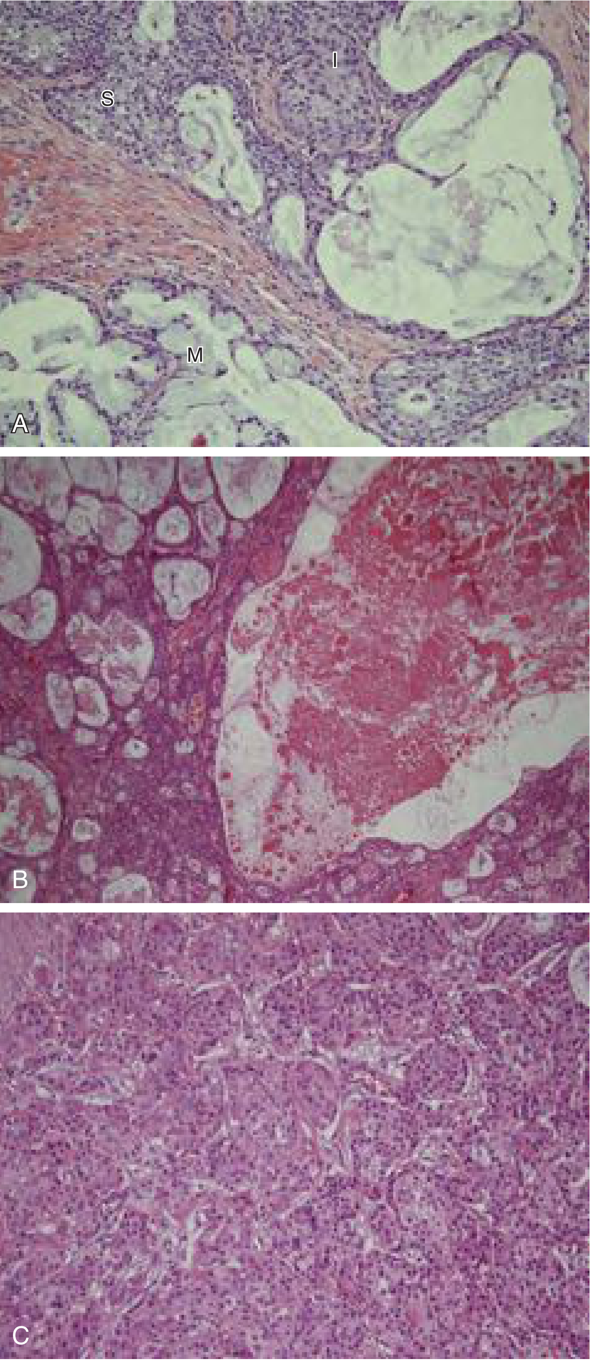
Definition: Mucoepidermoid Carcinoma (MEC) is the most common malignant salivary gland tumor. It is characterized histologically by three cell types: mucous cells, epidermoid (squamoid) cells, and intermediate cells.
Incidence: Most common salivary gland malignancy overall; most common salivary gland tumor in children.
Site:
- Parotid gland (most common major salivary gland site)
- Palate (most common minor salivary gland site)
- Mandible and maxilla (central/intraosseous MEC - odontogenic origin)
Age and Sex: Mean age ~45 years; slightly more common in females; also occurs in children.
Clinical Features:
- Painless, slow-growing mass
- May have bluish discoloration (cystic areas with mucinous content mimicking mucocele)
- Low-grade: soft, cystic consistency
- High-grade: firm, infiltrative, may involve facial nerve (parotid)
- Surface may ulcerate in high-grade tumors
Radiographic Features (for central MEC in jaw):
- Multilocular or unilocular radiolucency
- May show bony destruction in high-grade
Histopathological Features:
Three hallmark cell types:
- Mucous cells: Large, pale, vacuolated cytoplasm with peripheral nuclei; contain mucin (stain with PAS, mucicarmine, Alcian blue positive)
- Squamoid/Epidermoid cells: Large with abundant pink cytoplasm; resemble squamous cells but NO true keratinization (if keratinization present, consider adenosquamous carcinoma)
- Intermediate cells: Small, ovoid; modest pink or clear cytoplasm; predominate in most tumors
Architecture: Mixture of cystic and solid areas; mucous cells typically line cystic spaces.
Grading (AFIP grading system - Goode et al.):
| Grade | Features | Behavior |
|---|
| Low-grade | Predominantly cystic, many mucous cells, minimal atypia | Favorable |
| Intermediate-grade | Mix of cystic and solid | Intermediate |
| High-grade | Predominantly solid, few mucous cells, marked atypia, necrosis | Aggressive |
Molecular Marker: MAML2 gene rearrangement (t(11;19) translocation) - diagnostic marker
Treatment:
- Superficial parotidectomy for low-grade parotid tumors
- Total parotidectomy with nerve preservation for high-grade
- Neck dissection for high-grade or clinically N+ neck
- Postoperative radiotherapy for high-grade tumors
- Prognosis: Low-grade - excellent; High-grade - poor
Mucoepidermoid Carcinoma: (A) Three cell types - Intermediate (I), Mucous (M), Squamoid (S) cells (B) Tumor with cystic change (C) High-grade solid pattern
E. CANDIDIASIS (Oral Candidiasis)
Definition: Oral Candidiasis is a fungal infection of the oral mucosa caused primarily by Candida albicans, an opportunistic pathogen that is part of normal oral flora.
Etiology: Candida albicans (90%), also C. tropicalis, C. glabrata, C. krusei (in immunocompromised)
Predisposing Factors:
- Local: Xerostomia, denture wearing (especially ill-fitting), high-carbohydrate diet, smoking, poor oral hygiene, inhaled corticosteroids
- Systemic: Diabetes mellitus, immunosuppression (HIV/AIDS, chemotherapy), prolonged antibiotic therapy, infancy/old age, malnutrition, pregnancy
- Drugs: Corticosteroids, immunosuppressants, broad-spectrum antibiotics
Classification (Lehner 1966 / Holmstrup & Axell 1990):
PRIMARY (confined to oral mucosa):
- Acute forms:
- Acute pseudomembranous candidiasis (Thrush)
- Acute atrophic (erythematous) candidiasis
- Chronic forms:
- Chronic pseudomembranous candidiasis
- Chronic atrophic candidiasis (Denture stomatitis)
- Chronic hyperplastic candidiasis (Candidal leukoplakia)
- Candida-associated lesions:
- Denture stomatitis (Type I, II, III)
- Angular cheilitis (perlèche)
- Median rhomboid glossitis
SECONDARY (manifestation of systemic candidiasis):
- Oral lesions in systemic/mucocutaneous candidiasis
Clinical Features:
Acute Pseudomembranous (Thrush):
- White creamy, cottage-cheese-like plaques
- Can be wiped off to reveal erythematous/bleeding base
- Sites: Buccal mucosa, tongue, palate, oropharynx
- Burning sensation
Acute Atrophic (Antibiotic sore mouth):
- Erythematous, painful, depapillated tongue/mucosa
- Red, sore, burning; no white plaques
- After antibiotic therapy
Chronic Atrophic (Denture stomatitis):
- Erythematous mucosa under denture
- Three types (Newton): Simple hyperemia → Granular/papillary hyperplasia → Inflammatory papillary hyperplasia
- Usually asymptomatic
Chronic Hyperplastic (Candidal Leukoplakia):
- White patches that cannot be wiped off (unlike thrush)
- Firm, leathery texture
- Most common site: Commissures of mouth, anterior tongue
- Higher malignant potential (~15%) - especially speckled type
Angular Cheilitis:
- Erythema, fissuring, crusting at angles of mouth
- Often associated with Staph. aureus and C. albicans
Histopathological Features:
- PAS stain or Gram stain reveals Candida hyphae (pseudohyphae and blastospores/yeast forms) penetrating the upper layers of epithelium
- Hyphae are perpendicular to the epithelial surface
- Hyperparakeratosis
- Intraepithelial and subepithelial neutrophilic microabscesses (Munro's microabscesses in the parakeratotic layer)
- Subepithelial chronic inflammatory infiltrate (lymphocytes, plasma cells)
- In hyperplastic candidiasis: acanthosis, dysplasia may be present
Diagnosis:
- Smear (KOH/PAS stain): Shows hyphae and spores
- Culture on Sabouraud's dextrose agar
- Biopsy (especially for hyperplastic type)
Treatment:
- Topical: Nystatin (suspension, lozenges), Clotrimazole
- Systemic: Fluconazole (first choice for systemic/resistant cases), Itraconazole, Amphotericin B
- Treat predisposing factors
- For denture stomatitis: Clean and soak dentures in antifungal solution; Correct ill-fitting dentures
F. HYPERPARATHYROIDISM
Definition: Hyperparathyroidism is a condition characterized by excessive secretion of parathyroid hormone (PTH), leading to hypercalcemia and metabolic bone disease.
Types:
- Primary: Adenoma (most common, 85%), hyperplasia, or carcinoma of parathyroid gland
- Secondary: Compensatory hyperparathyroidism due to chronic renal failure or malabsorption (hypocalcemia)
- Tertiary: Autonomous hyperparathyroidism developing from long-standing secondary HPT
Effects of Excess PTH:
- Bone resorption (via osteoclastic activity) → hypercalcemia
- Increased renal calcium reabsorption
- Increased phosphate excretion (hypophosphatemia)
- Increased intestinal calcium absorption (via 1,25-dihydroxyvitamin D)
Clinical Features:
"Bones, Stones, Groans, Psychic Moans"
- Bones: Bone pain, pathological fractures, osteitis fibrosa cystica (von Recklinghausen's disease of bone)
- Stones: Renal calculi (nephrolithiasis)
- Groans: GI symptoms (nausea, vomiting, constipation, peptic ulcer)
- Psychic Moans: Psychiatric manifestations (depression, anxiety, psychosis)
Oral/Jaw Manifestations:
- Loss of lamina dura - early sign; lamina dura becomes thin, indistinct, or absent on radiograph
- Brown tumor (Giant cell tumor of HPT): Osteoclast-rich lesion; appears as radiolucency in jaw; histologically identical to giant cell granuloma
- Jaw enlargement due to multiple brown tumors
- "Ground glass" appearance of bone on radiograph
- Tooth mobility due to bone loss
- Generalized osteoporosis of jaw bones
Brown Tumor (Central Giant Cell Lesion/Osteoclastoma in HPT):
- Not a true neoplasm but represents focal accumulation of osteoclasts
- Radiograph: Multilocular radiolucency ("soap bubble"); may be unilocular
- Histology: Multinucleated giant cells (osteoclasts) on hemorrhagic fibrovascular stroma; hemosiderin deposits, new bone formation at periphery
- Distinguished from Giant Cell Granuloma by serum PTH levels
Lab Findings:
- Serum calcium: Elevated (hypercalcemia) - KEY FINDING
- Serum phosphate: Low (hypophosphatemia)
- Serum PTH: Elevated
- Serum alkaline phosphatase: Elevated (indicates bone turnover)
- Urine calcium: Elevated (hypercalciuria)
Radiographic Features of Jaw:
- Loss of lamina dura
- Ground glass bone (generalized)
- Brown tumors (multilocular/unilocular radiolucencies)
- Subperiosteal bone resorption (radial cortex of fingers is classic)
- "Salt and pepper" skull
Treatment:
- Primary: Parathyroidectomy (removal of adenoma/hyperplastic glands)
- Secondary: Treat underlying cause (renal failure); calcium/vitamin D supplementation
- Brown tumors regress after correction of hyperparathyroidism
G. REGRESSIVE ALTERATIONS OF TEETH
Definition: Regressive changes/alterations of teeth refer to the group of conditions characterized by loss or regression of the hard tissue of teeth, occurring as a result of physiological aging, functional wear, or pathological processes.
Classification:
1. ATTRITION
- Def: Physiological wearing away of tooth structure as a result of tooth-to-tooth contact during mastication and parafunction
- Types: Occlusal attrition, proximal attrition
- Features: Flat, shiny, polished wear facets; affects occlusal/incisal surfaces; dentin exposed gives yellow-brown stain; pulp may recede (secondary dentin formation)
- Histology: Increased secondary dentin, pulp recession, reparative dentin
2. ABRASION
- Def: Pathological wearing away of tooth structure by friction of a foreign substance
- Causes: Toothbrush abrasion (most common), bruxism with abrasive foods, pipe smoking, occupational habits
- Features: V-shaped/wedge-shaped notches at cervical region (toothbrush); groove-like defects; affects labial and buccal surfaces preferentially
- Histology: Smooth, polished surface; dentin exposed; secondary dentin formation
3. EROSION (Corrosion)
- Def: Irreversible loss of tooth structure by chemical process without bacterial involvement
- Causes:
- Extrinsic: Acidic food/drinks (carbonated beverages, citrus fruits, wine), acidic occupation
- Intrinsic: Gastroesophageal reflux disease (GERD), chronic vomiting (bulimia), rumination
- Features: Cup-shaped depressions on occlusal surfaces (cupping); smooth, glossy appearance; no plaque deposits; affects palatal surface of maxillary anteriors in intrinsic erosion
- Histology: Loss of enamel prisms, exposed dentin, smooth surface without scratches
4. ABFRACTION
- Def: Loss of tooth structure from mechanical loading forces causing flexion and fatigue of enamel prisms at the cervical region
- Mechanism: Lateral occlusal forces → tooth flexure → stress concentration at cervical fulcrum → enamel fracture
- Features: V-shaped notches at cervical region (like abrasion); occurs in absence of toothbrush use; subgingival location possible
- Related to parafunction (bruxism, clenching)
5. RESORPTION OF TEETH
Types:
- Internal Resorption (Pink spot): Odontoclastic resorption from within the pulp chamber; presents as pink spot through the crown; radiograph shows enlarged pulp canal/chamber
- External Resorption:
- External apical root resorption (orthodontic treatment, periapical inflammation)
- External cervical/lateral root resorption
- Inflammatory external resorption (trauma, infection)
- Replacement resorption (ankylosis - root replaced by bone)
6. HYPERCEMENTOSIS
- Excessive deposition of cementum on root surface
- Causes: Paget's disease, periapical inflammation, occlusal trauma, idiopathic
- Radiograph: Thickened root, bulbous root apex, PDL space maintained
7. PULPAL CHANGES (Regressive)
- Secondary dentin deposition (most common protective response)
- Pulp stones (denticles) - true and false
- Diffuse calcification ("pulp canals obliterated")
- Pulp atrophy (reduced cellularity and vascularity with age)
H. VERRUCOUS CARCINOMA
Definition: Verrucous Carcinoma is a low-grade variant of oral squamous cell carcinoma characterized by its exophytic, warty (verrucous), slowly growing nature, local invasion without metastasis (rare), and distinct histological features.
Synonym: Ackermann's tumor (described by Ackermann, 1948); "Oral florid papillomatosis" (older term)
Etiology:
- Tobacco use (especially smokeless/chewing tobacco - most common)
- Alcohol
- Betel nut chewing
- HPV infection (especially HPV-16 and HPV-18) - debated role
- Chronic irritation
Epidemiology:
- Age: 6th-7th decade; rare before 50 years
- Sex: Males predominantly (tobacco habit)
- Site: Buccal mucosa (most common), gingiva, alveolar mucosa, hard palate, tongue
Clinical Features:
- Exophytic, slow-growing, warty/papillary/pebbly white-gray mass
- Well-circumscribed borders
- Rough, corrugated surface with multiple white projections
- Broad base (sessile)
- "Shaggy," cauliflower-like surface
- Painless initially; may cause mild discomfort
- Bleeds easily on contact
- Does NOT typically ulcerate (vs conventional SCC)
- Locally destructive (invades soft tissue and bone)
- Lymph node metastasis is RARE (distinguishes from conventional SCC)
- May undergo transformation to conventional SCC (hybrid verrucous carcinoma) - especially after radiotherapy
Radiographic Features (when bone invasion present):
- Ill-defined bone destruction
- May mimic osteomyelitis
Histopathological Features:
KEY - requires FULL BIOPSY (not just superficial biopsy) for diagnosis:
- Exophytic, papillary/verrucous surface with thick, broad, blunt-ended, pushing rete ridges (not thin, infiltrative pegs)
- Parakeratin plugging between surface projections (crypts filled with keratin) - "church-spire"-like projections
- Well-differentiated squamous epithelium with minimal cytological atypia and NO significant nuclear pleomorphism
- Pushing/compressive border rather than infiltrative margin - "pushing invasion"
- Dense chronic inflammatory infiltrate at the base (tumor-stroma interface)
- NO single cell infiltration or desmoplasia (unlike conventional SCC)
- Broad, blunt invasive front extending into connective tissue like "elephant feet"
- Basement membrane usually intact
Diagnostic Challenge: Superficial biopsy may show only verrucous hyperplasia or hyperkeratosis; deep biopsy including the base is essential.
Differential Diagnosis:
- Verrucous hyperplasia (no invasion)
- Papilloma
- Conventional OSCC (cytological atypia, infiltrative borders, metastasis)
- Proliferative verrucous leukoplakia
Treatment:
- Wide surgical excision (treatment of choice)
- Radiotherapy: Controversial (risk of "anaplastic transformation" to conventional SCC post-radiation is debated)
- Chemotherapy: Used in unresectable cases or in combination
- Prognosis: Excellent if adequately excised; 5-year survival ~85-90%
I. IRON DEFICIENCY ANEMIA
Definition: Iron Deficiency Anemia (IDA) is the most common type of anemia worldwide, characterized by decreased hemoglobin due to inadequate iron stores, resulting in microcytic, hypochromic red blood cells.
Etiology:
- Inadequate intake (dietary deficiency, malnutrition)
- Increased demand (pregnancy, childhood growth)
- Malabsorption (celiac disease, gastrectomy, achlorhydria)
- Chronic blood loss (GI bleeding - most common cause in adults: peptic ulcer, colorectal cancer, hookworm; menorrhagia in women)
Clinical Features:
General:
- Fatigue, weakness, pallor
- Tachycardia, exertional dyspnea
- Headache, dizziness, irritability
- Cold intolerance
- Pica (craving for non-food substances: clay, ice - pagophagia)
Specific Signs:
- Koilonychia (spoon-shaped nails) - pathognomonic
- Angular cheilitis (angular stomatitis) - fissuring at corners of mouth
- Pallor of conjunctiva, skin, mucous membranes
Oral Manifestations (Important for BDS):
- Pallor of oral mucosa - pale, washed-out appearance
- Atrophic glossitis (Bald/Smooth tongue): Depapillation of filiform and fungiform papillae; smooth, shiny, red/pale tongue
- Burning sensation of tongue (glossodynia, glossopyrosis)
- Angular cheilitis - bilateral fissuring at mouth corners
- Recurrent aphthous ulcers - more frequent
- Candidal infections - increased susceptibility due to mucosal atrophy
- Oral mucosa pale and atrophic
Plummer-Vinson Syndrome (Patterson-Kelly Syndrome):
- Triad of: Iron deficiency anemia + Dysphagia + Postcricoid web (esophageal web)
- "Sideropenic dysphagia"
- Predominantly women in middle age
- Precancerous: Associated with development of post-cricoid carcinoma and oral SCC
- Additional features: Koilonychia, atrophic glossitis, angular cheilitis
Laboratory Findings:
| Parameter | IDA |
|---|
| Hemoglobin | Decreased |
| MCV (Mean Cell Volume) | Decreased (Microcytic) |
| MCH | Decreased (Hypochromic) |
| MCHC | Decreased |
| Serum Iron | Decreased |
| Serum Ferritin | Decreased (earliest marker) |
| TIBC (Total Iron Binding Capacity) | Increased |
| Peripheral smear | Microcytic, hypochromic RBCs, pencil cells, target cells |
Histopathology of Oral Mucosa:
- Atrophic squamous epithelium (reduced thickness)
- Loss of epithelial rete ridges
- Loss of papillae (tongue)
- Subepithelial chronic inflammatory infiltrate
- Vascular dilation
Treatment:
- Oral ferrous sulfate (most common: 325 mg TDS for 3-6 months)
- Treat underlying cause
- Dietary iron supplementation
- Parenteral iron (IV iron) if oral not tolerated
- Blood transfusion in severe cases
J. HISTOPATHOLOGY WITH DIAGRAMS
J(i). CALCIFYING ODONTOGENIC CYST (COC) - Gorlin Cyst
Definition: Calcifying Odontogenic Cyst (COC) is a rare developmental odontogenic cyst characterized by the presence of ghost cells (calcified cells that have lost their nuclei but retain their outlines) and calcification within the cyst lining.
Synonym: Gorlin cyst (described by Gorlin, 1962); Dentinogenic Ghost Cell Tumor (neoplastic form)
Classification (WHO 2022):
- Cystic variant: COC (Calcifying Odontogenic Cyst)
- Solid/neoplastic variant: Dentinogenic Ghost Cell Tumor (DGCT)
Clinical Features:
- Age: Bimodal distribution (2nd decade and over 70 years)
- Sex: Equal; slight male predilection
- Site: Mandible > Maxilla; anterior jaw (incisor/canine region most common)
- Painless, slow-growing swelling
- May be associated with unerupted tooth (30%)
- May be associated with other odontogenic tumors (especially odontoma, 24%)
Radiographic Features:
- Well-defined unilocular (occasionally multilocular) radiolucency
- Calcification within the cyst - variable; from very faint flecks to extensive opacities
- May show mixed radiolucent-radiopaque pattern
- Associated tooth displacement or root divergence
HISTOPATHOLOGICAL FEATURES (Most Important for Exam):
-
Cyst Lining: Stratified squamous epithelium resembling ameloblastomatous epithelium
- Basal layer: Palisaded, columnar cells resembling ameloblasts (tall cells with reverse polarity)
- Suprabasal cells: Loosely arranged, similar to stellate reticulum
-
GHOST CELLS (Pathognomonic Feature):
- Large, pale eosinophilic cells with distinct cell outlines but no nuclei (nucleus has dissolved - "ghost")
- May be present singly or in clusters
- Represent aberrant keratinization (abortive enamel matrix formation)
- May undergo calcification (become mineralized ghost cells)
- May form sheets/masses of ghost cells
-
Calcification:
- Ghost cells may calcify (dystrophic calcification)
- Dentinoid (dysplastic dentin/tubular dentin matrix) may be found adjacent to ghost cell masses
- Calcification may project into connective tissue wall
-
Fibrous Connective Tissue Wall:
- Dense fibrous wall
- Foreign body giant cell reaction around ghost cells that penetrate the wall
- Inflammatory cells (lymphocytes, plasma cells)
- Odontogenic epithelial rests may be present
-
Associated Structures:
- Enamel, dentin, or odontoma-like structures may be found (compound or complex odontoma in 24%)
SCHEMATIC DIAGRAM OF COC HISTOLOGY:
LUMEN OF CYST
        ↑
Ghost cells (pale, anucleate, eosinophilic)
        |
Stellate reticulum-like cells (loosely arranged)
        |
Palisaded columnar basal cells (like ameloblasts)
        |
Basement membrane
        ↓
FIBROUS CONNECTIVE TISSUE WALL
(with calcifications, dentinoid, foreign body giant cells)
Treatment:
- Enucleation and curettage (for cystic form - low recurrence)
- The neoplastic DGCT form requires wider excision
- Recurrence rate: Low for COC (< 5%)
J(ii). ADENOID CYSTIC CARCINOMA (AdCC) - Histopathology
Definition: Adenoid Cystic Carcinoma is a malignant salivary gland tumor characterized by slow, relentless progression, perineural invasion, late metastasis, and high recurrence. Histologically defined by three growth patterns.
Site: Equal distribution across all salivary gland sites; parotid, submandibular, palate (minor salivary glands)
Age/Sex: Peak 50-60 years; equal sex distribution
HISTOPATHOLOGICAL FEATURES (Three Growth Patterns):
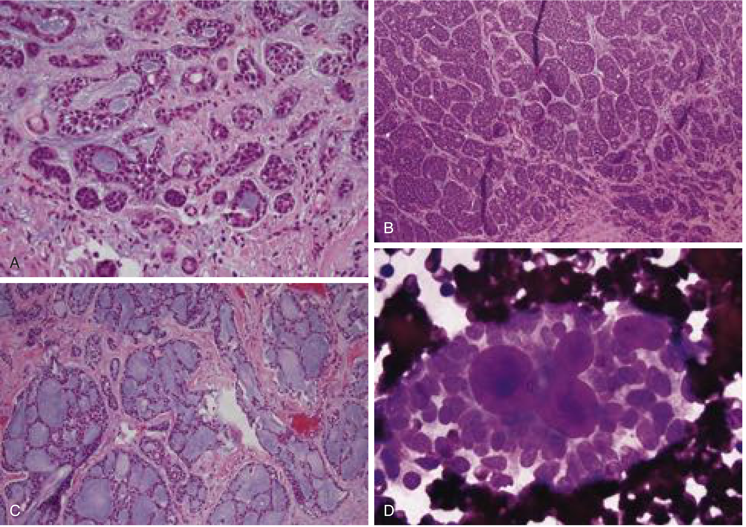
1. CRIBRIFORM PATTERN (Most Common/Classic):
- "Swiss cheese" appearance
- Nests/islands of basaloid cells arranged around multiple cylindrical pseudocysts (gland-like spaces)
- The spaces contain PAS-positive blue or pink material (replicated basement membrane/ground substance - NOT true mucin)
- This is extracellular matrix, NOT epithelial mucin
- Cells: Small, dark, hyperchromatic nuclei; scant cytoplasm; uniform, monotonous
2. TUBULAR PATTERN (Grade I - Best Prognosis):
- Small tubular/duct-like structures
- Double-layered tubules: Inner true luminal cells (small glandular spaces) + outer myoepithelial cells
- Stroma: Pink, hyalinized, hypocellular
- Cells: Small, basaloid; dark, hyperchromatic nuclei; minimal cytoplasm
3. SOLID PATTERN (Grade III - Worst Prognosis):
- Rounded lobules/nests of basaloid cells with NO gland-like structures
- High mitotic activity
- Necrosis may be present
- No characteristic architecture
- Worst prognosis; highest recurrence and metastasis
Grading:
- Grade I: Predominantly tubular (+ some cribriform); best prognosis
- Grade II: Predominantly cribriform (+ some tubular)
- Grade III: Predominantly solid (>30% solid); worst prognosis
KEY Histological Features:
- Basaloid cells with dark, hyperchromatic nuclei
- Scant cytoplasm
- Minimal nuclear pleomorphism ("deceptively bland")
- Perineural invasion (hallmark of AdCC - present in >60% cases)
- PAS-positive pseudo-luminal spaces (ground substance)
- Hyalinized stroma
- Myoepithelial cell component (clear cells at periphery of tubules)
SCHEMATIC DIAGRAM:
CRIBRIFORM PATTERN:
[Basaloid cells] — [Pseudo-lumen (PAS+ basement membrane material)] — [Basaloid cells]
Swiss-cheese appearance

TUBULAR PATTERN:
[Myoepithelial cells] → [True lumen (tiny glandular space)] → [Luminal cells]
                            Hyalinized pink stroma

SOLID PATTERN:
[Sheets of basaloid cells] — [Necrosis] — [High mitoses]
Perineural Invasion (Histological Feature):
- Tumor cells surround and infiltrate nerve fibers
- Responsible for: Pain, facial nerve paralysis (parotid), late recurrence along nerve track
Special Stains:
- PAS: Positive in pseudocysts (ground substance)
- Vimentin, S-100: Positive (myoepithelial cells)
- CD117 (c-KIT): Strongly positive - used in diagnosis
- MYB-NFIB gene fusion: Molecular marker (t(6;9) translocation)
Clinical Behavior:
- Slow but relentless growth
- Late hematogenous metastasis (to lung, bone, liver) - often years to decades later
- High local recurrence (even after complete excision)
- Perineural invasion leads to skip lesions and late recurrence
Prognosis:
- Despite slow growth, 10-year survival is only ~50%
- Grade III solid pattern: Significantly worse
- Submandibular gland AdCC: More aggressive than other sites
Treatment:
- Wide local excision with adequate margins
- Neck dissection for clinically positive nodes
- Postoperative radiotherapy (improves local control)
- Systemic therapy for metastatic disease (limited efficacy)
Adenoid Cystic Carcinoma: (A) Tubular pattern with hyalinized stroma (B) Solid pattern - rounded nests of basaloid cells (C) Cribriform (Swiss-cheese) pattern with PAS-positive pseudocysts (D) FNA showing acellular "cylinders" of basement membrane material
---All answers are complete. Here is a summary of what has been covered:
Quick Reference Summary for Your Exam
Long Essays:
| Q | Topic | Key Examiner Focus Points |
|---|
| Q1 | Odontogenic Cysts + Radicular Cyst | WHO classification, Rushton bodies, cholesterol clefts, corticated radiolucency at apex of non-vital tooth |
| Q2 | OPMDs + Oral Submucous Fibrosis | Arecoline mechanism, juxta-epithelial hyalinization, 4 histological stages, 7-13% malignant transformation |
Short Notes - Exam Tips:
| Topic | Must-Know Point |
|---|
| Bite Marks | ABFO scale, UV photography, DNA swabs first |
| Rootless Teeth | Dentin Dysplasia Type I; thistle-tube pulp on radiograph |
| Unicystic Ameloblastoma | Ackermann 3 types; Type 3 (mural) = worst prognosis |
| Mucoepidermoid Carcinoma | Most common salivary malignancy; 3 cell types; MAML2 translocation |
| Candidiasis | Pseudohyphae on PAS; Munro's microabscesses in hyperplastic type |
| Hyperparathyroidism | Bones/Stones/Groans/Moans; loss of lamina dura; brown tumor |
| Regressive Alterations | Attrition/Abrasion/Erosion/Abfraction/Resorption; wedge = abrasion, cupping = erosion |
| Verrucous Carcinoma | Ackermann's tumor; pushing border; parakeratin plugs; wide excision |
| Iron Deficiency Anemia | Plummer-Vinson syndrome (dysphagia triad); serum ferritin = earliest marker |
| COC | Ghost cells = PATHOGNOMONIC; PAS+ dentinoid; low recurrence |
| Adenoid Cystic Carcinoma | Swiss-cheese cribriform; perineural invasion; tubular = best, solid = worst |