
Internal capsule neuroanatomy 5m answer
Reading File
Finding Sources
Searching Images
internal capsule anatomy diagram limbs anterior posterior genu
Finding Sources
Reading File
Internal Capsule - Neuroanatomy (5-Minute Review)
What It Is
A V-shaped band of white matter that carries nearly all fibers passing between the cerebral cortex and the brainstem/spinal cord. It sits wedged between the major grey matter masses of the forebrain:
- Medial to it: caudate nucleus (head) and thalamus
- Lateral to it: lentiform nucleus (putamen + globus pallidus)
(Neuroanatomy through Clinical Cases, 3rd ed.)
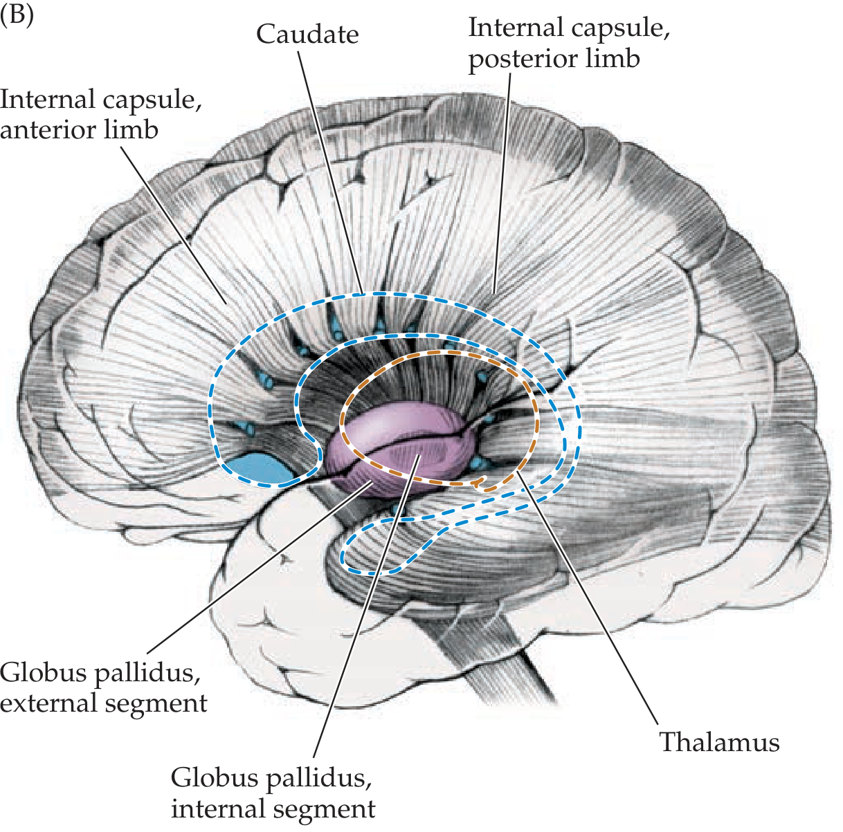
The 5 Parts (anterior to posterior)
| Part | Location | Key fibers carried |
|---|---|---|
| Anterior limb | Between caudate head (medial) and putamen (lateral) | Frontopontine fibers, anterior thalamic peduncle (thalamus <-> frontal lobe) |
| Genu | The "bend," at the level of the interventricular foramen | Corticobulbar tract (to cranial nerve motor nuclei) |
| Posterior limb | Between thalamus (medial) and putamen/globus pallidus (lateral) | Corticospinal tract (motor), corticorubral fibers, thalamocortical sensory radiations (touch/proprioception to parietal lobe) |
| Retrolenticular part | Behind the lentiform nucleus | Optic radiations (visual), parieto-occipito-pontine fibers |
| Sublenticular part | Below the lentiform nucleus | Auditory radiations, temporopontine fibers |
- Corticospinal fibers occupy roughly the posterior 2/3 to posterior half of the posterior limb, with a somatotopic organization (face fibers most anterior, leg fibers most posterior) - Localization in Clinical Neurology, 8e.
Blood Supply (why it matters clinically)
- Anterior limb + genu: lenticulostriate branches of the middle cerebral artery (MCA), and the recurrent artery of Heubner (branch of ACA) for part of the anterior limb.
- Posterior limb: lateral lenticulostriate arteries (MCA) and the anterior choroidal artery.
These are small, deep perforating vessels prone to lipohyalinosis/microatheroma, which is why the internal capsule is a classic site for lacunar infarcts.
Clinical Correlation
- A small lacunar stroke in the posterior limb can cause a dense pure motor hemiparesis (face, arm, leg all involved) with no cortical signs (no aphasia, neglect, or visual field cut) - because all descending motor fibers are compressed into this small space.
- Larger capsular hemorrhages (classically from small vessel rupture) produce contralateral hemiplegia and can progress to decorticate rigidity - Ganong's Review of Medical Physiology, 26th ed.
- Thalamic hemorrhage extending into the posterior limb can also produce contralateral ataxia by disrupting thalamocortical/cerebellar-relay fibers - Localization in Clinical Neurology, 8e.
- Because fibers are so tightly packed, a lesion the size of a pea in the internal capsule causes far more extensive deficits than an equivalent-sized cortical lesion.
High-Yield One-Liner
"Motor fibers in front (genu = face, posterior limb = body), sensory fibers behind them, visual/auditory fibers at the very back (retro/sublenticular) - and it's fed by tiny vessels that love to clot or bleed."
This is a shared conversation. Sign in to Orris to start your own chat.