
Hi, Ares. How are you?
Uh, Aris, can you do me a favor? Like, I want something, a PDF that should be made of approximately 200 to 400 pages, which will include basic and advanced mind maps, mnemonics, uh, mind maps with a creative color printing, and a lot of images, presentation to make that PDF very, very much interesting. And it's a PDF of... Based upon the current USMLE book, First Aid 1 and 2, including all the 19 subjects, basically, I want a PDF regarding the PYQs, PYTs of NEET PG, as well as INEICET, covering all the important topics with a proper explanation, but not in a very broad way, based upon the mind maps, based upon mnemonics, based upon, uh, very attractive images, and, uh, uh... Very, very attractive and presentable PDF of approximately 200 to 400 pages that will consider it a very good PDF, so that it will be helpful in NEET PG as well as INEICET pattern, based upon PYQs and PYT topics, make it so, so attractive, so that I can get a clear image of each and every part of 19 subject in a very good way, including the better and the best images in a way, including the labeling. Everything should be presentable in a proper mind maps, tables, flow charts, mnemonics that is necessary wherever you feel required. Uh, taking some good, uh... Perception of the first aid, step one and step two book. That is very, very good in this regard, mnemonics and flowcharts and table. Or you can use based upon, uh, previous year topics and, uh, questions of NEET PG and INESET, uh, for my 2026, uh, NEET PG and INESET so that... Uh, it will be more presentable and I can read it as many as times because it is looking so attractive based upon seeing that PDF with the images, mnemonics, flowcharts, mind maps, everything you find it good. Uh, can you do that?
Given the huge scope (19 subjects, 200-400 pages), how do you want to proceed? · Which subject should I build FIRST as the pilot chapter, to set the mind-map/mnemonic/image style before we scale to the rest? · What depth level do you want per topic?
-
Approach: Given the huge scope (19 subjects, 200-400 pages), would you rather I (a) build one subject at a time as a pilot first then continue across sessions, (b) attempt a condensed single-pass covering all 19 subjects now with shorter depth per subject, or (c) start with just a few high-yield subjects you pick?
-
Pilot subject: Which subject should I build first to set the mind-map/mnemonic/image style (e.g., Pharmacology, Pathology, Microbiology, Physiology, Anatomy, Biochemistry)?
-
Depth level: Do you want (a) ultra high-yield/crisp with max visuals, (b) moderate - short explanation + mnemonic + mind map + image per topic, or (c) detailed with mechanism/clinical correlation plus visuals?
Create a condensed single-pass covering all 19 subjects now
brachial plexus anatomy diagram labeled
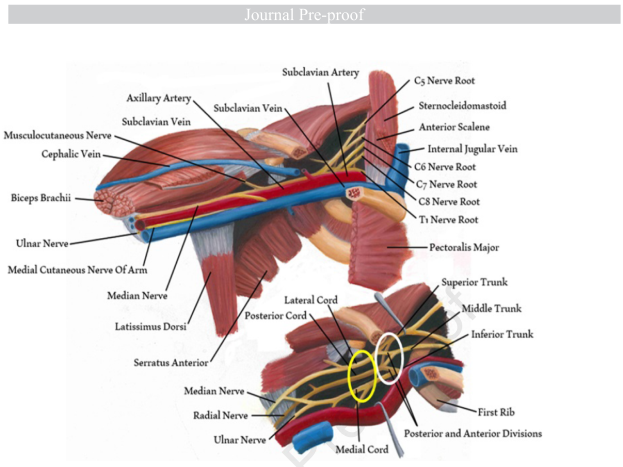
Summary : This anatomical illustration shows the brachial plexus and its relationship to surrounding muscles, arteries, and veins in the upper thoracic and shoulder region. It is a labeled diagram highlighting the major nerves, blood vessels, and muscle groups relevant to the anatomy of the brachial plexus.
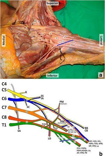
This composite educational resource details the anatomy and innervation of the brachial plexus. Panel (a) is a cadaveric dissection of the left axillary region. Key structures are labeled numerically: the median nerve (1), lateral and medial roots of the median nerve (2, 3), ulnar nerve (4), axillary artery (5), and axillary vein (6), alongside the medial cutaneous nerves of the arm (7) and forearm (8). In the supraclavicular region, the upper (9), middle (10), and lower (11) trunks are identified. Red and blue lines indicate proximal and distal bounds of a clinical lesion site. Panel (b) is a color-coded schematic diagram illustrating the brachial plexus architecture from roots (C5-T1) to terminal branches. It maps specific nerves to their target muscles, including the serratus anterior, deltoid, biceps brachii, and the intrinsic muscles of the hand. This content is designed for medical students and surgical residents to understand the relationship between neuroanatomical structures, their physiological territories, and the clinical correlation of axillary nerve injuries.
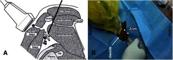
This dual-panel medical figure illustrates the anatomical and procedural approach for an ultrasound-guided posterior infraclavicular brachial plexus block. Panel A is a labeled anatomical diagram depicting a parasagittal section of the shoulder medial to the coracoid process. Key structures include the clavicle, scapula, and ribs, with muscular landmarks such as the trapezius, supraspinatus, subscapularis, deltoid, and pectoralis major. The brachial plexus nerves (N) are shown clustered around the axillary artery (A) and vein (V), superficial to the pleura. An ultrasound probe is positioned anteriorly on the chest wall, while a block needle follows a posterior-to-anterior trajectory, passing between the clavicle and scapula towards the neurovascular bundle. Panel B is a clinical photograph showing the surface anatomy and procedural setup. It indicates the ideal needle insertion point located 2 cm posterior to the clavicle, serving as a landmark to ensure the needle clears the inferior surface of the bone to reach the infraclavicular space. This content is designed for anesthesiology training, focusing on regional anesthesia techniques and ultrasound-guided nerve blocks.
myocardial infarction gross pathology heart specimen
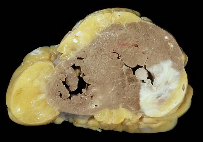
Gross autopsy photograph of a fixed human heart in cross-section, illustrating healed myocardial infarction involving the posterior and lateral left ventricular walls. The specimen demonstrates biventricular concentric hypertrophy with robust thickening of the left and right ventricles. The posterior papillary muscle shows scar tissue consistent with prior ischemic injury. The coronary arteries reveal atherosclerotic stenoses affecting the right coronary artery and the left coronary artery’s anterior interventricular branch (LAD), concordant with ischemic heart disease. Myocardial fibrosis replaces necrotic myocardium in infarcted regions, producing firm, pale areas against darker viable myocardium. The overall morphology supports a history of long-standing hypertensive cardiomyopathy with accompanying valvular insufficiency of the tricuspid and mitral valves, contributing to chronic hemodynamic burden. The image highlights key pathology: postinfarction scarring, papillary muscle involvement, and hypertrophic remodeling, which together predispose to functional mitral/tricuspid regurgitation and heart failure. This specimen is valuable for teaching gross anatomy, correlating macroscopic changes with coronary distribution and prior infarct patterns. Clinically, findings correlate with past hypertension, ischemic heart disease, and valvular disease; with autopsy context aiding diagnosis confirmation, research of myocardial remodeling, and education in cardiovascular pathology.
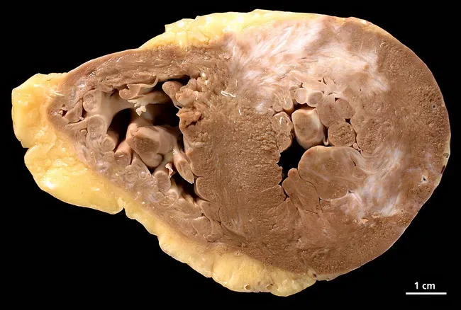
Gross pathology image of a human heart cross-section (short-axis/transverse) from a 73-year-old female with a history of arterial hypertension, chronic smoking, and three-vessel coronary artery disease. The specimen reveals chronic myocardial infarction scar throughout the myocardium with areas of subacute necrosis on both ventricles. The ventricles show concentric hypertrophy characterized by thickened ventricular walls and reduced cavity size, consistent with long-standing pressure overload. On cut surface, extensive fibrous replacement tissue is seen in the LV and RV walls, with pale, firm areas indicating scar tissue; endocardial surfaces may show thinning in infarcted zones. The presence of bilateral ventricular involvement suggests diffuse ischemic injury, possibly past infarcts with replacement fibrosis. The gross morphology corresponds to ischemic cardiomyopathy spectrum; pathophysiology includes atherosclerotic CAD, hypertension-induced hypertrophy, and chronic subendocardial ischemia. This combination results in remodeling and scar formation, predisposing to arrhythmias and heart failure. The image is from pathorama.ch, illustrating classic post-infarction changes: myocardial scar, subacute necrosis, and hypertrophic remodeling. Clinically, such pathology would correlate with prior chest pain episodes, elevated troponin in the past, and radiographic evidence of cardiomegaly; autopsy or pathology reporting would emphasize chronic infarct burden, LV dysfunction, and risk stratification for sudden cardiac death. Suitable for education on myocardial infarction progression and necrosis patterns.
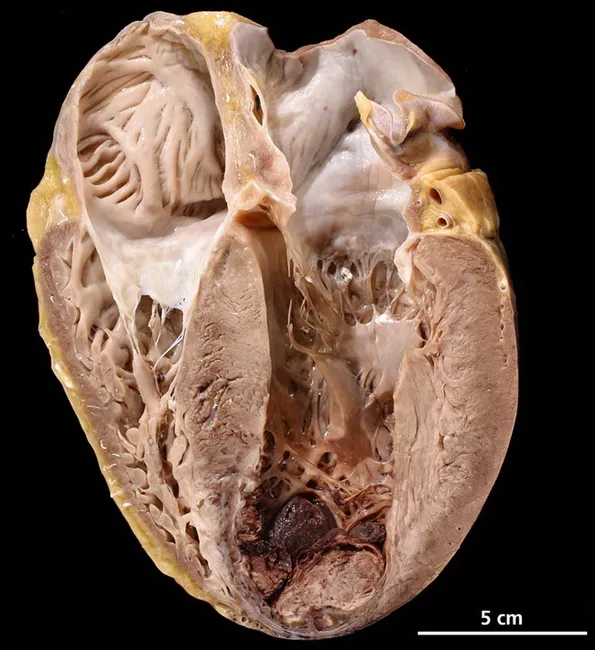
This gross pathology photograph documents an autopsy heart specimen with a longitudinal, apical view illustrating severe cardiovascular pathology underlying sudden death. Imaging modality: gross anatomy/necropsy photograph of a human heart, procured at autopsy, with a longitudinal/apical cross-section displaying the left ventricular apex. The heart exhibits marked biventricular hypertrophy with thickened LV walls and prominent endocardial surfaces. The anterior–apical septal region demonstrates features compatible with acute myocardial infarction, including pallor to dark discoloration and regional softening of myocardial tissue. A mural thrombus is evident at the LV apex, adherent to endocardial surface, presenting a risk for embolization. The coronary vasculature shows severe atherosclerotic narrowing in major epicardial vessels; although not visible in this specimen, the distribution is consistent with ischemic injury in the antero-apical-septal territory. The specimen is a postmortem evaluation, thus no in vivo contrast or staining was used. The scale bar indicates 5 cm for size reference. Clinical context includes obesity, diabetes mellitus type II, and tobacco use as major risk factors contributing to diffuse atherosclerosis and acute coronary occlusion. Diagnostic significance: confirms lethal ischemic heart disease with multivessel disease, supports sudden death due to acute MI with mural thrombus formation. Potential clinical applications include education, autopsy pathology reference, and radiology correlation.
gram positive gram negative bacteria stain classification
psoriasis skin lesion clinical photo
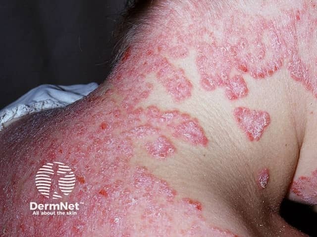
Clinical photography of facial skin demonstrates classic psoriatic plaques involving the malar region and surrounding cheeks. The lesions are erythematous to violaceous, well-demarcated, and markedly scaly with silvery-white scale, consistent with plaque-type psoriasis (psoriasis vulgaris). Involved areas show surface coarseness, thickening (acanthosis) and mild induration, while adjacent skin remains relatively spared. Perilesional inflammation creates a ring of erythema, and some plaques exhibit superficial fissuring. The distribution is facial rather than scalp, trunk, or limbs, suggesting involvement limited to the head-and-face region in this image. No vesiculation or purulence is evident. The appearance may mimic seborrheic dermatitis, eczema, or contact dermatitis; however, the well-circumscribed plaques with thick scale and the typical red-silver presentation strongly favor psoriasis. Skin photo shows high-contrast textural differences between lesional and non-lesional skin. Clinically, facial psoriasis can be challenging due to cosmetic and functional concerns, including itching and social impact. This image supports differential diagnosis discussions in dermatology education and serves as a reference for recognizing facial plaque psoriasis, assessing severity, and guiding treatment planning (topical steroids, vitamin D analogs, or phototherapy). It is useful for teaching photodocumentation, lesion morphology, and pattern recognition in psoriasis spectrum. This image thus enhances reasoning for diagnosis, prognosis, and management planning.
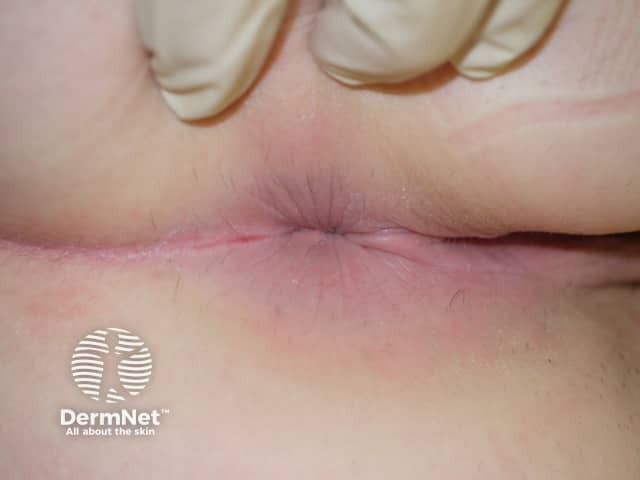
Clinical photograph of external genital skin showing an erythematous, well-demarcated plaque in an intertriginous fold. The lesion is pink to red, with a smooth surface and minimal peripheral scaling within moist skin areas; a slight fissure is seen along the crease. The surrounding skin is mildly inflamed but otherwise unremarkable. This appearance is characteristic of genital psoriasis, most compatible with inverse psoriasis in moist genital folds where scaling is less prominent than on extensor surfaces. Differential diagnoses include candidal balanitis or vulvitis, irritant contact dermatitis, lichen sclerosus, eczema, and less commonly erosive lesions. Clinical context such as past or concurrent psoriasis elsewhere, absence of itch or burning, and response to topical steroids can help confirmation. Histopathology would typically show epidermal hyperplasia with parakeratosis, regular elongation of rete ridges, and a dermal perivascular lymphocytic infiltrate if biopsy were performed; in practice, biopsy is not required for typical genital psoriasis. The diagnostic significance lies in recognizing genito-pubic psoriasis to guide therapy and avoid misdiagnosis as infection. This image is useful for education on genital psoriasis recognition, differential diagnosis, and management considerations including topical corticosteroids, vitamin D analogs, and calcineurin inhibitors, with referrals for systemic therapy if extensive disease. Clinical photo for reference.
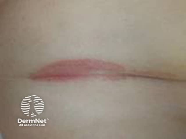
Clinical photography of the antecubital fossa region shows a single erythematous plaque within a flexural skin fold. The lesion is well-demarcated, with a smooth pink-to-erythematous surface and minimal scales evident, reflecting inverse/ flexural psoriasis features in moist environments where scaling is less conspicuous. The plaque lies centrally within the fold, with intact surrounding skin and no obvious crusting or excoriation. Lighting emphasizes a uniform coloration without effusion or ulceration. The regional skin may appear slightly edematous or glistening from moisture, a common appearance in intertriginous psoriasis. In clinical practice, such lesions are often symmetric or multipart and may co-occur with plaques on extensor surfaces; history of chronic plaque psoriasis or family history supports the diagnosis. Differential considerations include irritant dermatitis, candidal intertrigo, or seborrheic dermatitis; biopsy would reveal epidermal hyperplasia with parakeratosis and neutrophilic microabscesses typical of plaque psoriasis, though histology is not demonstrated here. The image is intended for dermatology education, aiding recognition of flexural psoriasis, differentiation from dermatitis, and guiding topical therapy, including high-potency corticosteroids or vitamin D analogs with occlusion, while avoiding excessive moisture. This photo supports image-based teaching, clinical pattern recognition, and correlation with patient history in practice and research.
chest x-ray pneumothorax
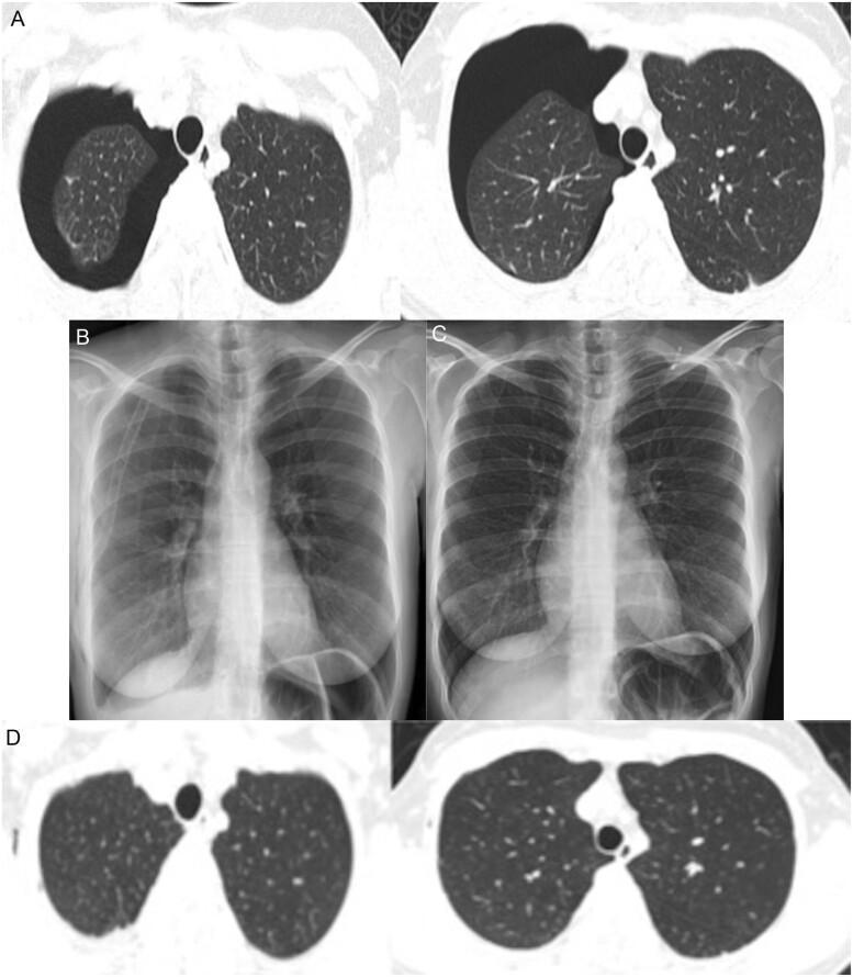
A multi-panel medical image sequence documenting the progression and resolution of a right-sided pneumothorax. Panel A presents axial Thorax Computed Tomography (TCT) lung window slices showing a significant right pneumothorax, characterized by a large, dark, air-filled pleural space (radiolucency) causing collapse of the right lung toward the mediastinum; the left lung remains fully expanded with normal density. Panel B is a Posteroanterior (PA) chest X-ray taken after tube thoracostomy, showing the right lung successfully re-expanded with the chest tube in situ. Panel C displays a follow-up chest X-ray after removal of the chest tube on the fifth day, maintaining full lung expansion and clear costophrenic angles. Panel D shows a follow-up axial TCT image two weeks post-discharge, confirming complete resolution of the pneumothorax with bilateral symmetric lung expansion and normal lung parenchyma. This sequence illustrates the clinical management of a spontaneous pneumothorax in the context of COVID-19 infection, from acute presentation to complete recovery.
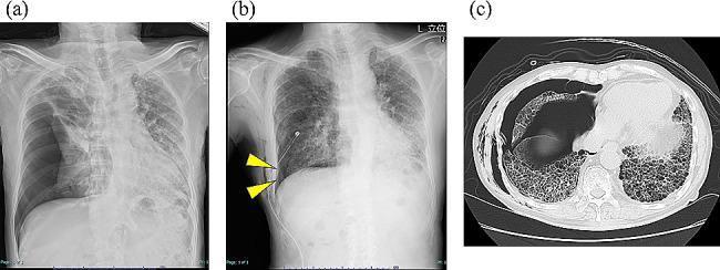
Educational clinical image set documenting a secondary pneumothorax in a patient with chronic hypersensitivity pneumonitis. Panel (a) is a posterior-anterior (PA) chest X-ray showing a large right-sided tension pneumothorax with significant mediastinal shift to the left and complete collapse of the right lung. Panel (b) is a follow-up chest X-ray post-thoracic drainage showing a visible chest tube and a persistent, mild residual pneumothorax cavity (indicated by yellow arrowheads) along the right lateral chest wall. Both radiographs reveal underlying diffuse reticular opacities consistent with interstitial lung disease. Panel (c) is an axial non-contrast chest CT scan (lung window) demonstrating extensive bilateral subpleural honeycombing and ground-glass opacities characteristic of advanced pulmonary fibrosis. The CT also captures the collapse of the right middle and lower lobes and an associated pleural air space. The series demonstrates the complication of intractable air leak in the context of advanced fibrotic lung disease.
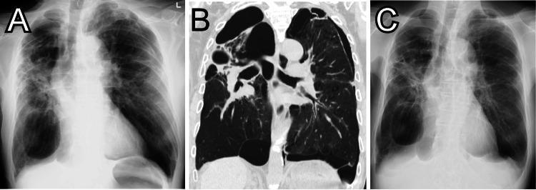
This composite figure demonstrates diagnostic imaging findings of bullous lung disease and pneumothorax in a patient with COPD. Image A is a frontal chest X-ray on presentation showing multiple large, thin-walled, hyperlucent areas consistent with giant bullae, particularly prominent in the lower zones, and a concomitant mild left-sided pneumothorax identified by a peripheral lucency and lack of lung markings. Image B is a coronal reformatted CT scan of the chest, providing high-resolution visualization of the lung parenchyma. It reveals extensive emphysematous changes and multiple, well-defined, thin-walled air-filled bullae of varying sizes occupying significant portions of both hemithoraces, with clear demarcation between the bullae and remaining compressed lung tissue. Image C is a follow-up chest X-ray taken 10 days later, showing stable appearance of the giant bullae but interval improvement and resolution of the previously seen left-sided pneumothorax, with better expansion of the underlying lung. The series highlights the importance of distinguishing between giant bullae and pneumothorax in patients with advanced obstructive lung disease.
diabetic retinopathy fundus photo
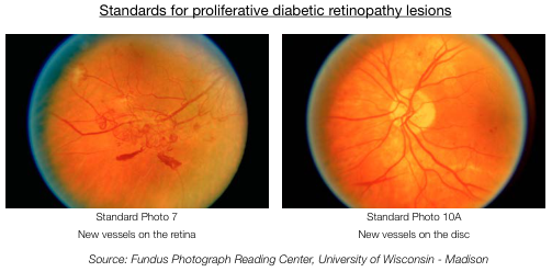
Summary : This figure presents standard fundus photographs used to identify proliferative diabetic retinopathy lesions, specifically highlighting new vessel formation on the retina and optic disc.
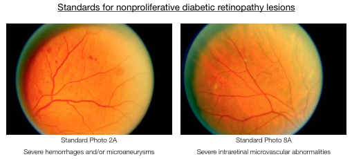
Summary : This figure presents two standard retinal photographs used to define nonproliferative diabetic retinopathy lesions, specifically severe hemorrhages/microaneurysms and severe intraretinal microvascular abnormalities.
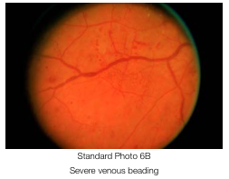
Summary : This image shows a retinal photograph demonstrating severe venous beading, used as a standard reference in ophthalmology.
tympanic membrane perforation otoscopy
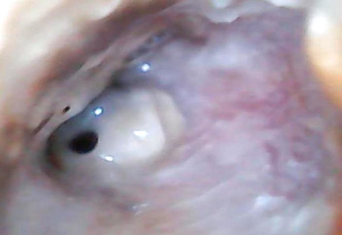
This clinical photograph displays an otoscopic view of the right ear, highlighting pathological findings of the external auditory canal (EAC) and tympanic membrane. The EAC shows significant inflammatory changes, characterized by diffuse erythema and edema of the canal walls. Visible purulent secretions are present within the lumen, suggesting an active infectious process such as chronic suppurative otitis media or acute exacerbation. The tympanic membrane is clearly visualized and exhibits a single, well-defined perforation located in the anterior-inferior quadrant. The perforation appears as a dark, circular opening amidst the remaining translucent, light-reflective portions of the membrane. Normal landmarks like the handle of the malleus are obscured by the surrounding inflammatory tissue and exudate. This image serves as a clinical example of suppurative middle ear disease with associated tympanic membrane rupture and secondary external canal inflammation, relevant for otolaryngology training in diagnostic otoscopy.
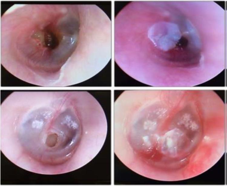
A comparison chart consisting of four video-otoscopy clinical photographs organized into two rows. The left column displays pre-treatment views of the tympanic membrane in two different patients, each exhibiting a clear central perforation. In the top left image, a medium-sized irregular perforation is visible with mildly inflamed margins. In the bottom left image, a smaller, circular, well-defined central perforation is seen in a membrane showing signs of myringosclerosis (white calcific plaques). The right column shows the same ears post-procedure, demonstrating the temporary closure of the defects using a translucent Bionext® cellulose film. The film is visible as an opaque-to-translucent patch covering the previous perforation sites, effectively sealing the middle ear space from the external auditory canal. This visual comparison illustrates a conservative clinical approach to managing symptomatic tympanic membrane perforations to assess immediate functional and symptomatic improvement before considering permanent surgical repair like myringoplasty.
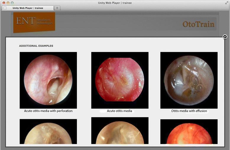
A comparison chart of otoscopic diagnostic images from a medical simulator, focusing on pathologies of the tympanic membrane (TM). The visual contains three labeled primary circular otoscopic views: 1) 'Acute otitis media with perforation,' showing an inflamed, erythematous TM with a large, irregular central perforation through which the middle ear space is visible; 2) 'Acute otitis media,' depicting an intensely hyperemic, opaque, and bulging TM with loss of normal landmarks and light reflex; and 3) 'Otitis media with effusion,' illustrating a translucent, amber-colored TM with visible air-fluid levels and bubbles in the middle ear space. These clinical photographs serve as educational examples for ENT trainees to differentiate between infectious stages (AOM) and non-purulent fluid accumulation (OME). The interface includes secondary supportive images below each category to demonstrate variability in presentation. This content is designed for medical students and residents to master diagnostic otoscopy and middle ear pathology recognition.
colles fracture wrist x-ray
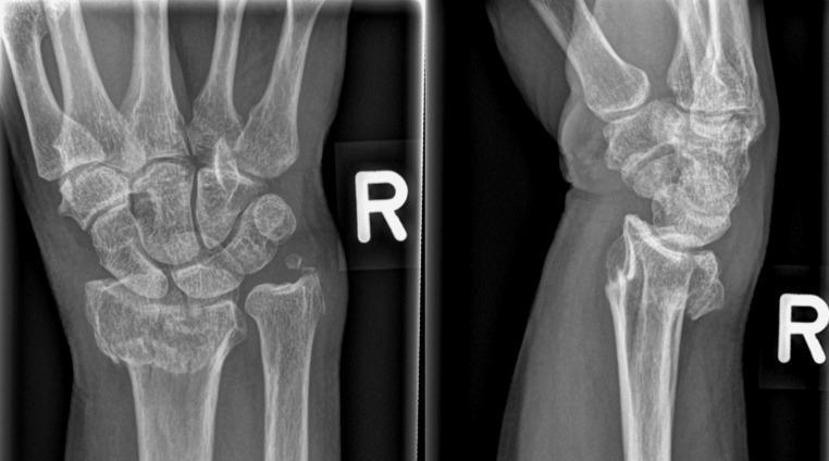
This diagnostic image consists of a two-view conventional radiograph (X-ray) of the right wrist, presenting both anteroposterior (AP) and lateral views. The image demonstrates a distal radius fracture with significant displacement. In the AP view (left), a transverse fracture line is visible across the distal metaphysis of the radius, accompanied by an associated fracture of the ulnar styloid process. The lateral view (right) clearly illustrates dorsal (posterior) displacement and dorsal angulation of the distal radial fragment, characteristic of a Colles-type fracture pattern. There is a disruption of the normal radiocarpal alignment, with the carpal bones following the dorsally displaced distal radius segment. Key anatomical structures visible include the distal radius and ulna, the proximal and distal carpal rows, and the bases of the metacarpals. This visual material is primarily used in orthopedic and emergency medicine education to teach fracture identification, terminology for displacement and angulation, and the assessment of wrist joint congruity.
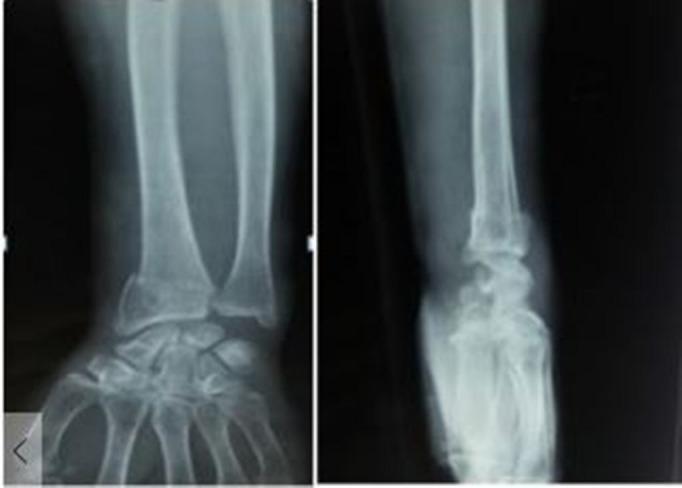
This diagnostic image consists of two X-ray views (anteroposterior and lateral) of a human wrist, demonstrating a displaced distal radius fracture. In the anteroposterior (AP) view, there is a clear transverse fracture line at the distal metaphysis of the radius with lateral displacement of the distal fragment and associated comminution. The ulnar styloid also appears involved, suggesting a potential associated injury. The lateral view reveals significant dorsal displacement and dorsal angulation of the distal radial fragment (characteristic of a Colles-type fracture pattern), along with impaction resulting in radial shortening. The radiocarpal joint alignment is disrupted due to the fracture angulation. This imaging is clinically significant for orthopedic evaluation, planning of surgical reduction, and assessing the degree of articular involvement in common distal forearm injuries.
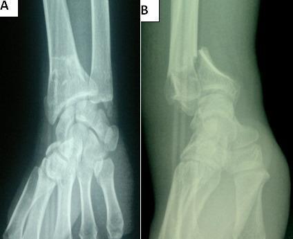
This diagnostic image consists of two preoperative X-ray views of the wrist, illustrating an unstable intra-articular distal radius fracture. View A is an anteroposterior (AP) radiograph showing comminution and a clear sagittal fracture line extending into the radiocarpal joint space. View B is a lateral radiograph demonstrating a Goyrand-Smith fracture, characterized by significant volar (palmar) displacement and angulation of the distal fracture fragment relative to the radial shaft. The alignment between the distal radius, carpal bones, and distal ulna is severely disrupted, with the carpus following the volar displacement of the distal radial fragment. This case highlights key orthopedic findings for surgical planning, specifically the involvement of the articular surface and the direction of fragment displacement, which distinguishes the Smith fracture from the more common dorsal displacement seen in Colles fractures.
fetal presentation diagram obstetrics
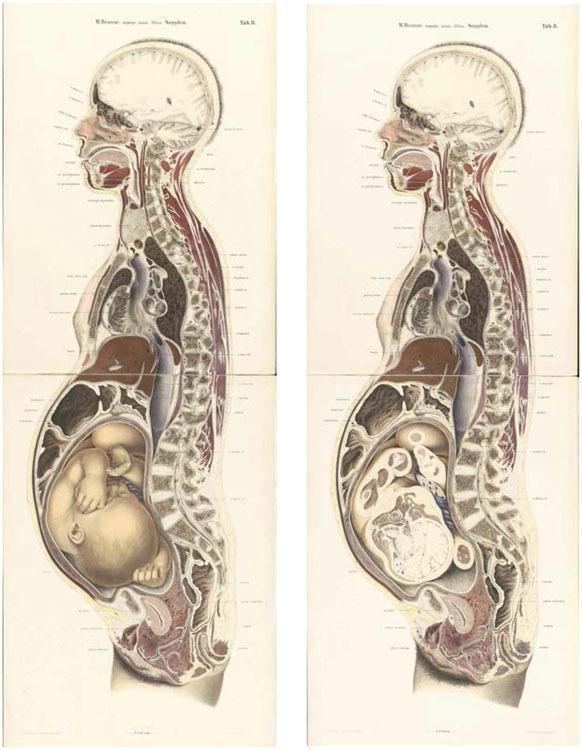
This historical anatomical diagram consists of two full-body mid-sagittal sections of a pregnant woman at full term, based on 19th-century cryosection techniques. The illustrations provide a comprehensive view of maternal and fetal topography. Key maternal structures include the brain, spinal column, esophagus, heart, diaphragm, liver, and a gravid uterus. The left illustration displays the fetus in situ within the uterus, shown in a cephalic presentation (head-down) and flexed 'fetal position' with visible limbs and facial features. The right illustration represents the same sagittal plane but with the fetus removed, revealing the empty uterine cavity, amniotic membranes, and the relationship between the uterus, bladder, and rectum in the pelvic cavity. This comparison highlights the displacement of maternal abdominal viscera (liver and intestines) by the distended uterus. The diagram serves as an educational resource for obstetrics, demonstrating fetal orientation, maternal-fetal anatomical relationships, and the physiological changes in late-stage pregnancy.
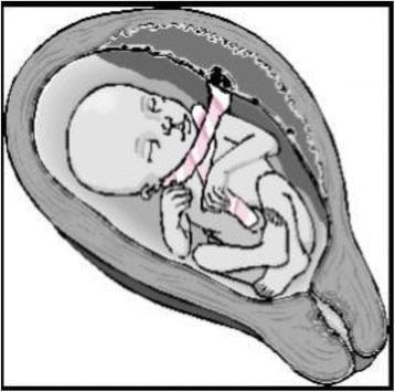
This medical illustration depicts an anatomical diagram of a fetus in utero demonstrating a nuchal cord with a 'free sliding pattern'. The fetus is shown in a flexed, cephalic presentation within the uterine cavity. The umbilical cord is visibly coiled around the fetal neck in at least two loose loops. These loops are characterized by a lack of constriction or indentation of the fetal soft tissues, representing a 'Type A' nuchal cord configuration where the cord can move freely and potentially undo itself spontaneously. The placenta is positioned at the fundal/posterior aspect of the uterus, with the cord extending toward the neck before reaching the umbilicus. This diagram serves as an educational tool in obstetrics to differentiate between non-constricting (Type A) and locked/constricting (Type B) nuchal cord patterns, highlighting the clinical relevance of cord entanglement and its impact on fetal well-being during gestation and labor.
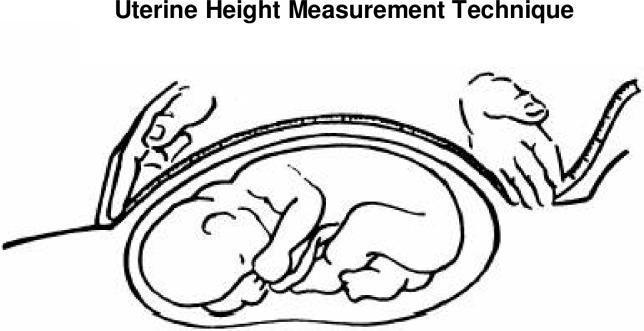
This medical illustration demonstrates the standardized technique for measuring symphysis-fundal height (SFH) during pregnancy. The diagram shows a sagittal-style cross-section of a gravid uterus containing a fetus in a cephalic presentation. A flexible, non-stretchable measuring tape is used to quantify the uterine height. One hand of the clinician is shown anchoring the 'zero' mark of the tape at the upper border of the symphysis pubis. The second hand is positioned at the uterine fundus, the highest point of the uterus, ensuring the tape follows the longitudinal curve of the abdominal wall. This clinical measurement is a primary screening tool in obstetrics used to monitor fetal growth, estimate gestational age, and screen for conditions such as intrauterine growth restriction (IUGR) or macrosomia. The visual emphasizes the proper placement of hands and the orientation of the measurement tool relative to maternal anatomical landmarks and the internal fetal position.
autonomic nervous system receptors diagram pharmacology
mkdir -p /tmp/workspace/neetpg-highyield-pdf/assets /tmp/workspace/neetpg-highyield-pdf/output && ls -la /tmp/workspace/
~/neetpg-highyield-pdf/data.py
# -*- coding: utf-8 -*-
# High-yield NEET-PG / INI-CET condensed content dataset
# 19 subjects x 6 topics, each with: exam pearl, mnemonic, mind-map branches, optional table, optional image
IMAGES = {
"anatomy": "https://cdn.orris.care/cdss_images/pmc_clinical_VQA_4228404f2d2e5a2fd501ff9156a4009148ff07cde4289fa58b1daf97dad26b2e.jpg",
"pathology": "https://cdn.orris.care/cdss_images/Pathology_1760051516527_c8a64a3a-6c12-41f5-8302-14cb7e183323.jpg",
"dermatology": "https://cdn.orris.care/cdss_images/DermNetNZ_1760035401252_c6bb8e5a-fce1-470a-a40a-5658e8580d67.jpg",
"radiology": "https://cdn.orris.care/cdss_images/pmc_clinical_VQA_14b2e996d38769225319c1263e6f78dba002c63db1ed31afdb40d26201c7bcbe.jpg",
"ophthalmology": "https://cdn.orris.care/cdss_images/GLGCA_16413_1763300025767_0197cef3-6d01-41f1-876f-75e78c510bf0.png",
"ent": "https://cdn.orris.care/cdss_images/pmc_clinical_VQA_48f074c8509cad55afd2e1fa010a862f18848f65d881dcdf929ef5a03537efce.jpg",
"orthopedics": "https://cdn.orris.care/cdss_images/pmc_clinical_VQA_6def68b239a90873bcce414e971c0810b94cbf5bfd79a61578d80757efbdc63c.jpg",
"obg": "https://cdn.orris.care/cdss_images/pmc_clinical_VQA_45d296e7018baf8d5320e3d99c4de7721aab8c33ae217e0910f35fdc30f08b14.jpg",
}
SUBJECTS = [
{"key":"anatomy","name":"Anatomy","color":"#E63946","image":"anatomy","topics":[
{"name":"Brachial Plexus","pearl":"Erb's palsy (C5-C6) = waiter's tip; Klumpke's (C8-T1) = claw hand.",
"mnemonic":("Randy Travis Drinks Cold Beer","Roots-Trunks-Divisions-Cords-Branches"),
"branches":[("Roots",["C5-T1","Between scalenus ant & med"]),
("Trunks",["Upper C5-C6","Middle C7","Lower C8-T1"]),
("Cords",["Lateral, Posterior, Medial","Named by relation to axillary artery"]),
("Key Injuries",["Erb's - upper trunk - waiter's tip","Klumpke's - lower trunk - claw hand"]),
("Branches",["Musculocutaneous, Axillary","Radial, Median, Ulnar"])],
"table":{"headers":["Injury","Level","Clinical Sign"],
"rows":[["Erb-Duchenne","C5-C6 upper trunk","Waiter's tip deformity"],
["Klumpke's","C8-T1 lower trunk","Claw hand, Horner's may occur"],
["Wrist drop","Radial nerve","Saturday night palsy"],
["Winging of scapula","Long thoracic nerve","Serratus anterior palsy"]]},
"image":"anatomy"},
{"name":"Femoral Triangle","pearl":"NAVEL from lateral to medial: Nerve, Artery, Vein, Empty space, Lymphatics.",
"mnemonic":("NAVEL","Nerve-Artery-Vein-Empty space-Lymphatics (lateral to medial)"),
"branches":[("Boundaries",["Base: Inguinal ligament","Lateral: Sartorius","Medial: Adductor longus"]),
("Contents (lateral to medial)",["Femoral Nerve","Femoral Artery","Femoral Vein","Empty space","Lymph nodes (Cloquet)"]),
("Clinical",["Femoral hernia - medial to vein","Femoral pulse - mid-inguinal point"]),
("Roof",["Fascia lata, superficial fascia"]),
("Floor",["Iliopsoas, Pectineus, Adductor longus"])],
"table":None,"image":None},
{"name":"Circle of Willis","pearl":"Most common site of berry aneurysm: Anterior communicating artery.",
"mnemonic":("ACA-PCA-ICA","Anterior, Posterior communicating link anterior & posterior circulation"),
"branches":[("Anterior circulation",["ICA -> ACA + MCA","Anterior communicating artery links two ACAs"]),
("Posterior circulation",["Vertebral -> Basilar -> PCA","Posterior communicating links ICA-PCA"]),
("Aneurysm sites",["ACOM - most common overall","PCOM - 2nd most common, CN III palsy"]),
("Watershed zones",["ACA-MCA border - hypotension infarct"]),
("Clinical",["PCOM aneurysm compresses CN III - ptosis, dilated pupil"])],
"table":None,"image":None},
{"name":"Inguinal Canal","pearl":"Direct hernia - medial to inferior epigastric vessels; Indirect - lateral.",
"mnemonic":("MALT","Direct hernia through weak conjoint tendon medially"),
"branches":[("Boundaries",["Ant: Ext oblique aponeurosis","Post: Transversalis fascia","Roof: Internal oblique+transversus"]),
("Rings",["Deep ring - mid-inguinal point","Superficial ring - pubic tubercle"]),
("Direct Hernia",["Medial to inferior epigastric","Through Hesselbach triangle","Acquired, elderly"]),
("Indirect Hernia",["Lateral to inferior epigastric","Through deep ring, patent processus vaginalis","Congenital, young"]),
("Contents",["Spermatic cord (M) / Round ligament (F)","Ilioinguinal nerve"])],
"table":{"headers":["Feature","Direct","Indirect"],
"rows":[["Relation to vessels","Medial","Lateral"],
["Origin","Hesselbach triangle","Deep inguinal ring"],
["Age","Elderly","Young/congenital"],
["Covering sac","Rarely reaches scrotum","Can reach scrotum"]]},
"image":None},
{"name":"Heart Chambers & Great Vessels","pearl":"Right heart border - RA; Left border - LV, LAA, pulmonary trunk.",
"mnemonic":("SAM stands, VAN sleeps","Femoral sheath lateral to medial ordering aid"),
"branches":[("Right Atrium",["SVC, IVC, coronary sinus drain here","Crista terminalis"]),
("Right Ventricle",["Most anterior chamber","Moderator band"]),
("Left Atrium",["Most posterior chamber","4 pulmonary veins drain"]),
("Left Ventricle",["Thickest wall","Apex of heart"]),
("Great Vessels",["Aorta - posterior to pulmonary trunk at base","Pulmonary trunk bifurcates T4-5"])],
"table":None,"image":None},
{"name":"Cranial Nerves Overview","pearl":"CN III, IV, VI - eye movement; CN X - vagus, widest distribution.",
"mnemonic":("Some Say Marry Money But My Brother Says Big Brains Matter More",
"Sensory-Sensory-Motor-Motor-Both-Motor-Both-Sensory-Both-Both-Motor-Motor"),
"branches":[("Olfactory-Optic-Oculomotor",["I Smell","II See","III moves eye+pupil"]),
("IV-V-VI",["Trochlear - superior oblique","Trigeminal - face sensation+mastication","Abducens - lateral rectus"]),
("VII-VIII",["Facial - expression+taste ant 2/3","Vestibulocochlear - hearing+balance"]),
("IX-X-XI-XII",["Glossopharyngeal - taste post 1/3","Vagus - parasympathetic to viscera","Accessory - SCM/trapezius","Hypoglossal - tongue muscles"]),
("Clinical",["CN III palsy - ptosis, mydriasis, down-out eye","Bell's palsy - CN VII, forehead involved"])],
"table":None,"image":None},
]},
{"key":"physiology","name":"Physiology","color":"#F4A261","image":None,"topics":[
{"name":"Cardiac Cycle (Wiggers Diagram)","pearl":"S1 = mitral+tricuspid closure; S2 = aortic+pulmonary closure.",
"mnemonic":("Lub-Dub","S1 at start of systole, S2 at start of diastole"),
"branches":[("Isovolumetric contraction",["All valves closed","Pressure rises, no volume change"]),
("Ejection",["Aortic valve opens","Rapid then reduced ejection"]),
("Isovolumetric relaxation",["All valves closed","Pressure falls sharply"]),
("Rapid filling",["Mitral valve opens","Passive ventricular filling"]),
("Atrial systole",["Active filling, 'atrial kick'","a wave on JVP"])],
"table":{"headers":["Heart Sound","Cause","Timing"],
"rows":[["S1","Mitral+Tricuspid closure","Onset of systole"],
["S2","Aortic+Pulmonic closure","Onset of diastole"],
["S3","Rapid ventricular filling","Early diastole, normal in kids/HF"],
["S4","Atrial contraction into stiff ventricle","Late diastole, HTN/LVH"]]},
"image":None},
{"name":"Renal Clearance & GFR","pearl":"Inulin/Creatinine clearance estimates GFR; PAH clearance estimates renal plasma flow.",
"mnemonic":("Creatinine Clears at GFR","Filtration marker = freely filtered, not reabsorbed/secreted"),
"branches":[("GFR markers",["Inulin - gold standard","Creatinine - clinically used, slightly overestimates"]),
("RPF marker",["PAH - filtered + secreted completely"]),
("Filtration Fraction",["GFR/RPF, normally ~20%"]),
("Clearance formula",["C = (U x V)/P"]),
("Autoregulation",["Myogenic + tubuloglomerular feedback","Maintains GFR over MAP 80-180 mmHg"])],
"table":None,"image":None},
{"name":"Nerve Action Potential","pearl":"Absolute refractory period = Na+ channels inactivated, no stimulus can excite.",
"mnemonic":("Na in, K out","Depolarization = Na+ influx; Repolarization = K+ efflux"),
"branches":[("Resting potential",["~-70mV","Maintained by Na-K ATPase + leak channels"]),
("Depolarization",["Voltage-gated Na+ channels open","Rapid influx of Na+"]),
("Repolarization",["Na+ channels inactivate","K+ channels open, efflux"]),
("Refractory periods",["Absolute - no stimulus works","Relative - stronger stimulus needed"]),
("Propagation",["Saltatory conduction in myelinated axons","Faster in larger diameter fibers"])],
"table":None,"image":None},
{"name":"Lung Volumes & Spirometry","pearl":"FEV1/FVC <70% = obstructive; reduced TLC = restrictive.",
"mnemonic":("IRV+TV+ERV+RV=TLC","Vital capacity = IRV+TV+ERV (excludes RV)"),
"branches":[("Static volumes",["Tidal Volume ~500mL","Residual Volume - cannot be measured by spirometry"]),
("Capacities",["FRC = ERV+RV","TLC = VC+RV"]),
("Obstructive pattern",["Low FEV1/FVC","Increased RV, TLC - COPD, asthma"]),
("Restrictive pattern",["Normal/high FEV1/FVC","Decreased TLC - fibrosis"]),
("Measuring RV/FRC/TLC",["Helium dilution or body plethysmography (not spirometry)"])],
"table":{"headers":["Pattern","FEV1/FVC","TLC","Example"],
"rows":[["Obstructive","Decreased","Normal/Increased","Asthma, COPD"],
["Restrictive","Normal/Increased","Decreased","Pulmonary fibrosis"]]},
"image":None},
{"name":"Hypothalamic-Pituitary Axis","pearl":"Prolactin is under tonic inhibition by dopamine; only pituitary hormone that increases when stalk is cut.",
"mnemonic":("FLAT PIG","FSH LH ACTH TSH Prolactin GH (Ant pituitary hormones)"),
"branches":[("Anterior pituitary",["FSH, LH, ACTH, TSH, GH, Prolactin","Under hypothalamic releasing/inhibiting hormones"]),
("Posterior pituitary",["Stores ADH + Oxytocin","Made in hypothalamus, transported down axons"]),
("Negative feedback",["Long-loop feedback on hypothalamus+pituitary"]),
("Prolactin unique",["Dopamine inhibits release","Stalk section -> increased prolactin"]),
("Clinical",["Prolactinoma - most common pituitary adenoma","Sheehan syndrome - postpartum pituitary necrosis"])],
"table":None,"image":None},
{"name":"Muscle Contraction (Sliding Filament)","pearl":"Ca2+ binds Troponin C, moves tropomyosin, exposes myosin binding site on actin.",
"mnemonic":("Ca binds Troponin, Cross-bridge cycles","Excitation-contraction coupling sequence"),
"branches":[("Excitation",["Action potential -> T-tubule -> DHP receptor"]),
("Ca release",["Ryanodine receptor on SR releases Ca2+"]),
("Cross-bridge cycle",["Ca-Troponin C -> tropomyosin shift -> myosin binds actin"]),
("Power stroke",["ATP hydrolysis powers myosin head movement"]),
("Relaxation",["Ca2+ pumped back into SR by SERCA"])],
"table":None,"image":None},
]},
{"key":"biochemistry","name":"Biochemistry","color":"#2A9D8F","image":None,"topics":[
{"name":"Glycolysis & Regulation","pearl":"Rate limiting enzyme: Phosphofructokinase-1 (PFK-1); inhibited by ATP, citrate.",
"mnemonic":("PFK is the Pacemaker","Committed, irreversible, most regulated step"),
"branches":[("Key enzymes",["Hexokinase/Glucokinase","PFK-1 (rate limiting)","Pyruvate kinase"]),
("Regulation",["PFK-1 inhibited by ATP, citrate","Activated by AMP, F2,6BP"]),
("Net yield",["2 ATP, 2 NADH, 2 Pyruvate per glucose (aerobic)"]),
("Enzyme deficiency",["Pyruvate kinase deficiency - hemolytic anemia"]),
("Fate of pyruvate",["Aerobic - Acetyl CoA (PDH)","Anaerobic - Lactate (LDH)"])],
"table":None,"image":None},
{"name":"Urea Cycle","pearl":"Carbamoyl phosphate synthetase I deficiency = most common urea cycle disorder presenting with hyperammonemia.",
"mnemonic":("Ordinarily, Careless Crappers Are Also Frivolous About Urination",
"Ornithine-Carbamoyl phosphate-Citrulline-Aspartate-Argininosuccinate-Fumarate-Arginine-Urea"),
"branches":[("Location",["First 2 steps mitochondria, rest cytosol"]),
("Key enzymes",["CPS-I (rate limiting, needs N-acetylglutamate)","Ornithine transcarbamylase (OTC)"]),
("OTC deficiency",["X-linked, most common urea cycle disorder","Increased orotic acid, no megaloblastic anemia"]),
("Hyperammonemia Rx",["Limit protein, Benzoate/Phenylbutyrate, Lactulose"]),
("Nitrogen sources",["NH4+ and Aspartate contribute N atoms to urea"])],
"table":None,"image":None},
{"name":"Vitamin Deficiencies","pearl":"B1 (thiamine) deficiency - Wernicke-Korsakoff, Beriberi; B12 - subacute combined degeneration.",
"mnemonic":("All-B-Complex","B1 Beriberi, B2 Angular stomatitis, B3 Pellagra, B6 Neuropathy, B12 SCD"),
"branches":[("Fat soluble (ADEK)",["A - night blindness","D - rickets/osteomalacia","E - ataxia, hemolysis","K - bleeding, PT prolonged"]),
("B1 Thiamine",["Beriberi, Wernicke-Korsakoff","Alpha-ketoglutarate DH, pyruvate DH cofactor"]),
("B3 Niacin",["Pellagra - Diarrhea, Dermatitis, Dementia"]),
("B9 Folate",["Megaloblastic anemia, NTDs in pregnancy"]),
("B12 Cobalamin",["Megaloblastic anemia + subacute combined degeneration","Methylmalonic acid raised (unlike folate)"])],
"table":{"headers":["Vitamin","Deficiency Disease","Key Feature"],
"rows":[["B1","Beriberi/Wernicke's","Ophthalmoplegia, confusion, ataxia"],
["B2","Ariboflavinosis","Angular stomatitis, cheilosis"],
["B3","Pellagra","3 D's - Diarrhea, Dermatitis, Dementia"],
["B12","Megaloblastic anemia + SCD","Raised methylmalonic acid + homocysteine"]]},
"image":None},
{"name":"Lipoproteins & Lipid Profile","pearl":"Chylomicrons carry dietary TG; LDL is 'bad' cholesterol carrier; HDL removes cholesterol.",
"mnemonic":("Chylomicron Carries Chow","Exogenous pathway - dietary fat transport"),
"branches":[("Chylomicrons",["Carry dietary TG from gut","ApoB-48"]),
("VLDL",["Carry endogenous TG from liver","ApoB-100"]),
("LDL",["Cholesterol delivery to tissues","Major atherogenic particle"]),
("HDL",["Reverse cholesterol transport","ApoA-1 activates LCAT"]),
("Key enzymes",["Lipoprotein lipase - clears chylomicrons/VLDL","LCAT - esterifies cholesterol on HDL"])],
"table":None,"image":None},
{"name":"Genetic Code & Mutations","pearl":"Frameshift mutations (insertion/deletion not in multiples of 3) are usually more deleterious than point mutations.",
"mnemonic":("SIN of mutation","Silent, missense, nonsense point mutation types"),
"branches":[("Point mutations",["Silent - no AA change","Missense - AA changed","Nonsense - premature stop codon"]),
("Frameshift",["Insertion/deletion not multiple of 3","Alters reading frame downstream"]),
("Trinucleotide repeat",["CAG repeat - Huntington's","CGG repeat - Fragile X"]),
("Genetic code features",["Degenerate, non-overlapping, universal","Wobble hypothesis at 3rd codon position"]),
("Splice site mutation",["Affects mRNA processing, exon skipping"])],
"table":None,"image":None},
{"name":"Enzyme Kinetics (Km & Vmax)","pearl":"Competitive inhibitor - increased Km, same Vmax; Noncompetitive - same Km, decreased Vmax.",
"mnemonic":("Km is the KardashIan of affinity","Low Km = high affinity for substrate"),
"branches":[("Km",["Substrate conc at half Vmax","Inversely related to affinity"]),
("Vmax",["Max reaction velocity at enzyme saturation"]),
("Competitive inhibition",["Increased Km, unchanged Vmax","Overcome by increasing substrate"]),
("Noncompetitive inhibition",["Unchanged Km, decreased Vmax","Cannot be overcome by substrate"]),
("Lineweaver-Burk plot",["1/v vs 1/[S] - straight line analysis"])],
"table":None,"image":None},
]},
{"key":"pathology","name":"Pathology","color":"#8B0000","image":"pathology","topics":[
{"name":"Acute vs Chronic Inflammation","pearl":"Neutrophils dominate acute inflammation; lymphocytes+macrophages dominate chronic.",
"mnemonic":("Neutrophils Now, Macrophages Much Later","Cell type sequence in inflammation"),
"branches":[("Acute inflammation",["Neutrophils predominant","Vascular changes - vasodilation, permeability"]),
("Chronic inflammation",["Lymphocytes, plasma cells, macrophages","Fibrosis and angiogenesis"]),
("Chemical mediators",["Histamine - immediate vascular permeability","Prostaglandins, Leukotrienes - pain/chemotaxis"]),
("Granulomatous inflammation",["TB, Sarcoidosis, Fungal - epithelioid cells + giant cells"]),
("Cardinal signs",["Rubor, Calor, Tumor, Dolor, Functio laesa"])],
"table":None,"image":None},
{"name":"Neoplasia - Grading & Tumor Markers","pearl":"Grading = degree of differentiation; Staging (TNM) is more prognostically important than grading.",
"mnemonic":("CEA-Colon, AFP-Liver/Germ cell, PSA-Prostate, CA125-Ovary, CA19-9-Pancreas",""),
"branches":[("Grading vs Staging",["Grade - differentiation","Stage (TNM) - spread, more prognostic"]),
("Tumor markers",["AFP - HCC, yolk sac tumor","CEA - colorectal","CA-125 - ovarian","PSA - prostate","CA19-9 - pancreatic","hCG - choriocarcinoma/germ cell"]),
("Metastasis route",["Carcinoma - lymphatic first","Sarcoma - hematogenous first"]),
("Common mets to bone",["Breast, Prostate, Lung, Kidney, Thyroid (BPLKT)"]),
("Paraneoplastic syndromes",["SIADH/ACTH - small cell lung ca","Hypercalcemia - PTHrP squamous cell lung ca"])],
"table":{"headers":["Marker","Associated Tumor"],
"rows":[["AFP","Hepatocellular carcinoma, yolk sac tumor"],
["CEA","Colorectal carcinoma"],
["CA-125","Ovarian carcinoma"],
["PSA","Prostate carcinoma"],
["CA19-9","Pancreatic carcinoma"],
["Beta-hCG","Choriocarcinoma, germ cell tumors"]]},
"image":None},
{"name":"Myocardial Infarction - Timeline","pearl":"Coagulative necrosis peaks with neutrophils at 1-3 days; danger of rupture at 4-7 days (macrophage/granulation weakness).",
"mnemonic":("1 day none, 1-3 days Neutrophils, 3-7 macrophages, weeks fibrosis",""),
"branches":[("0-24 hours",["Coagulative necrosis begins","Gross - no visible change early, dark mottling later"]),
("1-3 days",["Neutrophil infiltration","Risk of fibrinous pericarditis"]),
("4-7 days",["Macrophages, yellow softening","Highest risk of free wall rupture"]),
("1-3 weeks",["Granulation tissue formation"]),
("Months",["Dense fibrous scar (grey-white)"]),
("Biomarkers",["Troponin - rises 4h, peaks 24h, lasts 7-10 days","CK-MB - useful for reinfarction detection"])],
"table":None,"image":"pathology"},
{"name":"Anemia Classification","pearl":"Microcytic - Iron deficiency, Thalassemia, Sideroblastic, Chronic disease (late); Macrocytic - B12/Folate.",
"mnemonic":("TICS for Microcytic","Thalassemia, Iron deficiency, Chronic disease, Sideroblastic"),
"branches":[("Microcytic (MCV<80)",["Iron deficiency - low ferritin, high TIBC","Thalassemia - target cells","Sideroblastic - ring sideroblasts"]),
("Normocytic",["Hemolytic, Aplastic, Chronic disease (early)"]),
("Macrocytic",["Megaloblastic - B12/Folate deficiency","Non-megaloblastic - liver disease, hypothyroid"]),
("Hemolytic clues",["Increased reticulocytes, LDH, indirect bilirubin","Decreased haptoglobin"]),
("Iron studies",["Fe deficiency: low Fe, low ferritin, high TIBC","Chronic disease: low Fe, high ferritin, low TIBC"])],
"table":None,"image":None},
{"name":"Amyloidosis","pearl":"AA amyloid - chronic inflammation; AL amyloid - plasma cell dyscrasia; stains apple-green birefringence with Congo red.",
"mnemonic":("Congo Red = Apple Green under polarized light",""),
"branches":[("AL amyloid",["Light chain, plasma cell dyscrasia/myeloma"]),
("AA amyloid",["Serum amyloid A, chronic inflammation (RA, TB)"]),
("ATTR amyloid",["Transthyretin, senile systemic/familial cardiomyopathy"]),
("Staining",["Congo red - apple-green birefringence","H&E - amorphous eosinophilic material"]),
("Organ involvement",["Kidney - nephrotic syndrome","Heart - restrictive cardiomyopathy"])],
"table":None,"image":None},
{"name":"Granulomatous Diseases","pearl":"Caseating granuloma = TB (central necrosis); Non-caseating = Sarcoidosis.",
"mnemonic":("TB Caseates, Sarcoid doesn't",""),
"branches":[("Caseating",["TB - central necrosis, Langhans giant cells","Fungal infections"]),
("Non-caseating",["Sarcoidosis - no necrosis","Crohn's disease"]),
("Key cells",["Epithelioid macrophages","Langhans giant cells (horseshoe nuclei)"]),
("Sarcoidosis clues",["Bilateral hilar lymphadenopathy","Raised ACE, hypercalcemia"]),
("Foreign body granuloma",["Around suture material/talc, no immune-mediated response"])],
"table":None,"image":None},
]},
{"key":"pharmacology","name":"Pharmacology","color":"#457B9D","image":None,"topics":[
{"name":"Autonomic Receptors","pearl":"Alpha-1 - vasoconstriction (Gq); Beta-1 - heart (Gs); Beta-2 - bronchodilation (Gs); M3 - bronchoconstriction (Gq).",
"mnemonic":("Qiss and Qiq Kiss and Cry (Gq-Gs-Gi pattern)","Odd numbered Gq, others vary"),
"branches":[("Alpha-1",["Gq - vasoconstriction, mydriasis"]),
("Alpha-2",["Gi - decreased NE release (presynaptic)"]),
("Beta-1",["Gs - increased HR, contractility, renin"]),
("Beta-2",["Gs - bronchodilation, vasodilation, uterine relaxation"]),
("Muscarinic M3",["Gq - bronchoconstriction, increased secretions, miosis"])],
"table":{"headers":["Receptor","G-protein","Action"],
"rows":[["Alpha-1","Gq","Vasoconstriction"],
["Alpha-2","Gi","Inhibits NE release"],
["Beta-1","Gs","Increases heart rate/contractility"],
["Beta-2","Gs","Bronchodilation, vasodilation"],
["M3","Gq","Bronchoconstriction, secretions"]]},
"image":None},
{"name":"Antimicrobial Mechanisms","pearl":"Cell wall inhibitors (beta-lactams, vancomycin) are bactericidal; protein synthesis inhibitors mostly bacteriostatic (except aminoglycosides).",
"mnemonic":("Buy AT 30, CCEL at 50","30S - Aminoglycosides/Tetracyclines; 50S - Chloramphenicol/Clindamycin/Erythromycin/Linezolid"),
"branches":[("Cell wall synthesis",["Beta-lactams (PBP), Vancomycin (D-ala-D-ala)"]),
("30S ribosome",["Aminoglycosides (irreversible), Tetracyclines (reversible)"]),
("50S ribosome",["Macrolides, Chloramphenicol, Clindamycin, Linezolid"]),
("DNA/RNA synthesis",["Fluoroquinolones - Topoisomerase/DNA gyrase","Rifampin - RNA polymerase"]),
("Folate synthesis",["Sulfonamides - PABA analog","Trimethoprim - DHFR inhibitor"])],
"table":None,"image":None},
{"name":"Anti-TB Drugs & Toxicities","pearl":"Isoniazid - peripheral neuropathy (give Pyridoxine); Rifampin - orange secretions, potent CYP450 inducer; Ethambutol - optic neuritis.",
"mnemonic":("RIPE","Rifampin-Isoniazid-Pyrazinamide-Ethambutol"),
"branches":[("Isoniazid",["Peripheral neuropathy, hepatotoxicity","Give B6 (pyridoxine) to prevent neuropathy"]),
("Rifampin",["Red-orange body secretions","CYP450 inducer - reduces OCP efficacy"]),
("Pyrazinamide",["Hepatotoxicity, hyperuricemia"]),
("Ethambutol",["Optic neuritis - red-green color blindness"]),
("Streptomycin",["Ototoxicity, nephrotoxicity (2nd line injectable)"])],
"table":{"headers":["Drug","Key Toxicity"],
"rows":[["Isoniazid","Peripheral neuropathy, hepatotoxicity, SLE-like"],
["Rifampin","Orange secretions, CYP450 induction, flu-like"],
["Pyrazinamide","Hepatotoxicity, hyperuricemia/gout"],
["Ethambutol","Optic neuritis, red-green color blindness"]]},
"image":None},
{"name":"Anticoagulants","pearl":"Heparin - immediate, monitor aPTT, antidote Protamine; Warfarin - delayed, monitor INR, antidote Vit K.",
"mnemonic":("PTT-Heparin (both have T's); PT-Warfarin",""),
"branches":[("Heparin (UFH)",["Activates Antithrombin III","Monitor aPTT","Antidote Protamine sulfate"]),
("Warfarin",["Inhibits Vit K epoxide reductase","Monitor INR/PT","Antidote Vit K / FFP"]),
("DOACs",["Dabigatran - direct thrombin inhibitor","Rivaroxaban/Apixaban - factor Xa inhibitor"]),
("HIT",["Heparin-induced thrombocytopenia - Type II immune-mediated","Switch to non-heparin anticoagulant"]),
("Pregnancy",["Warfarin contraindicated (teratogenic)","Heparin/LMWH safe in pregnancy"])],
"table":None,"image":None},
{"name":"Antihypertensives - Classes","pearl":"ACE inhibitors - dry cough, hyperkalemia, contraindicated in pregnancy & bilateral renal artery stenosis.",
"mnemonic":("CAPTOPRIL causes Cough",""),
"branches":[("ACE Inhibitors",["Dry cough, hyperkalemia, angioedema","Avoid in pregnancy, bilateral RAS"]),
("ARBs",["Similar to ACEI, no cough (no bradykinin)"]),
("CCBs",["Dihydropyridines - peripheral edema","Non-DHP - bradycardia (Verapamil/Diltiazem)"]),
("Beta blockers",["Avoid abrupt withdrawal - rebound tachycardia","Caution in asthma (non-selective)"]),
("Diuretics",["Thiazide - hypokalemia, hyponatremia, hyperglycemia","Loop - ototoxicity, hypokalemia"])],
"table":None,"image":None},
{"name":"Chemotherapy Drug Toxicities","pearl":"Doxorubicin - cardiotoxicity; Cisplatin - nephro/ototoxicity; Vincristine - peripheral neuropathy; Bleomycin - pulmonary fibrosis.",
"mnemonic":("Doxo-Heart, Cis-Kidney/Ear, Vinc-Nerve, Bleo-Lung",""),
"branches":[("Doxorubicin",["Dilated cardiomyopathy (dose-dependent)","Prevent with Dexrazoxane"]),
("Cisplatin",["Nephrotoxicity, ototoxicity, peripheral neuropathy"]),
("Vincristine",["Peripheral neuropathy (microtubule inhibitor)"]),
("Bleomycin",["Pulmonary fibrosis"]),
("Methotrexate",["Myelosuppression, mucositis","Rescue with Leucovorin"])],
"table":None,"image":None},
]},
{"key":"microbiology","name":"Microbiology","color":"#6A4C93","image":None,"topics":[
{"name":"Gram Stain Classification","pearl":"Gram-positive - thick peptidoglycan retains crystal violet (purple); Gram-negative - thin wall, takes counterstain (pink/red).",
"mnemonic":("Purple=Positive","Crystal violet retained by thick peptidoglycan wall"),
"branches":[("Gram Positive Cocci",["Staph (clusters), Strep (chains)"]),
("Gram Negative Cocci",["Neisseria - diplococci"]),
("Gram Positive Rods",["Bacillus, Clostridium, Listeria, Corynebacterium"]),
("Gram Negative Rods",["E.coli, Klebsiella, Pseudomonas, Salmonella"]),
("Stain-resistant organisms",["Mycobacteria - acid fast (Ziehl-Neelsen)","Mycoplasma - no cell wall","Treponema - too thin, needs dark-field"])],
"table":None,"image":None},
{"name":"TORCH Infections","pearl":"Congenital Rubella - triad: cataract, deafness, PDA/cardiac defects. Toxoplasma - ring-enhancing brain lesions + chorioretinitis.",
"mnemonic":("TORCH","Toxoplasma-Others(syphilis,varicella)-Rubella-CMV-HSV"),
"branches":[("Toxoplasmosis",["Ring-enhancing brain lesions","Chorioretinitis, intracranial calcifications"]),
("Rubella",["Cataract, deafness, PDA (classic triad)"]),
("CMV",["Most common congenital infection","Periventricular calcifications, microcephaly"]),
("HSV",["Vesicular skin lesions, encephalitis"]),
("Syphilis",["Snuffles, saber shins, Hutchinson teeth"])],
"table":None,"image":None},
{"name":"Hepatitis Viruses Serology","pearl":"HBsAg = active infection; Anti-HBs = immunity; IgM anti-HBc = window period marker.",
"mnemonic":("Core window, Surface shield","Anti-HBc IgM fills serologic window when HBsAg gone, anti-HBs not yet risen"),
"branches":[("Acute Hep B",["HBsAg +, IgM anti-HBc +"]),
("Window period",["HBsAg negative, anti-HBs negative","IgM anti-HBc only marker positive"]),
("Immunity (vaccinated)",["Anti-HBs positive only"]),
("Chronic Hep B",["HBsAg + beyond 6 months","HBeAg indicates active replication/infectivity"]),
("Hepatitis routes",["A,E - fecal-oral","B,C,D - blood/sexual/vertical"])],
"table":{"headers":["Marker","Meaning"],
"rows":[["HBsAg","Current infection (acute or chronic)"],
["Anti-HBs","Immunity (vaccination or resolved infection)"],
["IgM anti-HBc","Recent/acute infection, window period"],
["HBeAg","Active viral replication, high infectivity"]]},
"image":None},
{"name":"Malaria Life Cycle","pearl":"P. falciparum - most severe, no hypnozoites, cerebral malaria; P. vivax/ovale - hypnozoites cause relapse.",
"mnemonic":("Vivax and Ovale hide (hypnozoites), Falciparum fights hardest",""),
"branches":[("Vector",["Female Anopheles mosquito"]),
("Liver stage",["Sporozoites infect hepatocytes","Vivax/Ovale form dormant hypnozoites"]),
("Blood stage",["Merozoites infect RBCs - clinical symptoms/fever"]),
("P. falciparum",["Most severe - cerebral malaria, blackwater fever","Every RBC size infected"]),
("Diagnosis",["Peripheral smear - gold standard","Rapid antigen test - HRP-2"])],
"table":None,"image":None},
{"name":"Mycobacterium Tuberculosis","pearl":"Ghon focus + hilar lymphadenopathy = Ghon complex (primary TB); reactivation favors apex (Simon focus).",
"mnemonic":("Ghon = Primary, Apex = Reactivation",""),
"branches":[("Primary TB",["Ghon focus - lower/mid lung zone","Ghon complex = focus + hilar nodes"]),
("Secondary/Reactivation",["Apical lung involvement (high oxygen)","Cavitation common"]),
("Diagnosis",["Ziehl-Neelsen acid-fast stain","Culture - Lowenstein-Jensen medium (gold standard)","GeneXpert/CBNAAT - rapid PCR"]),
("Immunity",["Cell-mediated (Th1), Type IV hypersensitivity","Mantoux test - delayed type hypersensitivity"]),
("Extrapulmonary",["Pott's disease - spine","Lymphadenitis - most common extrapulmonary"])],
"table":None,"image":None},
{"name":"Fungal Infections Classification","pearl":"Candida - pseudohyphae; Aspergillus - septate hyphae, acute angle branching; Mucor - broad aseptate, wide angle branching.",
"mnemonic":("Mucor is Massive and Messy (broad, aseptate, right-angle)",""),
"branches":[("Superficial mycoses",["Tinea/Dermatophytes - skin, hair, nails"]),
("Candidiasis",["Pseudohyphae + budding yeast","Oral thrush, vaginal candidiasis"]),
("Aspergillosis",["Septate hyphae, acute-angle branching","Fungal ball, allergic bronchopulmonary aspergillosis"]),
("Mucormycosis",["Broad aseptate hyphae, wide-angle branching","Diabetics/immunocompromised, rhino-orbital-cerebral"]),
("Cryptococcus",["India ink stain, soap-bubble lesions in brain","Common in HIV/AIDS"])],
"table":None,"image":None},
]},
{"key":"forensic","name":"Forensic Medicine & Toxicology","color":"#3D405B","image":None,"topics":[
{"name":"Postmortem Changes","pearl":"Rigor mortis - onset 1-2h, complete 12h, disappears 24-36h (Nysten's law - face to feet).",
"mnemonic":("Rigor: 1-2-12-24-36","Onset-Progression-Complete-Start disappear-Fully gone (hours)"),
"branches":[("Algor mortis",["Body cooling, ~0.5-1 degC/hr initially"]),
("Livor mortis (Hypostasis)",["Gravity-dependent purplish discoloration","Fixed after 6-8 hours"]),
("Rigor mortis",["Onset 1-2h, complete by 12h","Disappears in same order as onset (Nysten's law)"]),
("Putrefaction",["Greenish discoloration abdomen first (right iliac fossa)","Due to gas-forming bacteria"]),
("Adipocere/Mummification",["Adipocere - moist, cold environment","Mummification - dry, hot environment"])],
"table":None,"image":None},
{"name":"Types of Asphyxial Deaths","pearl":"Hanging - ligature mark above thyroid cartilage, oblique, non-continuous; Strangulation - below thyroid cartilage, horizontal, continuous.",
"mnemonic":("Hanging High and oblique, Strangulation Straight and low",""),
"branches":[("Hanging",["Ligature mark above thyroid cartilage","Oblique, non-continuous (inverted V)"]),
("Strangulation",["Ligature mark below/at thyroid cartilage","Horizontal, continuous"]),
("Throttling (manual)",["Fingernail marks, thumb bruises on neck"]),
("Smothering/Suffocation",["External airway obstruction - pillow, hand"]),
("Signs of asphyxia",["Tardieu spots, cyanosis, petechial hemorrhages","Congestion of face, subconjunctival hemorrhage"])],
"table":None,"image":None},
{"name":"Firearm Wounds","pearl":"Entry wound smaller with inverted margins; Exit wound larger, everted margins, no blackening/tattooing.",
"mnemonic":("Entry Inverted, Exit Everted",""),
"branches":[("Entry wound",["Inverted margins","Abrasion collar/contusion ring present"]),
("Exit wound",["Everted margins, usually larger","No blackening, tattooing, or searing"]),
("Range estimation",["Contact - muzzle imprint, soot in wound","Near - tattooing/stippling from unburnt powder","Distant - no soot/tattooing, entry only"]),
("Rifled vs Smooth bore",["Rifled - single projectile, lands & grooves","Smooth bore (shotgun) - multiple pellets, spread with distance"]),
("Medicolegal",["Distance estimation from pellet spread crucial"])],
"table":None,"image":None},
{"name":"Organophosphate Poisoning","pearl":"OPC inhibits acetylcholinesterase -> cholinergic crisis (SLUDGE); Antidote: Atropine + Pralidoxime.",
"mnemonic":("SLUDGE + Killer B's","Salivation Lacrimation Urination Defecation GI distress Emesis"),
"branches":[("Mechanism",["Irreversible AChE inhibition","Accumulation of acetylcholine"]),
("Muscarinic signs",["SLUDGE - Salivation Lacrimation Urination Defecation GI Emesis"]),
("Nicotinic signs",["Fasciculations, muscle weakness, paralysis"]),
("CNS signs",["Confusion, seizures, coma"]),
("Treatment",["Atropine - blocks muscarinic effects","Pralidoxime (2-PAM) - reactivates AChE if given early"])],
"table":None,"image":None},
{"name":"Medico-legal Autopsy","pearl":"Autopsy consent not required in medico-legal cases (police requisition suffices); purpose - establish cause & manner of death.",
"mnemonic":("Cause, Manner, Mechanism, Time of death - the 4 pillars",""),
"branches":[("Indications",["Unnatural/suspicious death","Sudden unexpected death","Custodial death"]),
("Legal requirement",["No consent needed - police requisition sufficient"]),
("Sequence",["External examination first","Internal - Y or I incision, organ examination"]),
("Time since death estimation",["Rigor, livor, putrefaction, stomach contents"]),
("Documentation",["Chain of custody, viscera preservation for toxicology"])],
"table":None,"image":None},
{"name":"Sexual Offences - Medicolegal Exam","pearl":"Section 375/376 IPC (now BNS) - medical exam should never be used to determine 'consent'; focus on injury documentation.",
"mnemonic":("2-finger test is banned - unscientific and unconstitutional",""),
"branches":[("Legal framework",["POCSO Act - minors","Medical exam within 24h ideally"]),
("Examination",["General + local + injury documentation","Sample collection for forensic evidence"]),
("Age estimation",["X-ray wrist/other joints, dental age when needed"]),
("Do NOT",["2-finger test / virginity testing - unscientific, prohibited"]),
("Documentation",["Detailed injury chart, consent, chain of custody of samples"])],
"table":None,"image":None},
]},
{"key":"psm","name":"Community Medicine (PSM)","color":"#43AA8B","image":None,"topics":[
{"name":"Epidemiological Study Designs","pearl":"RCT - highest level of evidence; Cohort - calculates relative risk; Case-control - calculates odds ratio.",
"mnemonic":("Cohort Counts forward (RR), Case-control Counts backward (OR)",""),
"branches":[("Cross-sectional",["Prevalence study, snapshot in time"]),
("Case-control",["Retrospective, starts with outcome","Measures Odds Ratio"]),
("Cohort",["Prospective/retrospective, starts with exposure","Measures Relative Risk"]),
("RCT",["Highest evidence, randomization eliminates confounding"]),
("Systematic review/Meta-analysis",["Highest in evidence pyramid","Synthesizes multiple studies"])],
"table":{"headers":["Study Design","Measure of Association"],
"rows":[["Cohort","Relative Risk (RR)"],
["Case-control","Odds Ratio (OR)"],
["Cross-sectional","Prevalence, Odds Ratio"],
["RCT","Relative Risk, Absolute Risk Reduction"]]},
"image":None},
{"name":"National Health Programs","pearl":"RNTCP/NTEP - DOTS strategy for TB; NVBDCP covers malaria, dengue, filaria, kala-azar, JE.",
"mnemonic":("NTEP DOTS, NVBDCP Vectors",""),
"branches":[("NTEP (TB)",["DOTS strategy, Nikshay portal","Daily regimen since 2021"]),
("NVBDCP",["Malaria, Dengue, Filaria, Kala-azar, JE, Chikungunya"]),
("RCH Program",["Maternal & child health, immunization, family planning"]),
("National Programs (recent)",["Ayushman Bharat - PMJAY + Health & Wellness centers","POSHAN Abhiyaan - malnutrition"]),
("Universal Immunization Program",["BCG, OPV, Pentavalent, Measles-Rubella, Rotavirus, PCV"])],
"table":None,"image":None},
{"name":"Screening Test Validity","pearl":"Sensitivity - correctly identifies diseased (rules OUT if negative, SnOUT); Specificity - correctly identifies healthy (rules IN if positive, SpIN).",
"mnemonic":("SnOUT and SpIN","High Sensitivity - negative rules OUT; High Specificity - positive rules IN"),
"branches":[("Sensitivity",["TP/(TP+FN)","High sensitivity - good for screening"]),
("Specificity",["TN/(TN+FP)","High specificity - good for confirmation"]),
("PPV",["TP/(TP+FP) - depends on prevalence"]),
("NPV",["TN/(TN+FN) - depends on prevalence"]),
("Prevalence effect",["Higher prevalence - higher PPV, lower NPV"])],
"table":{"headers":["Term","Formula","Use"],
"rows":[["Sensitivity","TP/(TP+FN)","Screening test"],
["Specificity","TN/(TN+FP)","Confirmatory test"],
["PPV","TP/(TP+FP)","Post-test probability if positive"],
["NPV","TN/(TN+FN)","Post-test probability if negative"]]},
"image":None},
{"name":"Immunization Schedule (NIS)","pearl":"BCG at birth (intradermal); OPV given at birth as zero dose + primary doses; Measles-Rubella first dose at 9-12 months.",
"mnemonic":("Birth: BCG+OPV0+HepB0","National immunization schedule birth doses"),
"branches":[("At Birth",["BCG, OPV-0, Hepatitis B-0"]),
("6-10-14 weeks",["Pentavalent (DPT+HepB+Hib), OPV, Rotavirus, PCV, IPV"]),
("9-12 months",["Measles-Rubella 1st dose, JE (endemic areas), Vit A"]),
("16-24 months",["DPT booster 1, OPV booster, MR 2nd dose"]),
("5-6 years / 10 & 16 years",["DPT booster 2 at 5-6yrs; TT at 10 & 16 yrs"])],
"table":None,"image":None},
{"name":"Nutrition Deficiency Disorders","pearl":"Kwashiorkor - protein deficiency with edema (adequate calories); Marasmus - total caloric deficiency, no edema.",
"mnemonic":("Kwashiorkor = edema, Marasmus = wasting, no edema",""),
"branches":[("Kwashiorkor",["Protein deficiency, adequate calories","Edema, flaky paint skin, hepatomegaly"]),
("Marasmus",["Total caloric deficiency","Severe wasting, no edema, 'old man' face"]),
("Vitamin A deficiency",["Bitot's spots, night blindness, xerophthalmia"]),
("Iodine deficiency",["Goiter, cretinism, IQ deficits"]),
("Assessment",["Mid-upper arm circumference (MUAC) for SAM screening","Weight for height - wasting; height for age - stunting"])],
"table":None,"image":None},
{"name":"Demography & Vital Statistics","pearl":"IMR = infant deaths <1yr / 1000 live births; MMR = maternal deaths / 100,000 live births.",
"mnemonic":("IMR per 1000, MMR per 100000",""),
"branches":[("Crude Birth/Death Rate",["Per 1000 population per year"]),
("IMR",["Deaths under 1 year / 1000 live births","Sensitive indicator of health status"]),
("MMR",["Maternal deaths / 100,000 live births"]),
("Total Fertility Rate",["Avg children per woman over reproductive age"]),
("Demographic transition",["High birth+death -> Low birth+death (4 stages)"])],
"table":None,"image":None},
]},
{"key":"ent","name":"ENT","color":"#F9844A","image":"ent","topics":[
{"name":"Otitis Media Spectrum","pearl":"CSOM tubotympanic (safe) - central perforation; Atticoantral (unsafe) - attic/marginal perforation with cholesteatoma risk.",
"mnemonic":("Safe Central, Unsafe Attic/marginal",""),
"branches":[("Acute Otitis Media",["Bulging, erythematous TM, common in kids"]),
("CSOM Tubotympanic (safe)",["Central perforation","Mucosal disease, no cholesteatoma"]),
("CSOM Atticoantral (unsafe)",["Attic/marginal perforation","Cholesteatoma - risk of complications"]),
("Complications",["Mastoiditis, labyrinthitis, facial palsy","Intracranial - meningitis, brain abscess"]),
("Otitis Media with Effusion",["Amber, air-fluid level, no perforation"])],
"table":None,"image":"ent"},
{"name":"CSOM Management","pearl":"Cholesteatoma requires surgical management (mastoidectomy); tubotympanic disease managed medically first.",
"mnemonic":("Attic disease = surgery, Central disease = medical first",""),
"branches":[("Diagnosis",["Otoscopy, pure tone audiometry, HRCT temporal bone"]),
("Tubotympanic Rx",["Aural toilet, topical/systemic antibiotics","Myringoplasty if dry perforation persists"]),
("Atticoantral Rx",["Mastoidectomy (canal wall up/down)"]),
("Complications to watch",["Facial nerve palsy, labyrinthine fistula"]),
("Hearing loss type",["Conductive hearing loss typical"])],
"table":None,"image":None},
{"name":"Nasal Polyp & Sinusitis","pearl":"Antrochoanal polyp - unilateral, arises from maxillary sinus; Ethmoidal polyps - usually bilateral, allergic.",
"mnemonic":("Antrochoanal = one-sided, Ethmoidal = both-sided",""),
"branches":[("Antrochoanal polyp",["Unilateral, from maxillary sinus","Common in children/young adults"]),
("Ethmoidal (allergic) polyp",["Bilateral, associated with allergic rhinitis/asthma"]),
("Sinusitis - most common",["Maxillary sinus most frequently affected"]),
("Complications",["Orbital cellulitis, cavernous sinus thrombosis"]),
("Samter's triad",["Asthma + Aspirin sensitivity + Nasal polyps"])],
"table":None,"image":None},
{"name":"Laryngeal Carcinoma","pearl":"Glottic carcinoma has best prognosis (early hoarseness, late nodal spread due to sparse lymphatics).",
"mnemonic":("Glottic = Good prognosis (early symptom, late spread)",""),
"branches":[("Glottic",["Early hoarseness, sparse lymphatics - late nodal spread","Best prognosis"]),
("Supraglottic",["Rich lymphatics - early nodal metastasis","Presents late (vague symptoms)"]),
("Subglottic",["Rare, presents with stridor"]),
("Risk factors",["Smoking, alcohol - synergistic effect"]),
("Management",["Early - radiotherapy/partial laryngectomy","Advanced - total laryngectomy + neck dissection"])],
"table":None,"image":None},
{"name":"Vertigo - BPPV vs Meniere's","pearl":"BPPV - positional, seconds, Dix-Hallpike positive, treated with Epley maneuver; Meniere's - episodic, hours, with tinnitus+hearing loss.",
"mnemonic":("BPPV = Brief Positional; Meniere = Minutes-hours with Hearing loss",""),
"branches":[("BPPV",["Otoliths in semicircular canal (usually posterior)","Positional, seconds duration","Dix-Hallpike test, Epley maneuver treatment"]),
("Meniere's Disease",["Endolymphatic hydrops","Triad - vertigo, tinnitus, sensorineural hearing loss","Episodes last minutes to hours"]),
("Vestibular neuritis",["Sudden severe vertigo, no hearing loss, follows viral illness"]),
("Labyrinthitis",["Vertigo + hearing loss, viral/bacterial"]),
("Central vs Peripheral vertigo",["Central - other neuro signs, no latency","Peripheral - latency present, fatigable nystagmus"])],
"table":None,"image":None},
{"name":"Tonsillitis & Peritonsillar Abscess (Quinsy)","pearl":"Peritonsillar abscess - 'hot potato' voice, trismus, uvula deviation away from abscess side.",
"mnemonic":("Quinsy = deviated Uvula away, trismus, hot potato voice",""),
"branches":[("Acute Tonsillitis",["Sore throat, fever, exudates","Group A strep - most common bacterial cause"]),
("Peritonsillar Abscess",["Trismus, muffled 'hot potato' voice","Uvula deviated to opposite side"]),
("Indications for Tonsillectomy",["Recurrent tonsillitis (>=7/yr), OSA, peritonsillar abscess recurrence"]),
("Complications of tonsillitis",["Rheumatic fever, glomerulonephritis (post-strep)"]),
("Management of Quinsy",["Needle aspiration/incision & drainage + antibiotics"])],
"table":None,"image":None},
]},
{"key":"ophtho","name":"Ophthalmology","color":"#277DA1","image":"ophthalmology","topics":[
{"name":"Glaucoma - Open vs Closed Angle","pearl":"Primary open-angle - gradual painless peripheral vision loss; Acute angle closure - painful red eye, fixed mid-dilated pupil, emergency.",
"mnemonic":("Closed = Crisis (painful emergency); Open = slOw painless",""),
"branches":[("Open-angle glaucoma",["Chronic, painless, peripheral field loss first","Optic disc cupping"]),
("Acute angle-closure",["Severe eye pain, headache, nausea/vomiting","Fixed mid-dilated pupil, hazy cornea","Ocular emergency"]),
("Risk factors (closure)",["Hyperopia, shallow anterior chamber, pupillary dilation"]),
("Treatment - acute",["IV Mannitol, Timolol, Pilocarpine, then laser iridotomy"]),
("Treatment - chronic open",["Prostaglandin analogs (first line) - Latanoprost","Beta-blockers, laser trabeculoplasty"])],
"table":None,"image":None},
{"name":"Cataract - Types","pearl":"Senile cataract - most common type overall; nuclear sclerotic cataract typical of aging lens.",
"mnemonic":("Senile = most common; Congenital rubella = classic pediatric cause",""),
"branches":[("Senile cataract",["Most common, nuclear sclerosis typical"]),
("Congenital cataract",["Rubella, galactosemia, Down syndrome"]),
("Complicated cataract",["Secondary to uveitis, diabetes, high myopia"]),
("Traumatic cataract",["Rosette/stellate shaped opacity"]),
("Management",["Phacoemulsification with IOL implant - standard of care"])],
"table":None,"image":None},
{"name":"Diabetic Retinopathy","pearl":"Proliferative DR - neovascularization, treated with Pan-Retinal Photocoagulation (PRP); NPDR - microaneurysms, hemorrhages.",
"mnemonic":("NPDR = No new vessels; PDR = Proliferating new vessels",""),
"branches":[("Non-proliferative (NPDR)",["Microaneurysms, dot-blot hemorrhages, hard exudates","Cotton wool spots - severe NPDR"]),
("Proliferative (PDR)",["Neovascularization - risk of vitreous hemorrhage"]),
("Diabetic Macular Edema",["Leading cause of vision loss in diabetics"]),
("Treatment - PDR",["Pan-retinal photocoagulation (PRP)"]),
("Treatment - Macular edema",["Anti-VEGF intravitreal injections"])],
"table":None,"image":"ophthalmology"},
{"name":"Refractive Errors","pearl":"Myopia - image focuses in front of retina, corrected with concave (diverging) lens; Hyperopia - behind retina, convex lens.",
"mnemonic":("MyOPIA = Minus lens (concave); HyperOPIA = Plus lens (convex)",""),
"branches":[("Myopia",["Eyeball too long / lens too strong","Corrected with concave (minus) lens"]),
("Hyperopia",["Eyeball too short","Corrected with convex (plus) lens"]),
("Astigmatism",["Unequal corneal curvature","Corrected with cylindrical lens"]),
("Presbyopia",["Age-related loss of accommodation (>40yrs)"]),
("Surgical correction",["LASIK - reshapes cornea"])],
"table":None,"image":None},
{"name":"Red Eye - Differential Diagnosis","pearl":"Conjunctivitis - diffuse redness, no pain/vision loss; Acute glaucoma/Uveitis/Keratitis - painful, vision-threatening.",
"mnemonic":("Conjunctivitis - Comfortable; Glaucoma/Uveitis - Grave",""),
"branches":[("Conjunctivitis",["Diffuse redness, discharge, no vision change"]),
("Keratitis",["Corneal involvement, pain, photophobia, fluorescein uptake"]),
("Uveitis/Iritis",["Ciliary flush (circumcorneal redness), pain, photophobia"]),
("Acute angle-closure glaucoma",["Severe pain, halos, fixed mid-dilated pupil"]),
("Subconjunctival hemorrhage",["Painless, no vision change, self-limiting"])],
"table":None,"image":None},
{"name":"Retinal Detachment","pearl":"Rhegmatogenous RD - most common, associated with retinal tear/high myopia; presents with flashes, floaters, curtain-like vision loss.",
"mnemonic":("Flashes and Floaters -> think Retinal Detachment/Tear",""),
"branches":[("Rhegmatogenous",["Most common, retinal break + vitreous traction","High myopia, trauma risk factors"]),
("Tractional",["Fibrovascular proliferation pulls retina - diabetic"]),
("Exudative",["Fluid accumulation, no break - tumors, inflammation"]),
("Symptoms",["Flashes, floaters, curtain/shadow vision loss"]),
("Management",["Urgent - laser photocoagulation/cryotherapy or vitrectomy"])],
"table":None,"image":None},
]},
{"key":"medicine","name":"General Medicine","color":"#D62828","image":None,"topics":[
{"name":"Heart Failure Classification","pearl":"HFrEF (EF<40%) - systolic dysfunction; HFpEF (EF>=50%) - diastolic dysfunction; NYHA class based on symptom severity.",
"mnemonic":("NYHA I-IV: No symptoms -> symptoms at rest",""),
"branches":[("HFrEF",["EF <40%, systolic dysfunction","Rx - ACEI/ARNI, beta-blocker, MRA, SGLT2i"]),
("HFpEF",["EF >=50%, diastolic dysfunction","Often HTN, elderly, diabetic"]),
("NYHA Classification",["I - no limitation","II - mild","III - marked","IV - symptoms at rest"]),
("BNP/NT-proBNP",["Elevated in heart failure, useful for diagnosis/prognosis"]),
("Acute decompensation Rx",["Diuretics, oxygen, vasodilators, inotropes if needed"])],
"table":None,"image":None},
{"name":"Diabetes Mellitus - Diagnosis","pearl":"Diagnostic criteria - FPG>=126, RPG>=200 with symptoms, OGTT 2hr>=200, HbA1c>=6.5%.",
"mnemonic":("126-200-200-6.5","FPG-RPG-OGTT-HbA1c cutoffs"),
"branches":[("Diagnostic criteria",["FPG >=126 mg/dL","2hr OGTT >=200 mg/dL","HbA1c >=6.5%","Random >=200 with symptoms"]),
("Prediabetes",["FPG 100-125 (IFG)","OGTT 140-199 (IGT)","HbA1c 5.7-6.4%"]),
("Microvascular complications",["Retinopathy, Nephropathy, Neuropathy"]),
("Macrovascular",["CAD, PAD, Stroke - leading cause of death in DM"]),
("First-line drug",["Metformin (unless contraindicated)"])],
"table":None,"image":None},
{"name":"CKD Staging (KDIGO)","pearl":"CKD defined as kidney damage/decreased GFR for >=3 months; Stage 5 (ESRD) - GFR<15, needs dialysis/transplant.",
"mnemonic":("G1-G5 by GFR, A1-A3 by Albuminuria",""),
"branches":[("Stage G1-G2",["GFR >=60, kidney damage present"]),
("Stage G3",["GFR 30-59, moderate decrease"]),
("Stage G4",["GFR 15-29, severe decrease"]),
("Stage G5 (ESRD)",["GFR <15, dialysis/transplant needed"]),
("Complications",["Anemia (low EPO), renal osteodystrophy, hyperkalemia, metabolic acidosis"])],
"table":None,"image":None},
{"name":"Acid-Base Disorders","pearl":"Winter's formula checks appropriate respiratory compensation in metabolic acidosis; MUDPILES for high anion gap acidosis.",
"mnemonic":("MUDPILES","Methanol-Uremia-DKA-Propylene glycol-Iron/Isoniazid-Lactic acidosis-Ethylene glycol-Salicylates"),
"branches":[("Metabolic acidosis",["High anion gap - MUDPILES causes","Normal anion gap - diarrhea, RTA"]),
("Metabolic alkalosis",["Vomiting, diuretics, hyperaldosteronism"]),
("Respiratory acidosis",["Hypoventilation - COPD, opioid overdose"]),
("Respiratory alkalosis",["Hyperventilation - anxiety, high altitude, PE"]),
("Compensation",["Winter's formula for expected PCO2 in metabolic acidosis"])],
"table":None,"image":None},
{"name":"Stroke - Ischemic vs Hemorrhagic","pearl":"tPA window - 4.5 hours from symptom onset for ischemic stroke; CT head first to rule out hemorrhage before thrombolysis.",
"mnemonic":("Time is Brain - CT first, then tPA if ischemic",""),
"branches":[("Ischemic stroke",["~85% of strokes","Thrombotic/embolic - large vessel, small vessel, cardioembolic"]),
("Hemorrhagic stroke",["~15%, worse prognosis","Hypertension most common cause of ICH"]),
("Initial workup",["Non-contrast CT head first - rules out bleed"]),
("Treatment window",["IV tPA - within 4.5h","Mechanical thrombectomy - up to 24h in select cases"]),
("Risk factors",["HTN, AFib, DM, smoking, dyslipidemia"])],
"table":None,"image":None},
{"name":"Anemia Workup - Approach","pearl":"Start with MCV to classify anemia, then reticulocyte count to assess marrow response.",
"mnemonic":("MCV first, then Retic count",""),
"branches":[("Step 1 - MCV",["Microcytic/Normocytic/Macrocytic classification"]),
("Step 2 - Reticulocyte count",["High - hemolysis/blood loss (marrow responding)","Low - production defect"]),
("Iron studies",["Ferritin, TIBC, transferrin saturation"]),
("B12/Folate levels",["For macrocytic anemia workup"]),
("Peripheral smear",["Key initial+confirmatory test in all anemia workup"])],
"table":None,"image":None},
]},
{"key":"surgery","name":"General Surgery","color":"#003049","image":None,"topics":[
{"name":"Acute Abdomen - Approach","pearl":"McBurney's point tenderness - appendicitis; Murphy's sign - cholecystitis; Rebound tenderness - peritonitis.",
"mnemonic":("Murphy-Gallbladder, McBurney-Appendix, Rovsing-Appendix",""),
"branches":[("Appendicitis",["McBurney's point, Rovsing's sign, psoas sign"]),
("Cholecystitis",["Murphy's sign - RUQ pain on inspiration"]),
("Peritonitis",["Rebound tenderness, guarding, rigidity"]),
("Pancreatitis",["Epigastric pain radiating to back, raised lipase/amylase"]),
("Imaging",["USG first line - gallbladder/appendix","CT for complicated/unclear cases"])],
"table":None,"image":None},
{"name":"Breast Lump - Differential","pearl":"Fibroadenoma - most common benign breast lump in young women; mobile, painless ('breast mouse').",
"mnemonic":("Fibroadenoma = mobile mouse in young",""),
"branches":[("Fibroadenoma",["Young women, mobile, painless - 'breast mouse'"]),
("Fibrocystic disease",["Cyclical pain, multiple lumps, premenopausal"]),
("Breast carcinoma",["Hard, fixed, irregular, skin/nipple retraction"]),
("Triple assessment",["Clinical exam + Imaging (USG/mammography) + FNAC/core biopsy"]),
("Red flags",["Peau d'orange, nipple discharge (bloody), axillary nodes"])],
"table":None,"image":None},
{"name":"Thyroid Nodule Workup","pearl":"FNAC is the investigation of choice for thyroid nodule; TSH first to rule out functioning nodule.",
"mnemonic":("TSH first, then FNAC",""),
"branches":[("Step 1",["Check TSH - rule out hyperfunctioning nodule"]),
("Step 2",["USG - characterize nodule (solid/cystic, suspicious features)"]),
("Step 3",["FNAC - investigation of choice for diagnosis"]),
("Suspicious features on USG",["Microcalcifications, irregular margins, hypoechoic"]),
("Papillary carcinoma",["Most common thyroid cancer, psammoma bodies, good prognosis"])],
"table":None,"image":None},
{"name":"Hernia - Types","pearl":"Richter's hernia - only antimesenteric wall involved, can present without obstruction (dangerous, can strangulate silently).",
"mnemonic":("Richter = partial wall, sneaky strangulation",""),
"branches":[("Inguinal (Direct/Indirect)",["Most common hernia overall"]),
("Femoral hernia",["More common in females, higher strangulation risk"]),
("Richter's hernia",["Only antimesenteric border involved","May not cause obstruction, high strangulation risk"]),
("Littre's hernia",["Contains Meckel's diverticulum"]),
("Obturator hernia",["Elderly thin women, Howship-Romberg sign"])],
"table":None,"image":None},
{"name":"Intestinal Obstruction","pearl":"Small bowel obstruction - adhesions most common cause (post-surgical); Large bowel - carcinoma most common cause.",
"mnemonic":("Small = Surgery(adhesions), Large = Lesion(cancer)",""),
"branches":[("Small bowel obstruction",["Adhesions - most common cause","Multiple air-fluid levels, step-ladder pattern"]),
("Large bowel obstruction",["Carcinoma - most common cause","Volvulus, diverticulitis other causes"]),
("Signs",["Colicky pain, distension, vomiting, absolute constipation"]),
("Strangulation signs",["Continuous pain, fever, tachycardia, peritonism"]),
("Management",["NG decompression, IV fluids, surgery if strangulated"])],
"table":None,"image":None},
{"name":"Burns Management","pearl":"Rule of Nines estimates % body surface area; Parkland formula = 4mL x %BSA x body weight(kg) for first 24h fluid.",
"mnemonic":("Parkland: 4 x %BSA x kg (half in first 8 hours)",""),
"branches":[("Rule of Nines",["Head 9%, each arm 9%, each leg 18%, trunk front/back 18% each"]),
("Parkland formula",["4mL x %BSA x weight(kg) - crystalloid over 24h","Half given in first 8 hours"]),
("Depth classification",["Superficial - epidermis, painful, no blister","Partial thickness - blisters, painful","Full thickness - painless, leathery, needs grafting"]),
("Airway concern",["Suspect inhalation injury - singed nasal hair, soot in mouth"]),
("Escharotomy",["Indicated in circumferential full-thickness burns compromising circulation"])],
"table":None,"image":None},
]},
{"key":"obg","name":"Obstetrics & Gynecology","color":"#E76F51","image":"obg","topics":[
{"name":"Fetal Presentation & Position","pearl":"Cephalic presentation most common (~95%); occiput anterior is the most favorable position for vaginal delivery.",
"mnemonic":("Vertex is best, Breech needs attention",""),
"branches":[("Cephalic (vertex)",["Most common, ~95% of term pregnancies","Best presentation for vaginal delivery"]),
("Breech",["Frank, complete, footling types","Higher C-section rate, cord prolapse risk"]),
("Transverse/Shoulder",["Requires C-section, high cord prolapse risk"]),
("Diagnosis",["Leopold's maneuvers, USG confirmation"]),
("Occiput position",["OA (occiput anterior) - most favorable","OP (occiput posterior) - prolonged labor risk"])],
"table":None,"image":"obg"},
{"name":"Partograph","pearl":"Alert line and action line on partograph monitor labor progress; crossing action line indicates need for intervention.",
"mnemonic":("Alert = Watch, Action = Act",""),
"branches":[("Components",["Cervical dilatation, descent of head, contractions","Fetal heart rate, liquor, moulding"]),
("Alert line",["Slower than expected progress - increase monitoring"]),
("Action line",["4 hours right of alert line - intervene (augmentation/LSCS)"]),
("Active phase",["Starts at 4-6cm dilation as per WHO 2018 updates"]),
("Use",["Early detection of prolonged/obstructed labor"])],
"table":None,"image":None},
{"name":"Postpartum Hemorrhage (PPH)","pearl":"Uterine atony - most common cause of PPH; first line drug - Oxytocin.",
"mnemonic":("4 T's","Tone-Tissue-Trauma-Thrombin"),
"branches":[("Tone",["Uterine atony - most common cause"]),
("Tissue",["Retained placenta/products"]),
("Trauma",["Cervical/vaginal lacerations, uterine rupture"]),
("Thrombin",["Coagulopathy - DIC, existing bleeding disorder"]),
("Management",["Oxytocin first line, then Methylergometrine/Carboprost/Misoprostol","Bimanual compression, balloon tamponade, surgery if refractory"])],
"table":None,"image":None},
{"name":"Preeclampsia & Eclampsia","pearl":"Preeclampsia = HTN + proteinuria after 20 weeks; Magnesium sulfate is drug of choice for seizure prophylaxis/treatment.",
"mnemonic":("Mg for seizures in Preeclampsia/Eclampsia",""),
"branches":[("Definition",["BP >=140/90 after 20 weeks + proteinuria/end-organ dysfunction"]),
("Severe features",["BP >=160/110, headache, visual disturbance, HELLP"]),
("Eclampsia",["Preeclampsia + seizures"]),
("Treatment",["MgSO4 - seizure prophylaxis/treatment (monitor for toxicity)","Antihypertensives - Labetalol, Nifedipine, Methyldopa"]),
("Definitive treatment",["Delivery of the placenta"])],
"table":None,"image":None},
{"name":"Menstrual Cycle Hormones","pearl":"LH surge triggers ovulation ~36 hours later; Progesterone dominant in luteal phase, maintains endometrium.",
"mnemonic":("FSH grows follicle, LH surge = ovulation, Progesterone maintains",""),
"branches":[("Follicular phase",["FSH stimulates follicle growth","Rising estrogen"]),
("Ovulation",["LH surge triggers ovulation (~36h later)"]),
("Luteal phase",["Corpus luteum secretes progesterone","Maintains secretory endometrium"]),
("No fertilization",["Corpus luteum regresses, progesterone falls, menstruation"]),
("If fertilization",["hCG from trophoblast maintains corpus luteum"])],
"table":None,"image":None},
{"name":"Contraception Methods","pearl":"Copper IUD - effective emergency contraception up to 5 days; combined OCPs contraindicated in smokers>35yrs, migraine with aura.",
"mnemonic":("Copper T = non-hormonal, longest emergency window",""),
"branches":[("Barrier methods",["Condoms - also prevent STIs"]),
("Hormonal - OCPs",["Combined - inhibit ovulation","Contraindicated - smoker>35, migraine with aura, VTE history"]),
("IUCDs",["Copper T - non-hormonal, up to 10yrs","LNG-IUS - hormonal, reduces menstrual bleeding"]),
("Emergency contraception",["Levonorgestrel - within 72h","Copper IUD - most effective, up to 5 days"]),
("Permanent",["Tubal ligation, Vasectomy"])],
"table":None,"image":None},
]},
{"key":"pediatrics","name":"Pediatrics","color":"#F7B801","image":None,"topics":[
{"name":"Developmental Milestones","pearl":"Social smile - 6 weeks; Sits without support - 6 months; Walks alone - 12-15 months; Speaks 2-word sentences - 2 years.",
"mnemonic":("6wk-Smile, 6mo-Sit, 12mo-Walk, 2yr-2words",""),
"branches":[("Gross motor",["Head control - 3-4mo","Sits without support - 6mo","Walks alone - 12-15mo"]),
("Fine motor",["Palmar grasp - 6mo","Pincer grasp - 9mo"]),
("Language",["Social smile - 6wks","Single words - 12mo","2-word phrases - 2yrs"]),
("Social",["Stranger anxiety - 6-9mo","Parallel play - 2-3yrs"]),
("Red flags",["No social smile by 3mo","No walking by 18mo needs evaluation"])],
"table":None,"image":None},
{"name":"Neonatal Jaundice","pearl":"Physiological jaundice appears after 24h, peaks day 3-5; Jaundice within first 24 hours is always pathological.",
"mnemonic":("Jaundice <24h = Pathological, always investigate",""),
"branches":[("Physiological jaundice",["Appears after 24h, peaks day 3-5, resolves by 2 weeks"]),
("Pathological jaundice",["Onset <24h, rapid rise, prolonged >2weeks","Causes - Rh/ABO incompatibility, sepsis, G6PD deficiency"]),
("Kernicterus",["Bilirubin-induced brain damage - unconjugated bilirubin crosses BBB"]),
("Treatment",["Phototherapy - based on bilirubin nomogram","Exchange transfusion if severe/failed phototherapy"]),
("Breast milk jaundice",["Prolonged unconjugated hyperbilirubinemia, benign"])],
"table":None,"image":None},
{"name":"Congenital Heart Disease","pearl":"VSD - most common CHD overall; Tetralogy of Fallot - most common cyanotic CHD presenting beyond infancy.",
"mnemonic":("5 T's of Cyanotic CHD","TOF-Transposition-Truncus arteriosus-Tricuspid atresia-TAPVC"),
"branches":[("Acyanotic",["VSD - most common overall","ASD, PDA - left to right shunts"]),
("Cyanotic",["TOF - most common cyanotic CHD (boot-shaped heart)","Transposition of great arteries - most common in neonates"]),
("TOF components",["Pulmonary stenosis + VSD + RVH + overriding aorta"]),
("Eisenmenger syndrome",["Chronic L-to-R shunt reverses to R-to-L (late cyanosis)"]),
("Tet spells",["Squatting relieves cyanotic spells (increases SVR)"])],
"table":None,"image":None},
{"name":"Immunization Schedule (Pediatric focus)","pearl":"Preterm infants immunized as per chronological age, not corrected/adjusted age.",
"mnemonic":("Chronological age for vaccines, always",""),
"branches":[("Birth",["BCG, OPV-0, Hep B-0"]),
("6-10-14 weeks",["Pentavalent, IPV/OPV, Rotavirus, PCV"]),
("9-12 months",["MR-1, JE (if endemic), Vitamin A"]),
("Boosters",["16-24mo DPT/OPV/MR-2","5-6yr DPT booster"]),
("Special cases",["Preterm - chronological age used","HIV-exposed - avoid live vaccines if symptomatic"])],
"table":None,"image":None},
{"name":"Growth Charts & Malnutrition","pearl":"WHO growth charts used for 0-5 years; weight for age below -3SD = severe underweight (SAM).",
"mnemonic":("Z-score below -3SD = Severe",""),
"branches":[("Weight for age",["Reflects both acute and chronic malnutrition"]),
("Height for age",["Stunting - chronic malnutrition"]),
("Weight for height",["Wasting - acute malnutrition"]),
("SAM criteria",["MUAC <11.5cm OR WFH <-3SD OR bilateral pitting edema"]),
("MUAC screening",["Simple field tool for children 6-59 months"])],
"table":None,"image":None},
{"name":"IMNCI - Danger Signs","pearl":"IMNCI danger signs in young infant - not feeding well, convulsions, fast/difficult breathing, fever/hypothermia, lethargy.",
"mnemonic":("Not feeding, Convulsions, Breathing fast, Fever/cold, Movement less",""),
"branches":[("General danger signs (2mo-5yr)",["Unable to drink/feed, vomits everything","Convulsions, lethargic/unconscious"]),
("Young infant (0-2mo) danger signs",["Not feeding well, fast breathing (>=60/min)","Severe chest indrawing, fever/hypothermia"]),
("Classification colors",["Pink - home care","Yellow - outpatient treatment","Red - urgent referral"]),
("Pneumonia classification",["Fast breathing thresholds vary by age"]),
("Purpose",["Standardize care at primary health level, reduce under-5 mortality"])],
"table":None,"image":None},
]},
{"key":"ortho","name":"Orthopedics","color":"#6D6875","image":"orthopedics","topics":[
{"name":"Colles Fracture","pearl":"Colles fracture - distal radius fracture with dorsal displacement/angulation, 'dinner fork' deformity; FOOSH mechanism.",
"mnemonic":("Colles = Dorsal (COLLES sounds like 'call up/back')",""),
"branches":[("Mechanism",["Fall on outstretched hand (FOOSH)"]),
("Deformity",["Dorsal displacement/angulation","Dinner fork deformity"]),
("Smith fracture",["Opposite - volar displacement (fall on flexed wrist)"]),
("Associated injury",["Median nerve involwc -l /tmp/workspace/neetpg-highyield-pdf/data.py && tail -20 /tmp/workspace/neetpg-highyield-pdf/data.py
~/neetpg-highyield-pdf/data.py
Successfully replaced 1 instance(s) of the string in '/tmp/workspace/neetpg-highyield-pdf/data.py'
Yes
Which of the 19 subjects is highest priority for your NEET-PG/INI-CET prep?