
Bethesda System (TBS) 2nd mbbs pathology ROBBINS AND COTRANS PATHOGENESIS OF DISEAS 2ND YEAR
Reading File
Bethesda System (TBS) in 2nd MBBS pathology is usually studied under two major reporting systems:
-
The Bethesda System for Reporting Thyroid Cytopathology
- Used for fine-needle aspiration cytology (FNAC) of thyroid nodules.
- Six diagnostic categories, risk of malignancy, and recommended clinical action.
-
The Bethesda System for Reporting Cervical Cytology
- Used for Pap smear reporting.
- Covers specimen adequacy, NILM, epithelial cell abnormalities, ASC-US, LSIL, HSIL, and glandular abnormalities.
-
Cervical intraepithelial lesion concepts
- Relation of LSIL and HSIL to HPV infection and cervical intraepithelial neoplasia (CIN).
-
Thyroid cytology concepts
- Why FNAC cannot distinguish follicular adenoma from follicular carcinoma.
- Role of capsular and vascular invasion in diagnosing follicular carcinoma on histology.
-
Exam approach
- Definitions
- Classification tables
- Risk of malignancy
- Clinical significance and follow-up
- Common viva questions
For Robbins and Cotran, the thyroid component links to thyroid neoplasia and cytopathology, while the cervical component links to HPV-associated cervical carcinoma and its precursor lesions.
Which do you want in detailed 2nd MBBS exam-note format?
- Thyroid Bethesda System
- Cervical Bethesda System
- Both, concise comparison
- Viva questions and answers
Explain the Cervical Bethesda System with Pap smear categories
Reading File
Finding Sources
Finding Sources
Searching PubMed
"cervical cancer screening" AND Bethesda
Searching the Web
NCI Bethesda System cervical cytology Pap test categories ASC-US LSIL HSIL
Reading File
Reading File
Searching Images
Pap smear cervical cytology LSIL HSIL koilocytes Bethesda System diagram
Cervical Bethesda System (TBS) for Pap Smear Reporting
The Bethesda System is a standardized format for reporting cervical cytology (Pap smear). It reports whether cervical cells are normal, show infection/reactive changes, have precancerous abnormalities, or suggest malignancy.
The system is clinically useful because it links cytological findings with the likely underlying lesion and guides further assessment, usually HPV testing and/or colposcopy.
Basic report format
A Pap smear report under TBS contains:
-
Specimen type
- Conventional Pap smear
- Liquid-based cytology
-
Specimen adequacy
- Satisfactory for evaluation
- Unsatisfactory for evaluation, for example because of scant cells, obscuring blood or inflammation, or poor fixation
- Presence or absence of transformation-zone/endocervical cells is documented
-
General categorization
- NILM: Negative for intraepithelial lesion or malignancy
- Epithelial cell abnormality
- Other findings, such as endometrial cells in women aged 45 years or above
-
Interpretation/result
- Detailed cytological category
1. NILM: Negative for Intraepithelial Lesion or Malignancy
This means there is no evidence of a precancerous lesion or cancer in the smear.
It may still report non-neoplastic findings such as:
- Normal cellular pattern
- Reactive cellular changes due to inflammation, repair, radiation, or an intrauterine contraceptive device
- Organisms:
- Candida species
- Trichomonas vaginalis
- Shift in flora suggestive of bacterial vaginosis
- Actinomyces-like organisms
- Herpes simplex virus-related changes
- Atrophic changes, especially after menopause
NILM does not mean the cervix is necessarily free from all disease. It means the smear does not show an intraepithelial lesion or malignancy.
Epithelial Cell Abnormalities
These are divided into squamous-cell abnormalities and glandular-cell abnormalities.
A. Squamous-cell abnormalities
1. ASC-US
Atypical Squamous Cells of Undetermined Significance
- Squamous cells are abnormal, but the changes are insufficient for a definite diagnosis of LSIL.
- It is the commonest equivocal Pap smear result.
- Causes may include transient HPV infection, inflammation, repair, atrophy, or an early low-grade lesion.
Meaning: Borderline abnormality. It is not equivalent to cancer.
2. ASC-H
Atypical Squamous Cells, cannot exclude HSIL
- Atypical squamous cells are present.
- The smear has features suspicious for a high-grade lesion, but is insufficient for a definite diagnosis of HSIL.
Meaning: More concerning than ASC-US because an underlying high-grade lesion must be excluded.
3. LSIL
Low-grade Squamous Intraepithelial Lesion
Includes:
- HPV-related cytopathic changes, especially koilocytosis
- CIN 1 or mild dysplasia
Typical cytological features include enlarged hyperchromatic nuclei and koilocytes, which have a perinuclear halo.
Biological significance
- Usually represents a productive HPV infection.
- Many lesions regress spontaneously.
- Only a minority progress to HSIL.
Robbins equates CIN 1 with LSIL, while CIN 2 and CIN 3 are grouped as HSIL. LSIL is often associated with koilocytic atypia due to HPV infection. Robbins, Cotran & Kumar Pathologic Basis of Disease, p. 923.
4. HSIL
High-grade Squamous Intraepithelial Lesion
Includes:
- CIN 2: Moderate dysplasia
- CIN 3: Severe dysplasia and carcinoma in situ
Cytological features
- Smaller immature cells
- High nuclear-to-cytoplasmic ratio
- Marked nuclear hyperchromasia
- Coarse chromatin
- Irregular nuclear contours
- Relatively scant cytoplasm
Biological significance
HSIL is a true precancerous lesion with a greater risk of progression to invasive squamous-cell carcinoma if persistent and untreated. It requires prompt diagnostic evaluation.
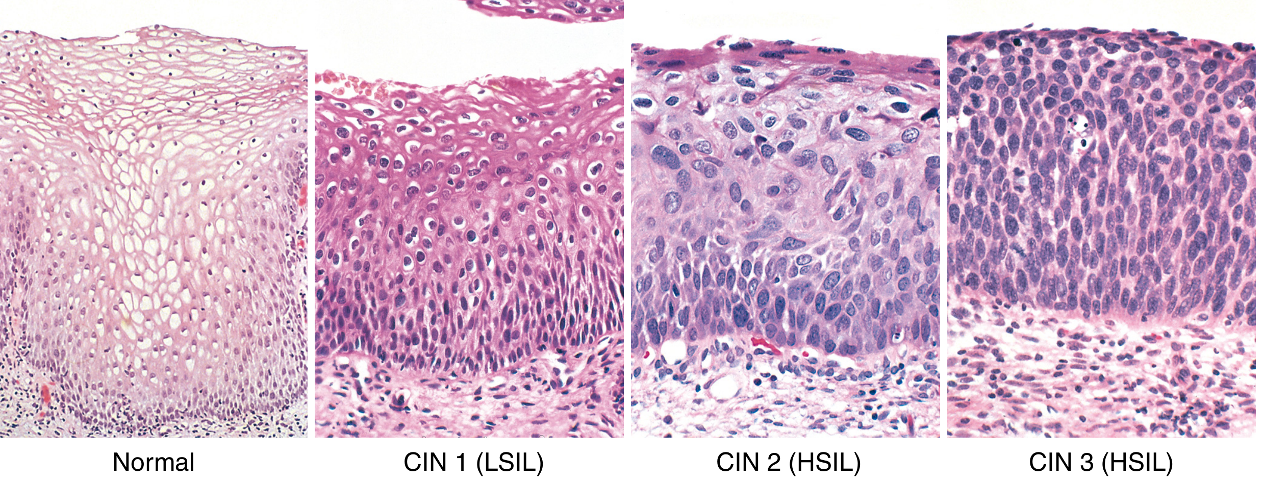
LSIL/CIN 1 has koilocytic atypia. In HSIL, immature basal-type cells expand beyond the lower third of epithelium in CIN 2 and extend toward the full epithelial thickness in CIN 3. Robbins, Cotran & Kumar Pathologic Basis of Disease, p. 923.
5. Squamous Cell Carcinoma
- Malignant squamous cells are seen in the Pap smear.
- Features can include marked pleomorphism, hyperchromasia, irregular nuclei, tumor diathesis, and abnormal keratinization in keratinizing tumors.
This category suggests invasive cervical squamous-cell carcinoma, though biopsy is required for confirmation.
B. Glandular-cell abnormalities
Glandular cells may arise from the endocervix or endometrium.
1. AGC
Atypical Glandular Cells
- Glandular cells show abnormalities beyond reactive changes.
- The report should specify, if possible:
- Atypical endocervical cells
- Atypical endometrial cells
- Atypical glandular cells, not otherwise specified
Meaning: Requires evaluation because it may be associated with significant cervical or endometrial pathology.
2. Atypical Endocervical Cells, Favour Neoplastic
- More abnormal than AGC-not otherwise specified.
- Suggests a possible glandular neoplasm.
3. Endocervical Adenocarcinoma in Situ (AIS)
- A preinvasive glandular neoplastic lesion.
- Often associated with high-risk HPV, particularly HPV 18.
4. Adenocarcinoma
- Malignant glandular cells are identified.
- May be endocervical, endometrial, or from another site.
High-yield correlation table
| Bethesda cytology term | Approximate histological correlation | Significance |
|---|---|---|
| ASC-US | Equivocal, may be reactive or HPV-related | Minor abnormality |
| ASC-H | Possible HSIL | HSIL must be excluded |
| LSIL | HPV effect and CIN 1 | Usually low-risk lesion; many regress |
| HSIL | CIN 2 and CIN 3 | Significant precancerous lesion |
| Squamous cell carcinoma | Invasive squamous carcinoma | Malignant |
| AGC | Possible cervical/endometrial glandular lesion | Requires evaluation |
| AIS | Preinvasive endocervical adenocarcinoma | Glandular precursor lesion |
| Adenocarcinoma | Invasive glandular malignancy | Malignant |
CIN versus Bethesda terminology
| Old histopathology terminology | Bethesda cytology terminology |
|---|---|
| Mild dysplasia | LSIL |
| CIN 1 | LSIL |
| Moderate dysplasia, CIN 2 | HSIL |
| Severe dysplasia, CIN 3 | HSIL |
| Carcinoma in situ | HSIL |
The two-tier terminology is used because clinical management broadly distinguishes lesions likely to regress from higher-grade precancerous lesions that need closer assessment or treatment. Berek & Novak's Gynecology, p. 844.
One-minute exam answer
The Bethesda System is a standardized reporting system for cervical Pap smears. A report comments on specimen adequacy and classifies findings as NILM or epithelial cell abnormality. Squamous abnormalities include ASC-US, ASC-H, LSIL, HSIL, and squamous-cell carcinoma. LSIL corresponds to HPV effect and CIN 1, whereas HSIL corresponds to CIN 2 and CIN 3. Glandular abnormalities include AGC, AIS, and adenocarcinoma. The system helps identify cervical precancerous lesions and guides further evaluation.
Create a one-page Bethesda classification table
Reading File
Finding Sources
Searching PubMed
"cervical cytology" AND Bethesda
Searching the Web
Bethesda System 2014 cervical cytology categories NILM ASC-US ASC-H LSIL HSIL AGC AIS
Cervical Bethesda System (TBS) Classification Table for Pap Smear
| Report category | Abbreviation / full form | Cytological meaning | Histological correlation / significance |
|---|---|---|---|
| Specimen unsatisfactory | - | Inadequate cells or smear obscured by blood, inflammation, poor fixation, etc. | Repeat sample is required. |
| Negative for intraepithelial lesion or malignancy | NILM | No precancer or malignancy detected. Reactive changes, inflammation, atrophy, or organisms may be noted. | Normal/benign cytology. |
| Atypical squamous cells of undetermined significance | ASC-US | Squamous atypia greater than reactive change, but insufficient for LSIL. | Equivocal finding; may be reactive, HPV-related, or early LSIL. |
| Atypical squamous cells, cannot exclude HSIL | ASC-H | Abnormal squamous cells suspicious for HSIL, but diagnostic criteria are incomplete. | Must exclude underlying high-grade lesion. |
| Low-grade squamous intraepithelial lesion | LSIL | HPV cytopathic effect, often with koilocytosis; mild nuclear atypia. | HPV infection + CIN 1 (mild dysplasia). Many regress spontaneously. |
| High-grade squamous intraepithelial lesion | HSIL | Marked atypia: high nuclear-cytoplasmic ratio, hyperchromasia, coarse chromatin, and irregular nuclear outline. | CIN 2 and CIN 3, including severe dysplasia/carcinoma in situ. True precancerous lesion. |
| Squamous cell carcinoma | SCC | Malignant squamous cells in smear. | Suggests invasive cervical squamous carcinoma. Confirm by biopsy. |
| Atypical glandular cells | AGC | Abnormal glandular cells, beyond reactive changes. May be specified as endocervical or endometrial. | May indicate significant endocervical or endometrial pathology. |
| Atypical glandular cells, favour neoplastic | AGC, favour neoplastic | Glandular atypia more suspicious for neoplasia than AGC-NOS. | Possible AIS or invasive adenocarcinoma. |
| Endocervical adenocarcinoma in situ | AIS | Neoplastic glandular cells without evidence of stromal invasion on cytology. | Preinvasive endocervical glandular neoplasia. |
| Adenocarcinoma | - | Malignant glandular cells, possibly endocervical, endometrial, or extrauterine. | Invasive glandular malignancy. |
Key correlation to remember
| Older terminology / CIN | Bethesda terminology |
|---|---|
| HPV changes / koilocytosis | LSIL |
| Mild dysplasia / CIN 1 | LSIL |
| Moderate dysplasia / CIN 2 | HSIL |
| Severe dysplasia / CIN 3 | HSIL |
| Carcinoma in situ | HSIL |
Mnemonic:
ASC-US -> ASC-H -> LSIL -> HSIL -> SCC
ASC-US -> ASC-H -> LSIL -> HSIL -> SCC
- LSIL = CIN 1
- HSIL = CIN 2 + CIN 3
- ASC-H is more serious than ASC-US because HSIL cannot be excluded.
Robbins describes LSIL as CIN 1 with HPV-associated koilocytic changes, while HSIL combines CIN 2 and CIN 3 because these lesions have higher risk of progression and similar clinical management. Robbins, Cotran & Kumar Pathologic Basis of Disease, p. 923.
Explain in brief for 5marks
Reading File
Finding Sources
Searching PubMed
"cervical cytology" AND Bethesda
Searching the Web
Bethesda System cervical cytology Pap smear categories ASC-US ASC-H LSIL HSIL AGC
Bethesda System for Cervical Cytology - 5 Marks
The Bethesda System (TBS) is a standardized method for reporting Pap smear (cervical cytology) findings. It helps detect HPV-associated precancerous lesions and cervical carcinoma.
Components of report
- Specimen adequacy: satisfactory or unsatisfactory.
- General categorization: NILM or epithelial cell abnormality.
- Interpretation: specific cytological category.
Pap smear categories
| Category | Meaning / correlation |
|---|---|
| NILM | Negative for intraepithelial lesion or malignancy. May show inflammation, infection, or atrophy. |
| ASC-US | Atypical squamous cells of undetermined significance. Changes are more than reactive but insufficient for LSIL. |
| ASC-H | Atypical squamous cells, cannot exclude HSIL. |
| LSIL | Low-grade squamous intraepithelial lesion. Includes HPV changes/koilocytosis and CIN 1 (mild dysplasia). |
| HSIL | High-grade squamous intraepithelial lesion. Includes CIN 2 and CIN 3, including severe dysplasia/carcinoma in situ. It is a significant precancerous lesion. |
| SCC | Squamous cell carcinoma. Suggests invasive cancer. |
| AGC | Atypical glandular cells. May arise from endocervix or endometrium. |
| AIS / Adenocarcinoma | Adenocarcinoma in situ or invasive adenocarcinoma. |
Important correlation
- LSIL = HPV infection + CIN 1
- HSIL = CIN 2 + CIN 3
- ASC-H is more serious than ASC-US, as HSIL must be ruled out.
Thus, TBS provides a uniform Pap smear report and identifies women requiring HPV testing, colposcopy, or biopsy. Robbins, Cotran & Kumar Pathologic Basis of Disease, p. 923.
This is a shared conversation. Sign in to Orris to start your own chat.