
I am very new fetal medicine fellow and i need to learn scanning but there are less chances so any other way? Like a game any exercise or anything for stable hand
ultrasound scanning simulation training fetal medicine fellows games exercises hand stability
ultrasound simulator for fetal medicine training 2024 2025
Ways to Learn Fetal Scanning Without a Real Patient
1. Ultrasound Simulators (Best Option)
-
SonoSim / SonoSimulator - A laptop-based system with a physical probe that mimics real probe movements (fanning, rocking, rotating). You practice on real patient cases stored in software. Has 55+ hours of OB/GYN content, fetal anomaly cases, placental pathology, and more. Ask your department if they have access.
-
Vimedix (Surgical Science) - Has a dedicated OB/GYN module called "Catherine" covering first trimester, second trimester fetal anomalies, fetal echo, multiple gestations, and placental conditions. It tracks your performance metrics over time. The 2025 version includes mixed reality (XR) features.
-
Ultrasound Mentor (Surgical Science) - A validated simulation platform specifically shown to improve basic obstetric ultrasound skills in residents (published in PLoS One, 2025).
2. Free / Low-Cost Apps and Online Tools
- Lumify (Philips) - Some training modules available.
- Ultrasound-Guide apps on iOS/Android - Many free quiz-based apps help with image recognition and plane identification (e.g., "SonoWorld," "Ultrasound Cases").
- Fetal Medicine Foundation (FMF) - Free online image library and certification courses. Excellent for learning standard planes and biometry.
- ISUOG - Has free webinars, image quizzes, and practice guidelines for fetal scanning.
3. Hands-on Hand Stability Exercises
- Video games with a controller - Genuinely useful. Games requiring fine joystick movements (e.g., racing sims, flight sims) train the same fine motor control and wrist stability you need for probe handling.
- Drawing/sketching - Freehand anatomical drawing trains hand steadiness and spatial awareness simultaneously.
- Origami or model building - Fine motor tasks that train tremor suppression.
- Steady-hand maze toy - The classic "Operation"-style steady-hand games directly target the still-hand skill.
- Practice with a probe on yourself or a water-filled glove/balloon - Put gel on your forearm or a balloon filled with water and practice slow, deliberate probe movements. Some fellows use a water bottle wrapped in cling film as a makeshift phantom.
4. Structured Learning Approaches
- Watch and narrate - Sit beside your senior during scans, narrate what you'd do next, and mentally rehearse your hand position. This builds the mental model before the motor skill.
- Video libraries - Watch recorded annotated scans on YouTube (search "fetal anomaly scan tutorial" or "ISUOG standard planes"). ISUOG has an official YouTube channel.
- Image interpretation practice - Review saved images daily, label planes, identify structures. This is a skill you can build independently without touching a probe.
- AIUM Courses - The AIUM "Essentials of Obstetric Ultrasound" is a hands-on 1-day course with live models and simulators.
5. Quick Tip on Probe Holding
Important questions asked in fetal medicine exam by icog
ICOG fetal medicine fellowship exam important questions syllabus India
https://icogonline.org/index.html@p=42
ICOG Fetal Medicine Fellowship - Important Exam Topics
SECTION 1: FIRST TRIMESTER SCREENING (Very Heavily Tested)
- NT (Nuchal Translucency) measurement - technique, normal values, cut-offs, quality standards (FMF criteria)
- Combined first trimester screening - NT + serum PAPP-A + free β-hCG + maternal age = risk calculation
- Additional first trimester markers:
- Nasal bone (absent = trisomy 21 risk ↑)
- Ductus venosus (absent/reversed a-wave)
- Tricuspid regurgitation
- Frontomaxillary facial angle
- Detection rates - T21 ~95% detection, 5% FPR with combined screening
- Cell-free DNA (cfDNA / NIPT) - how it works, sensitivity/specificity per chromosome, limitations, PPV vs NPV, what it cannot detect
- Contingent screening strategies (who gets cfDNA vs invasive testing)
SECTION 2: PRENATAL DIAGNOSIS - INVASIVE PROCEDURES
- Amniocentesis - timing (15-20 weeks), technique, risk (0.1-0.3% loss rate), indications
- Chorionic Villus Sampling (CVS) - timing (10-13 weeks), transabdominal vs transcervical, risk (~0.5-1%), confined placental mosaicism
- Cordocentesis (PUBS) - indications, technique, risks
- Chromosomal Microarray (CMA) - when to offer (structural anomaly found), advantages over karyotype, VUS (variants of uncertain significance)
- Exome/genome sequencing - emerging indications
SECTION 3: CHROMOSOMAL ABNORMALITIES
| Condition | Key Features to Know |
|---|---|
| Trisomy 21 (Down) | NT↑, nasal bone absent, short femur/humerus, echogenic bowel, EIF, DS markers |
| Trisomy 18 (Edwards) | Choroid plexus cysts, overlapping fingers, strawberry skull, AVSD, IUGR |
| Trisomy 13 (Patau) | Holoprosencephaly, proboscis, facial clefts, polydactyly, cardiac defects |
| Turner (45X) | Cystic hygroma, hydrops, coarctation, horseshoe kidney |
| Triploidy | Partial mole, severe IUGR, large placenta, skin syndactyly |
SECTION 4: FETAL ANOMALY SCAN (18-22 WEEKS - THE CORE EXAM TOPIC)
- Head: BPD, HC, lateral ventricles, cerebellum, cisterna magna, cavum septum pellucidum
- Face: lips (cleft), orbits, nasal bone, profile
- Spine: 3 ossification centers, neural tube defects
- Heart: 4-chamber view, LVOT, RVOT, 3-vessel view, 3-vessel-trachea view
- Abdomen: AC, stomach, kidneys, bladder, cord insertion, abdominal wall
- Limbs: femur, humerus, hands, feet
- Neural tube defects: anencephaly, spina bifida (banana sign, lemon sign, Arnold-Chiari)
- Ventriculomegaly: borderline vs severe, causes
- Dandy-Walker malformation vs variant vs mega cisterna magna
- Congenital heart defects: VSD, TOF, TGA, HLHS, AVSD
- Abdominal wall defects: gastroschisis vs omphalocele (key differences)
- Renal: multicystic dysplastic kidney, obstructive uropathies, renal agenesis
- Skeletal dysplasias: thanatophoric, achondroplasia, osteogenesis imperfecta
SECTION 5: DOPPLER IN FETAL MEDICINE (Always Examined)
- Umbilical artery (UA) Doppler - S/D ratio, RI, PI; absent end-diastolic flow (AEDF), reversed end-diastolic flow (REDF) - significance
- Middle cerebral artery (MCA) - PSV for fetal anemia, brain-sparing (low PI), cerebro-placental ratio (CPR)
- Ductus venosus (DV) - PIV, absent/reversed a-wave = late sign of compromise
- Uterine artery - notching, RI > 0.58 at 20-24 weeks = preeclampsia/IUGR risk
- Fetal surveillance sequence in FGR: UA → MCA → DV → biophysical profile
SECTION 6: FETAL GROWTH RESTRICTION (FGR)
- Early onset vs late onset FGR (key differences)
- SGA vs FGR (definitions)
- ISUOG/SMFM criteria for FGR diagnosis
- Placental causes vs fetal causes
- Management and timing of delivery (TRUFFLE trial, PORTO study)
- Biophysical Profile (BPP) scoring - 8 parameters, score interpretation
SECTION 7: TWIN PREGNANCIES
- Chorionicity determination - lambda sign (DCDA), T-sign (MCDA); must be done at 11-14 weeks
- MCDA complications: TTTS (Quintero staging I-V), TAPS, sIUGR (types I/II/III), TRAP sequence
- TTTS management: laser photocoagulation of anastomoses, amnioreduction, septostomy
- DCDA complications: discordant anomalies, growth discordance
- Quintero staging for TTTS - I through V
SECTION 8: FETAL HYDROPS
- Immune vs non-immune hydrops (causes)
- Rhesus isoimmunization - mechanism, MCA PSV monitoring (>1.5 MoM), intrauterine transfusion
- Non-immune: cardiac causes, infectious (parvovirus B19), metabolic, structural
- Investigation protocol for hydrops
SECTION 9: SPECIFIC HIGH-YIELD SHORT ANSWERS
- What is the ISUOG definition of ventriculomegaly? (atrial width ≥10 mm)
- Banana sign and lemon sign - what they mean and why they occur
- What is the Quintero staging of TTTS?
- MCA PSV 1.5 MoM - what it indicates and what to do
- Lambda vs T-sign - how to differentiate chorionicity
- What is TRAP sequence? (acardiac twin)
- Difference between gastroschisis and omphalocele (membrane, cord, associated anomalies)
- AEDF vs REDF in umbilical artery - clinical significance
- Soft markers for Down syndrome and their likelihood ratios
- When to offer NIPT vs invasive testing?
- What is confined placental mosaicism?
- Cerebro-placental ratio (CPR) - formula, significance
- IUGR vs SGA - definitions
- Ductus venosus waveform components (a, S, D waves)
- What is the diagnosis when NT >3.5mm with normal karyotype? (need cardiac echo, other structural survey, offer CMA)
SECTION 10: FETAL INTERVENTIONS
- IUT (Intrauterine Transfusion) - indications, technique, calculation of volume
- Laser ablation in TTTS - Solomon technique
- Fetal pleural shunting - pleural effusion, hydrothorax
- Vesico-amniotic shunting - lower urinary tract obstruction
- Radiofrequency ablation (RFA) - used in TRAP sequence, selective reduction
SECTION 11: MATERNAL CONDITIONS AFFECTING FETUS
- Pre-eclampsia: uterine artery Doppler, sFlt-1/PlGF ratio
- Gestational diabetes: macrosomia, polyhydramnios, surveillance
- Antiphospholipid syndrome: recurrent loss, IUGR
- SLE: NLE, heart block (anti-Ro/La antibodies), surveillance
- Thyroid disease and fetal effects
Recommended Books for ICOG Exam Preparation
- Creasy & Resnik's Maternal-Fetal Medicine (the standard comprehensive reference - also in your library)
- Nicolaides / FMF guidelines - free at fetalmedicine.org (first trimester screening, FGR, TTTS)
- ISUOG Practice Guidelines - free PDFs at isuog.org (anomaly scan, Doppler, fetal heart)
- The Fetal Medicine Foundation (FMF) 11-13+6 weeks scan book - essential for first trimester
Exam Pattern (ICOG)
- Written exam: MCQs + short answer questions
- Practical/OSCE: Image interpretation, scan station
- Viva: Structured oral examination on cases
Make a visual poster comparing trisomies 13, 18, and 21 key features
A clean, professional medical education poster comparing Trisomy 13 (Patau syndrome), Trisomy 18 (Edwards syndrome), and Trisomy 21 (Down syndrome). The poster has a dark navy blue background with three side-by-side columns, one for each trisomy, each with a distinct color header: red for Trisomy 13, orange for Trisomy 18, and blue for Trisomy 21. Each column includes the chromosome number prominently at the top, the syndrome name below it, and then clearly organized sections with icons or bullet points covering: Incidence/prevalence, Key facial features, CNS findings, Cardiac defects, Limb/skeletal findings, Other organ anomalies, Ultrasound soft markers, Survival/prognosis. Use medical illustration style with small anatomical icons next to each finding. Include a title at the top: "Autosomal Trisomies - Key Features Comparison". The layout is clean, easy to read, suitable for a fetal medicine study poster. White text on colored backgrounds for headings, light gray text for bullet points.
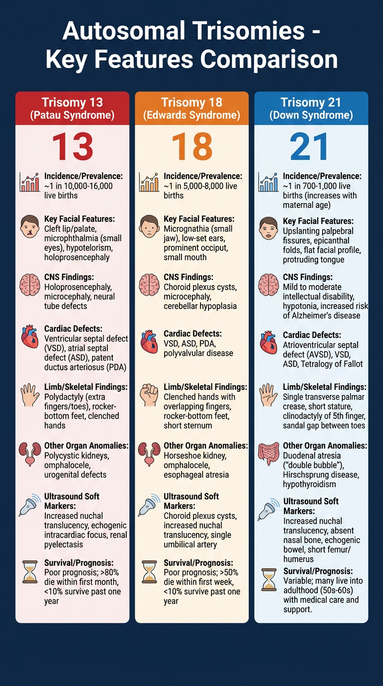
| Feature | Trisomy 13 (Patau) | Trisomy 18 (Edwards) | Trisomy 21 (Down) |
|---|---|---|---|
| Incidence | 1 in 10,000 | 1 in 6,000 | 1 in 700 |
| Face | Holoprosencephaly, proboscis, cyclopia, midline cleft lip/palate | Micrognathia, low-set ears, small mouth | Flat face, upslanting palpebral fissures, epicanthic folds, small ears |
| Brain/CNS | Holoprosencephaly, neural tube defects | Choroid plexus cysts, strawberry skull, SNHL | Mild ventriculomegaly, hypotonia |
| Heart | VSD, ASD, PDA, DORV | VSD, ASD, PDA, AVSD | AVSD (most specific), VSD, ASD |
| Hands/Limbs | Postaxial polydactyly, clubfoot | Overlapping fingers (index over 3rd), rocker bottom feet, short limbs | Short femur/humerus, single palmar crease |
| Other organs | Renal dysplasia, omphalocele | Omphalocele, renal anomalies, IUGR | Echogenic bowel, duodenal atresia ("double bubble"), EIF |
| Key US markers | Midline defects, polydactyly, heart | CPC, overlapping fingers, IUGR, heart | NT ↑, absent nasal bone, short nasal bone, EIF, soft markers |
| Prognosis | Median survival days-weeks; 90% die in 1st year | Median survival days-weeks; 5-10% survive 1 year | Variable; life expectancy ~60 years with support |
- T13 = 13 letters in "HOLOPROSENCEPHALY" - midline brain defects dominate
- T18 = "EDGE" - Edwards, overlapping fingers (index over 3rd), IUGR
- T21 = "DOWN = AVSD + nasal bone absent + soft markers" - the most survivable, with the most soft marker associations

Normal?
⚠️ This is NOT Normal
Patient Details
- Name: Snehlata, 61 years, Female
- Specimen: Endocervical scraping (Vial A) + Endometrial scraping (Vial B)
- Date: 20/07/2026
Vial A - Endocervix
Vial B - Endometrium ⚠️ THE CONCERNING ONE
| Finding | What it means |
|---|---|
| Hyperplastic stratified squamous epithelium in endometrium | Squamous epithelium should NOT be in the endometrium - this is abnormal squamous metaplasia or extension from cervix |
| Koilocytotic changes | Koilocytes = hallmark of HPV infection. These are cells with perinuclear cytoplasmic clearing (halo), nuclear enlargement, hyperchromasia, and irregular nuclear membranes. They are the microscopic signature of active HPV (Berek & Novak's Gynecology) |
| Few endometrial glands | The sample is fragmented and poorly representative |
| Advice: repeat biopsy | The pathologist is NOT satisfied - this sample is inadequate to fully characterize the lesion |
What Could This Mean?
- Cervical squamous pathology (CIN/SCC) extending upward - HPV-driven squamous lesion from the cervix involving or tracked into the endometrial cavity
- Primary squamous cell carcinoma of the endometrium (rare but possible - represents ~1% of uterine cancers)
- Ichthyosis uteri / benign squamous metaplasia - more benign, but must be excluded
- Poor sampling - squamous cells from cervix mixed into endometrial sample during scraping (contamination), giving a false impression
What Should Be Done Next?
- Colposcopy + directed cervical biopsy - to evaluate the cervix and transformation zone properly (the Vial A sample was inadequate)
- HPV genotyping - confirm high-risk HPV
- Pap smear / liquid-based cytology - if not already done
- Repeat fractional curettage - separately curettings from endocervix and endometrium to ensure no contamination
- Hysteroscopy + biopsy - gold standard to visualize and sample the endometrial cavity directly
- MRI pelvis - if a lesion is found, for staging
- Referral to gynecological oncologist - given the patient's age and these findings
Bottom Line
This report shows HPV-associated koilocytotic changes with squamous epithelium where it should not be, in a 61-year-old postmenopausal woman. This is not normal, the sample is inadequate to characterize fully, and the pathologist has correctly advised a repeat biopsy. This woman needs urgent gynae-oncology review. Do not delay.