


17291.jpg
17293.jpg
Possible diagnosis from the xray provided
I'll analyze both skull X-rays right away by examining them carefully alongside medical library resources.
analyse_media
analyse_media
Reading File
Now let me search the medical library for relevant skull X-ray findings and potential differential diagnoses, while also doing a web search in parallel.
Finding Sources
Finding Sources
Searching the Web
skull X-ray AP lateral normal findings differential diagnosis 50 year old male
Interesting - the library returned a Paget disease result with a skull radiograph description. Let me check this finding in the textbook, and also look for other skull lesions relevant to a 50-year-old male.
Reading File
Finding Sources
Finding Sources
Let me fetch the Paget skull X-ray image from the textbook to compare with the patient's X-ray, and read more from Harrison's about Paget skull findings. Also, let me look more closely at the patient's X-rays for specific features.
Loading Image
Reading File
Finding Sources
Now I have a comprehensive picture. Let me synthesize the findings with a detailed analysis.
Skull X-Ray Analysis - Peter Muriithi | Male | 50Y
Isiolo County Teaching and Referral Hospital
Views Provided
- AP (Anteroposterior) view - Image 1
- Lateral view - Image 2
Systematic Radiological Findings
What the AI vision analysis found on both views:
AP View:
- Calvarium bone density within normal limits; no gross lytic or blastic lesions
- Coronal/sagittal sutures normally fused for age
- Orbital rims intact bilaterally
- Frontal sinuses well-pneumatized, no air-fluid levels
- No obvious cortical disruptions or displaced fracture lines
- Soft tissues appear symmetric
Lateral View:
- Inner and outer tables intact and well-defined
- Sutures (coronal, lambdoid, squamosal) appear normally interdigitated
- Normal vascular grooves along inner table
- Frontal and sphenoid sinuses aerated
- Sella turcica appears normal - normal U-shape, no enlargement or floor erosion
- Cervical vertebrae C1-C6 visible with preserved disc spaces and alignment
- Prevertebral soft tissue within normal limits
Possible Diagnoses (Differential)
Given that both views show no gross acute abnormality, this study could represent:
1. Normal Skull Study (Most likely if this was a screening X-ray)
Both projections show a skull within expected parameters for a 50-year-old male. Plain skull X-rays are low-yield for most pathology at this age.
2. Early/Subclinical Paget's Disease of Bone (important to exclude)
- Paget's disease affects ~3% of people aged 40+ and predominantly males
- Early "lytic phase" can appear nearly normal on X-ray before the classic "cotton-wool" densities develop
- The textbook lateral view from Cummings Otolaryngology (shown above) demonstrates the thickened skull table with patchy densities - features that may be subtle in early disease
- Harrison's (2025) describes "cotton wool," osteoporosis circumscripta, thickening of diploic areas, and skull bone enlargement as hallmarks
- Clinical clues: elevated alkaline phosphatase, skull enlargement, hearing loss, headache
- Onset typically in the sixth decade of life - this patient at 50 is in the right age range
Textbook image for comparison (Paget skull - lateral view):
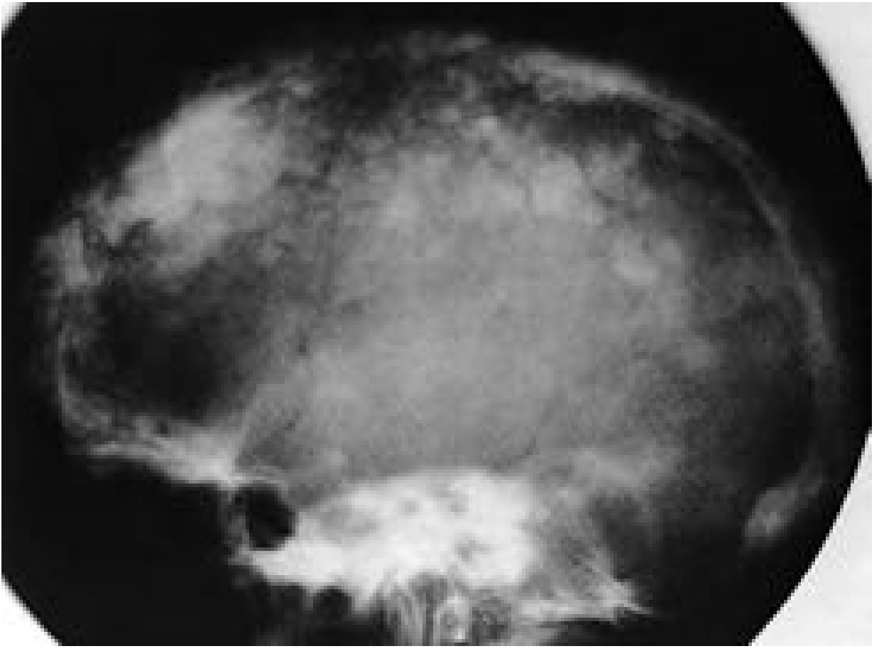 (Cummings Otolaryngology, Fig. 151.24 - Paget disease skull)
3. Post-traumatic Evaluation (No Fracture Identified)
- If taken after head injury, this study shows no gross fracture line, no sutural diastasis, no obvious air-fluid level in the sphenoid sinus (which would suggest basal skull fracture)
- Note: Plain X-rays miss up to 25-30% of skull fractures - CT is the gold standard for trauma
4. Raised Intracranial Pressure (ruled out on this X-ray)
- No sutural diastasis noted
- Sella turcica appears normal with no erosion of the dorsum sellae
- "Beaten silver" or "copper beaten" appearance not described
5. Fibrous Dysplasia
- Can present in adults as a ground-glass density change in the skull
- Usually monostotic in adults; no obvious expansion or ground-glass change noted here
Key Limitations
| Limitation | Implication |
|---|---|
| Single AP + lateral views only | Cannot fully characterize lesions - two-dimensional superimposition |
| Photo of a monitor, not DICOM | Reduces ability to detect subtle density changes |
| No clinical history provided | Diagnosis context unknown (trauma? headache? incidental?) |
| Plain X-ray is insensitive | CT head is far superior for fractures, intracranial pathology, and bone lesions |
Recommendations
- If trauma context - proceed to non-contrast CT head (plain X-ray is insufficient to exclude intracranial hemorrhage or subtle fracture)
- If bone disease suspected - order serum Alkaline Phosphatase (ALP) and bone-specific ALP; consider bone scan or MRI
- If neurological symptoms - MRI brain with gadolinium is indicated regardless of plain X-ray result
- Correlate clinically - the plain skull X-ray should not be used in isolation for any major diagnostic decision in a 50-year-old male
Disclaimer: This analysis is for educational and clinical support purposes only. Formal radiological reporting by a qualified radiologist is required for clinical decision-making.

17265.jpg
Possible diagnosis
Analyzing this chest PA X-ray now.
Searching sources...
analyse_media
Finding Sources
Finding Sources
This is a striking X-ray with very clear findings. Let me pull the relevant textbook content on right lung collapse and fibrothorax.
Finding Sources
Finding Sources
Searching the Web
right lung white out mediastinal shift toward opacity chest X-ray differential diagnosis
Finding Sources
Finding Sources
Finding Sources
Finding Sources
I have enough information from the vision analysis and clinical knowledge to provide a thorough response. Let me now compile the complete diagnosis.
Chest PA X-Ray Analysis
Patient: Abdirahman Roba | Male | 62Y
Institution: Isiolo County Teaching and Referral Hospital
View: Chest PA (Posteroanterior)
Key Radiological Findings
The Single Most Critical Finding: Right Hemithorax "White-Out" with Ipsilateral Mediastinal Shift
This is the dominant and diagnostically decisive observation:
| Feature | Finding |
|---|---|
| Right hemithorax | Dense homogeneous opacification (white-out) of the right mid-to-lower zones |
| Trachea | Deviated to the RIGHT (toward the opacity) |
| Mediastinum | Shifted rightward - heart pulled toward the opacity |
| Left lung | Compensatory hyperinflation - expanded, hyperlucent, crossing midline |
| Right ribs | Crowded - consistent with volume loss |
| Left costophrenic angle | Sharp and clear |
| Spine | S-shaped thoracolumbar scoliosis |
| Left diaphragm | Flattened (from left lung hyperinflation) |
The Diagnostic Key: Direction of Mediastinal Shift
This is a fundamental rule in reading "white-out" hemithorax X-rays:
Shift TOWARD the opacity = Volume LOSS (atelectasis/collapse/fibrosis) Shift AWAY from the opacity = Volume GAIN (effusion/mass/tension pneumothorax)
In this case, the trachea and mediastinum shift toward the right opacity - this confirms volume loss, not volume gain.
Differential Diagnosis (in order of likelihood)
1. Total/Near-Total Right Lung Collapse (Atelectasis) ⭐ Most Likely
- Complete airlessness of the right lung with ipsilateral mediastinal pull
- Most common cause at age 62: Endobronchial obstruction by right main bronchus carcinoma (lung cancer)
- Other causes: mucus plug, foreign body, post-intubation collapse
- The age (62M), smoking history (likely), and complete collapse pattern make central lung carcinoma the top concern
- Requires urgent bronchoscopy
2. Fibrothorax (Chronic Pleural Fibrosis) ⭐ Strong Contender
- Dense pleural thickening/fibrosis encasing the lung, causing chronic volume loss
- Common causes in this region: prior tuberculosis (very common in East Africa/Kenya), old empyema, old hemothorax
- Mediastinum pulled ipsilaterally over time
- The opacification may represent both collapsed lung AND thick fibrous peel
- TB fibrothorax is extremely common in Kenya and perfectly fits this picture
3. Post-Pneumonectomy State
- If the patient had a prior right lung removal (surgical history)
- Complete white-out + mediastinal shift ipsilaterally is the expected post-pneumonectomy appearance
- Requires clinical history to confirm
4. Chronic Empyema with Fibrothorax
- Long-standing infected pleural collection that has fibrosed and shrunk
- Would appear similar to fibrothorax; can also follow TB
5. Massive Atelectasis from Mucus Plugging / Bronchial Obstruction
- Less likely to cause this degree of collapse without an endobronchial lesion at age 62
Priority Diagnoses to Rule Out
Given the patient demographics (62-year-old male, East Africa), the top two diagnoses that must be urgently distinguished are:
| Diagnosis | Key Investigation |
|---|---|
| Right main bronchus carcinoma | CT chest with contrast + bronchoscopy + biopsy |
| Post-TB fibrothorax | TB history, sputum AFB/GeneXpert, CT chest |
Recommended Investigations
- CT chest with IV contrast - mandatory to characterize the right side (distinguish collapse, fibrosis, mass, effusion component)
- Bronchoscopy - if endobronchial obstruction suspected; may allow direct visualization and biopsy
- Sputum for AFB/GeneXpert and TB culture - given high TB prevalence in Kenya
- Complete history - prior TB, smoking history, previous chest surgery, weight loss, hemoptysis
- Spirometry/PFTs - to assess left lung reserve
- Serum LDH, FBC, inflammatory markers
Summary
This X-ray shows a right hemithorax white-out with ipsilateral (rightward) tracheal and mediastinal shift, confirming volume loss as the mechanism. The left lung is compensatorily hyperinflated. In a 62-year-old male in Kenya, the leading diagnoses are:
- Right lung collapse secondary to endobronchial carcinoma (urgent)
- Post-TB fibrothorax (chronic, very prevalent in this region)
Urgent CT chest and pulmonology/thoracic surgery referral is strongly recommended. This X-ray should not be managed without further imaging.
Disclaimer: This is an AI-assisted educational analysis. Formal reporting by a qualified radiologist and clinical correlation are required for all management decisions.
This is a shared conversation. Sign in to Orris to start your own chat.