
tragal sign in ent All about
Reading File
The tragal sign is a clinical sign in ENT used to distinguish between external otitis (otitis externa) and acute otitis media. Pressing the tragus causes pain in otitis externa but not in otitis media.
Searching sources...
Finding Sources
Searching the Web
tragal sign ENT otitis externa clinical significance
Reading File
Finding Sources
Reading File
Finding Sources
Reading File
Finding Sources
I have all the information needed for a thorough answer. Let me compile it now.
Tragal Sign in ENT - Complete Guide
What is the Tragus?
The tragus is the small, cartilaginous, tongue-shaped projection of the auricle that lies anterior to the external auditory canal (EAC) opening. It partially covers and protects the entrance to the ear canal.
Definition of the Tragal Sign
The tragal sign (also called tragal tenderness) refers to pain elicited upon applying direct pressure to the tragus. It is considered positive when this maneuver produces significant pain or discomfort.
- Simultaneously, traction of the pinna (pulling the outer ear upward and backward) also produces pain in the same condition.
- Together, tragal pressure and pinna traction are the two classic physical examination maneuvers for diagnosing otitis externa.
Clinical Significance - What Does It Indicate?
Primary Association: Acute Otitis Externa (AOE) - "Swimmer's Ear"
A positive tragal sign is the hallmark clinical finding of acute otitis externa, distinguishing it from other causes of ear pain.
When pressure is applied to the tragus, it compresses and transmits force to the inflamed walls of the external auditory canal. This is extremely painful because:
- The EAC skin overlies thinly padded periosteum (bone or cartilage)
- In otitis externa, the skin is intensely inflamed, edematous, and erythematous
- Even minimal mechanical deformation of the canal causes severe pain
As noted in KJ Lee's Essential Otolaryngology:
"Tragal sign - pressure on the tragus induces extreme pain" - this is listed as a cardinal sign of Acute Otitis Externa.
Harrison's Principles of Internal Medicine (2025) confirms: "Physical exam is notable for pain on movement of the auricle or tragus and an external auditory canal that is erythematous, edematous, inflamed."
Tragal Sign: Key Differentiating Value
| Feature | Otitis Externa | Acute Otitis Media |
|---|---|---|
| Tragal sign (tragal pressure pain) | Positive (hallmark) | Negative |
| Pinna traction pain | Positive | Negative |
| TM mobility | Normal | Reduced |
| Otorrhea | Purulent, canal-based | Through perforated TM |
| Fever | Rare (unless cellulitis) | Common |
| Canal appearance | Red, edematous, debris-filled | Often normal canal |
Harrison's notes: "AOM with tympanic membrane rupture can be associated with ear discharge and debris in the ear canal, but (unlike otitis externa) without sensitivity to movement of the auricle."
This makes the tragal sign one of the most practically useful bedside tests to distinguish between these two common causes of otalgia.
Full Clinical Picture of AOE (Context of Tragal Sign)
Symptoms:
- Rapid onset of severe ear pain (< 48 hours)
- Usually unilateral
- Pruritus
- Otorrhea (often purulent)
- Aural fullness and hearing loss (if canal is significantly edematous)
- Fever is rare (if present, suggests periauricular cellulitis or necrotizing OE)
Signs on Examination:
- Positive tragal sign (pathognomonic)
- Positive pinna traction sign
- Erythema and edema of EAC (may nearly occlude the canal)
- Purulent or serous debris in canal
- Normal TM mobility on pneumatic otoscopy (key differentiator from AOM)
- Preauricular or cervical lymphadenopathy
Grading of Otitis Externa (Severity Classification)
| Grade | Features |
|---|---|
| Mild | Pruritus, minimal edema, discomfort |
| Moderate | Pain, partial canal occlusion, drainage |
| Severe | Severe pain, complete canal occlusion, systemic symptoms (fever, fatigue), possible lymphadenopathy, cellulitis of auricle |
Pathogens Causing AOE
- Pseudomonas aeruginosa - most common (especially in swimmer's ear)
- Staphylococcus aureus - second most common
- Staphylococcus epidermidis, other gram-negative rods
- Bacterial infections account for ~90% of AOE cases
- Fungal (Aspergillus, Candida) - ~10%; typically causes pruritus and discharge without significant pain (so tragal sign may be less prominent in fungal OE)
Necrotizing (Malignant) Otitis Externa - Special Consideration
In malignant/necrotizing otitis externa, the tragal sign is positive but accompanied by additional alarming features:
- High fever
- Granulation tissue or necrotic tissue in the EAC floor
- Patient is usually diabetic or immunocompromised (elderly, HIV, AIDS)
- Risk of osteomyelitis of temporal bone
- Can extend to involve cranial nerves (CN VII most commonly), causing facial palsy
- Requires urgent CT/MRI imaging and IV antipseudomonal antibiotics (e.g., ciprofloxacin)
Referred Ear Pain and Tragal Sign
The tragal sign is negative in referred otalgia (pain referred to the ear from distant sources), which helps rule out otitis externa when the sign is absent despite severe ear pain. Referred otalgia can arise via:
- CN V - dental pathology, TMJ disorders
- CN VII - Ramsay Hunt syndrome (herpes zoster oticus), Bell's palsy
- CN IX - tonsillitis, pharyngitis
- CN X - GERD, myocardial infarction (rare)
- C2/C3 - cervical spine pathology
Diagnostic Criteria for AOE (AAO-HNS Guidelines)
A clinical diagnosis of AOE requires all three:
- Rapid onset (symptoms within 48 hours in the past 3 weeks)
- Symptoms of ear canal inflammation: ear pain, itching, or fullness (±hearing loss or jaw pain)
- Signs of ear canal inflammation: tenderness of tragus/pinna OR diffuse canal edema/erythema (±otorrhea, TM erythema, cellulitis, or lymphadenitis)
As cited from the AAO-HNS clinical practice guideline on acute otitis externa, tragal/pinna tenderness is listed as a primary sign sufficient to meet diagnostic criteria.
Treatment (Brief Overview)
| Severity | Treatment |
|---|---|
| Mild | Acidifying irrigation (dilute acetic acid/white vinegar) |
| Moderate-Severe | Topical antibiotics (ciprofloxacin, aminoglycosides, polymyxin B) ± topical steroids; aural toilet (debridement); otwick if canal nearly occluded |
| Necrotizing OE | IV antipseudomonal antibiotics, surgical debridement, imaging, hospitalization |
Key principles:
- Aural toilet (debridement) is essential to allow drops to penetrate
- Otwick (ear wick/ribbon gauze) placed when canal edema prevents drop penetration; replaced every 3-5 days
- Keep ear dry (no swimming, use ear plugs for bathing)
- Oral antibiotics only if significant cellulitis beyond the canal
Summary
| Point | Detail |
|---|---|
| Sign | Tragal sign = pain on pressing the tragus |
| Positive in | Acute Otitis Externa (hallmark sign) |
| Negative in | Acute Otitis Media, referred otalgia |
| Mechanism | Pressure transmitted to inflamed, periosteum-bound EAC skin |
| Paired maneuver | Pinna traction (also positive in OE) |
| Fungal OE | Less pain - tragal sign may be less prominent |
| Necrotizing OE | Positive + systemic features (fever, CN palsies, immunocompromised host) |
Sources: KJ Lee's Essential Otolaryngology; Harrison's Principles of Internal Medicine 22E (2025); Merck Manual - Acute External Otitis; CMAJ - Otitis Externa
reason for positive and steps
The tragal sign is positive because pressing the tragus mechanically compresses the inflamed external auditory canal skin, causing pain. The steps involve standing in front of the patient and pressing the tragus inward with your index finger.
Searching sources...
Finding Sources
Finding Sources
Reading File
Finding Sources
Reading File
Finding Sources
Now I have all the anatomical basis needed. Let me fetch the ear anatomy image too.
Loading Image
Loading Image
Here is a complete breakdown of the reason for a positive tragal sign and the steps to elicit it, fully grounded in anatomy.
Tragal Sign - Reason for Positive & How to Elicit It
Anatomy First (The Basis of Everything)
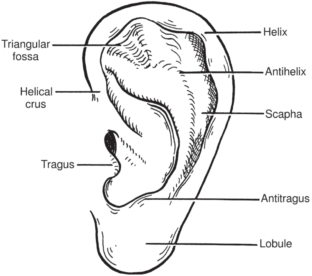
The tragus is a small cartilaginous flap of fibrocartilage (the same type as the cartilaginous EAC) sitting directly anterior to the entrance of the external auditory canal. It is structurally continuous with the canal.
The EAC has two distinct zones:
| Zone | Length | Skin Characteristics |
|---|---|---|
| Cartilaginous (outer 1/3) | ~8 mm | Has sebaceous glands, ceruminous glands, hair follicles, some subcutaneous fat |
| Bony (inner 2/3) | ~16 mm | Skin is extremely thin, tightly adherent to periosteum, no subcutaneous tissue |
"The skin over the bony canal is tight and has no subcutaneous tissue except periosteum." - KJ Lee's Essential Otolaryngology
This anatomical fact is the core reason for the pain.
Nerve Supply of the EAC (Why It Hurts So Much)
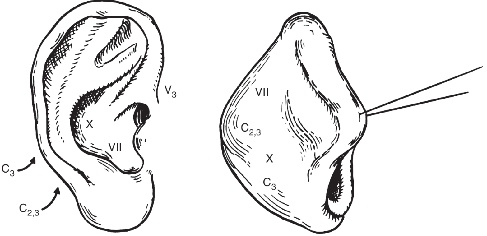
The EAC and auricle are supplied by multiple sensory nerves:
- V3 (auriculotemporal branch of mandibular nerve) - anterior EAC and tragus area
- CN VII (facial nerve, via posterior auricular branch) - posterior canal skin
- CN X (Arnold's nerve / auricular branch of vagus) - posterior/inferior canal floor (Arnold's reflex - cough when EAC touched)
- C2, C3 (via great auricular nerve and lesser occipital nerve) - auricle and periauricular skin
This rich, multi-nerve innervation of the canal means any inflammation there generates disproportionately severe pain - far more than the visible degree of swelling would suggest.
Why is the Tragal Sign POSITIVE in Otitis Externa?
The mechanism works through a direct mechanical pressure-pain pathway in 3 steps:
Step 1 - Inflammation of the EAC skin
In acute otitis externa (AOE), bacterial pathogens (typically Pseudomonas aeruginosa) invade the macerated skin of the EAC. The canal skin becomes:
- Intensely erythematous and edematous
- Infiltrated with inflammatory cells and mediators (prostaglandins, bradykinin, histamine)
- Already hypersensitive due to lowered pain threshold from inflammation
Step 2 - The tragus acts as a direct compressor of the canal
The tragus sits like a "lid" over the anterior EAC opening. When you press it inward:
- It physically deforms and compresses the anterior cartilaginous canal wall
- This compression is transmitted inward along the canal
- The inflamed, swollen canal skin - which has no cushion of subcutaneous tissue in the bony portion - is crushed between the pressing force and the rigid bony/cartilaginous canal walls
Step 3 - Stimulation of sensitized nerve endings
The already hyperalgesia-sensitized nerve fibers (V3, VII, X) within the inflamed periosteum and dermis are mechanically stimulated by this compression. The result is extreme, disproportionate pain - the positive tragal sign.
Why is it NEGATIVE in Otitis Media?
In acute otitis media (AOM), the inflammation is confined to the middle ear behind the tympanic membrane. The external auditory canal skin is normal - not inflamed, not edematous. Pressing the tragus compresses normal canal skin against the normal bony walls - no sensitized nerve endings, no pain. Hence negative tragal sign.
This is the key differentiating anatomical principle.
Steps to Elicit the Tragal Sign
Patient Positioning
- Patient seated comfortably, facing you
- Both ears should be tested (compare affected vs unaffected side)
- Explain the maneuver to the patient beforehand: "I am going to press gently in front of your ear - tell me if it hurts"
Step-by-Step Technique
Step 1: Stand or sit facing the patient, at the same level.
Step 2: With your index finger (or thumb), locate the tragus - the small cartilage flap just anterior to the ear canal opening.
Step 3: Apply firm but gentle inward pressure on the tragus, pressing it posteriorly toward the canal.
- Use the pad of the finger, not the fingernail
- Press for 1-2 seconds
- Do not jab - apply steady, controlled pressure
Step 4: Observe the patient's response:
- Positive: Patient winces, pulls away, or reports significant pain/tenderness
- Negative: No pain or only mild discomfort
Step 5 (Companion maneuver - Pinna Traction Test):
- Grip the pinna (upper helix) between thumb and index finger
- Pull it upward and backward (posterosuperiorly)
- This also compresses/stretches the cartilaginous canal
- Positive = pain in otitis externa; Negative in otitis media
Step 6: Compare both sides - the unaffected ear should have no tenderness on the same maneuver.
Summary Table
| Aspect | Detail |
|---|---|
| Anatomical basis | Tragus is fibrocartilage, structurally continuous with anterior EAC wall |
| Why bony canal causes severe pain | Skin tightly adherent to periosteum, zero subcutaneous cushion |
| Nerve supply | V3, CN VII, CN X, C2/C3 - rich multi-nerve supply |
| Mechanism | Pressure on tragus → compresses inflamed canal skin → stimulates sensitized nociceptors |
| Positive in | Acute otitis externa (hallmark sign) |
| Negative in | Otitis media, referred otalgia (normal canal skin) |
| Companion test | Pinna traction test (same mechanism, different direction of force) |
| Finger used | Index finger pad |
| Direction of pressure | Inward/posteriorly on the tragus |
| Compare | Always compare with opposite ear |
Sources: KJ Lee's Essential Otolaryngology; Harrison's Principles of Internal Medicine 22E (2025, McGraw Hill Medical); StatPearls - Otitis Externa
This is a shared conversation. Sign in to Orris to start your own chat.