
From Guyton and Hall Textbook of Medical Physiology Fourth, South Asia edition, writer, 10 - long mark answer on Sarcomere-define,diagram;Sliding Filament Theory-explain
Reading File
Finding Sources
Finding Sources
Searching PubMed
"skeletal muscle contraction" AND "sliding filament"
Searching the Web
Guyton Hall Textbook Medical Physiology South Asia edition Fourth sarcomere sliding filament theory
Finding Sources
Finding Sources
Searching Images
sarcomere relaxed and contracted sliding filament diagram actin myosin Z disc I band A band H zone
Reading File
Sarcomere and Sliding Filament Theory
Definition of sarcomere
A sarcomere is the structural and functional contractile unit of striated muscle. It is the part of a myofibril between two successive Z discs (Z lines).
It contains:
- Thin filaments: actin, with troponin and tropomyosin
- Thick filaments: myosin
- Supporting proteins, especially titin, which helps maintain the position and elasticity of thick filaments
During contraction, each sarcomere shortens. This causes shortening of the myofibrils, muscle fibres, and finally the whole muscle.
Guyton and Hall Textbook of Medical Physiology, Ch. 6, “Contraction of Skeletal Muscle”
Note: exact page numbering differs between printings of the Fourth South Asia edition.
Note: exact page numbering differs between printings of the Fourth South Asia edition.
Diagram of a sarcomere
Relaxed sarcomere

Z disc                                                      Z disc
  |                                                           |
  |---- I band ----|----------- A band -----------|---- I ----|
  |                |                              |     band   |
  |---- Actin ----->>>>>>>        <<<<<<<----- Actin ----|
                   \              H zone         /
                    \________ Myosin ___________/
                              M line

Key:
Z disc  = boundary of one sarcomere; attachment point for actin
I band  = actin only
A band  = entire length of thick myosin filament
H zone  = myosin only, at the centre of A band
M line  = middle line of the sarcomere; holds myosin filaments
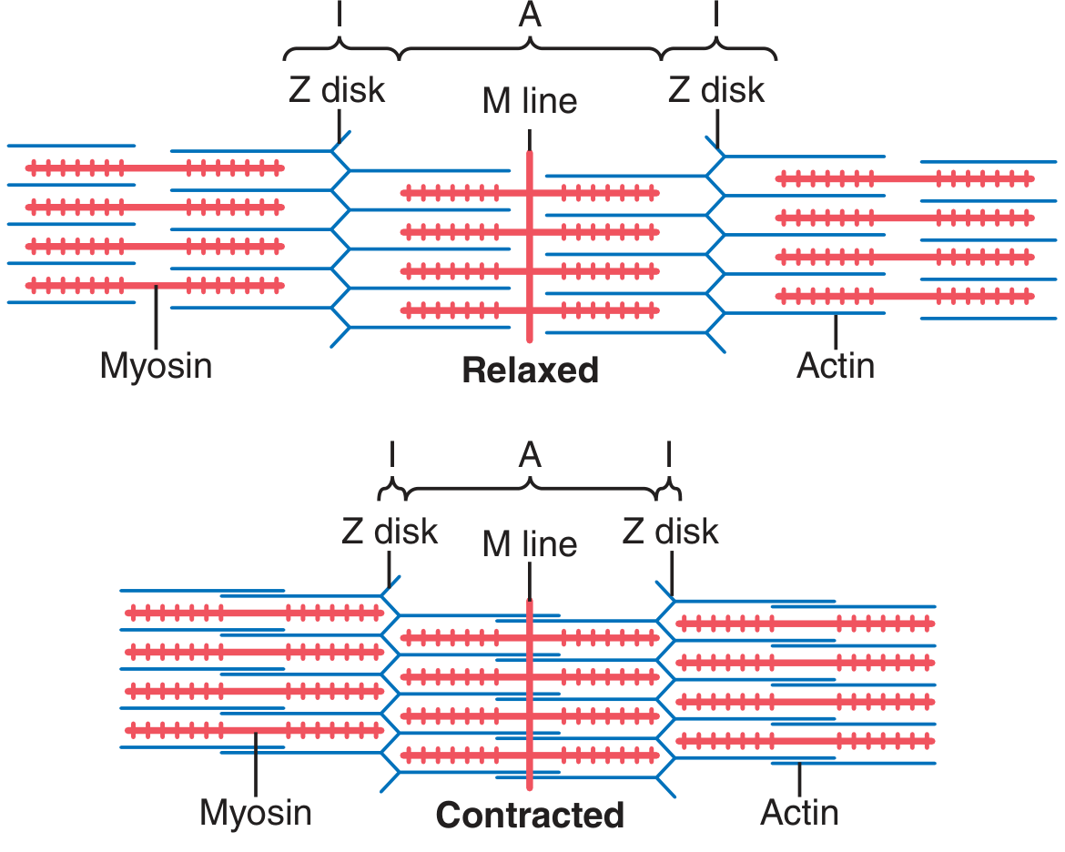
Important structural features
| Part | Composition / significance |
|---|---|
| Z disc | Forms either end of a sarcomere and anchors thin actin filaments |
| Actin filament | Thin filament that extends inward from the Z disc |
| Myosin filament | Thick filament situated centrally, with projecting cross-bridges |
| M line | Centre of sarcomere where thick filaments are held together |
| I band | Light band containing only thin actin filaments |
| A band | Dark band corresponding to the full length of thick myosin filaments |
| H zone | Central lighter part of A band where only myosin is present |
| Titin | Elastic protein connecting myosin to the Z disc and maintaining alignment |
Sliding Filament Theory of Muscle Contraction
Definition
The sliding filament theory states that muscle contraction occurs because the thin actin filaments slide inward between thick myosin filaments. The filaments themselves do not shorten. Rather, their overlap increases and the Z discs are pulled closer together.
This shortening of individual sarcomeres produces contraction of the entire skeletal muscle.
Explanation of the mechanism
1. Resting state
At rest:
- The active sites on actin are covered by tropomyosin.
- Tropomyosin is held in this blocking position by the troponin complex.
- Myosin cross-bridges cannot effectively bind to actin.
- The sarcomere remains relaxed.
2. Calcium release
When a nerve impulse reaches skeletal muscle:
- An action potential spreads over the sarcolemma and into the T tubules.
- The sarcoplasmic reticulum releases large amounts of Ca²⁺ into the sarcoplasm.
- Calcium binds with troponin C.
3. Exposure of actin active sites
Binding of Ca²⁺ to troponin causes a conformational change:
- Tropomyosin moves away from the active sites on actin.
- The myosin-binding sites on actin become exposed.
- Myosin heads can now attach to actin and form cross-bridges.
4. Cross-bridge formation
The energized myosin head, containing ADP and phosphate, attaches to an exposed active site on actin.
Myosin head + Actin active site → Actin-myosin cross-bridge
5. Power stroke
- Release of phosphate and then ADP causes the myosin head to bend.
- The attached actin filament is pulled toward the centre, that is, toward the M line.
- This is called the power stroke.
- As many cross-bridges act repeatedly, the thin filaments slide farther inward.
6. Detachment of myosin from actin
- A fresh ATP molecule binds to the myosin head.
- This reduces myosin's affinity for actin.
- The cross-bridge detaches.
ATP is necessary for separation of myosin from actin. In the absence of ATP, persistent actin-myosin attachment occurs, as in rigor mortis.
7. Reactivation of myosin head
- ATP is hydrolysed by myosin ATPase into ADP and phosphate.
- Energy released re-cocks or re-energizes the myosin head.
- The myosin head binds to another active site farther along the actin filament.
- The cycle repeats as long as Ca²⁺ and ATP are available.
Changes in bands during contraction
Relaxation                         Contraction
Z disc     Z disc                  Z disc   Z disc
 |----------|                       |------|
Sarcomere is long                  Sarcomere shortens

I band: wide                       I band: shortens
H zone: present                    H zone: shortens or disappears
A band: unchanged                  A band: unchanged
Actin and myosin: same length      Actin and myosin: same length
Overlap: less                      Overlap: increased
Thus, during contraction:
- Z discs move closer together
- Sarcomere length decreases
- I band decreases in length
- H zone becomes narrower and may disappear
- A band remains constant
- Length of actin and myosin filaments remains unchanged
- Degree of actin-myosin overlap increases
Conclusion
The sliding filament theory explains contraction as a cyclic interaction between actin and myosin cross-bridges. Calcium initiates cross-bridge formation by exposing actin active sites, while ATP supplies energy for detachment and repeated cycling of myosin heads. Repeated inward sliding of actin filaments pulls Z discs together, shortens sarcomeres, and produces skeletal muscle contraction.
This is a shared conversation. Sign in to Orris to start your own chat.