
pyogenic granuloma
pyogenic granuloma clinical appearance
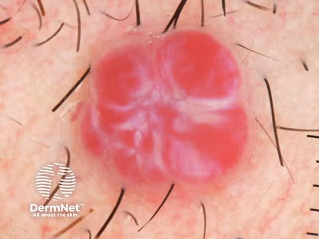
Clinical dermatology image of a solitary cutaneous pyogenic granuloma (lobular capillary hemangioma) on the skin. The lesion is a small, raised, pedunculated nodule with a bright red, vascular surface. It appears lobulated, with pale, intervening septa or lines separating conspicuous vascular lobules. The surface is smooth but friable, often bleeding with minor trauma; in this image there is no active bleeding at capture. Surrounding skin is normal and hair-bearing, indicating a non-mucosal site. The lesion is well-circumscribed, with a slightly translucent to erythematous appearance, consistent with superficial dermal capillary proliferation. Clinically, pyogenic granuloma presents as a rapidly growing, painless to tender vascular lesion that bleeds easily. The vascular lobules are arranged in a lobular architecture within a fibromucinous stroma; histology would show capillary channels lined by flattened endothelial cells. Differential diagnoses include cherry angioma, Kaposi sarcoma, amelanotic melanoma, squamous cell carcinoma, and vascular malformations; however, the rapid growth, friability, and classic lobular pattern strongly support pyogenic granuloma. Diagnostic significance lies in distinguishing benign vascular proliferation from malignancy or infection. Management may involve conservative destruction via curettage or laser, topical therapy, or surgical excision with biopsy for histopathology. Clinical correlation with trauma history is helpful. No systemic involvement is expected.
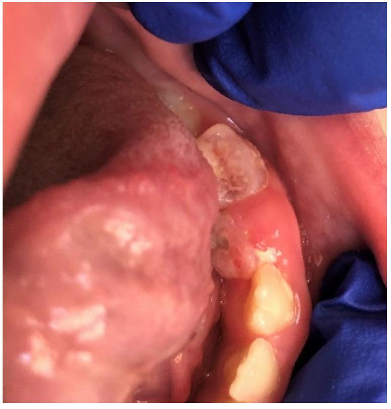
This clinical photograph displays a pyogenic granuloma in the oral cavity of a pediatric patient, specifically located on the gingiva adjacent to deciduous teeth 74 and 75. The lesion presents as a smooth, elevated, and sessile mass characterized by a broad base of attachment. The surface of the growth exhibits a pink-to-reddish hue, which is characteristic of highly vascularized reactive inflammatory tissue. The surrounding gingival tissue shows clinical signs of localized inflammation, including erythema and swelling, with a loss of the normal stippled texture. The teeth in the immediate vicinity appear to have significant dental plaque and debris, suggesting poor oral hygiene as a likely precipitating factor for this reactive hyperplasia. The image illustrates the typical appearance of a non-neoplastic, hyperplastic lesion often occurring in response to chronic low-grade irritation or trauma. This material is useful for dental and oral pathology education regarding the differential diagnosis of localized gingival enlargements.
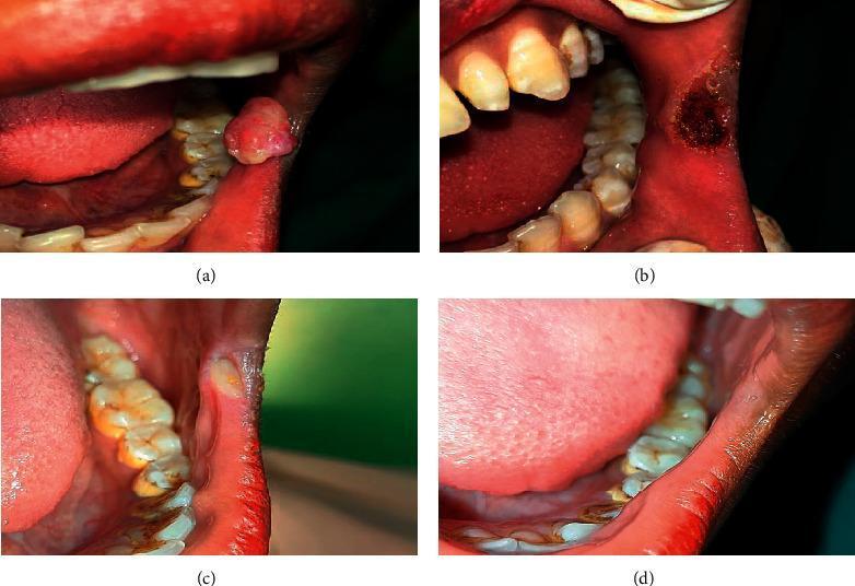
This set of four intraoral clinical photographs documents the management and healing progression of a pyogenic granuloma in the retrocommissural region. Image (a) presents the initial clinical finding: a reddish-pink, sessile, exophytic growth with a rounded morphology located on the buccal mucosa near the oral commissure. Image (b) shows the immediate postoperative appearance following surgical excision using a 980 nm diode laser; the site exhibits a charred, dark-pigmented circular wound with no active bleeding, characteristic of laser ablation. Image (c) demonstrates a follow-up stage showing significant regression and early secondary intention healing, with a small area of granulation tissue or resolving ulceration visible. Image (d) displays complete clinical resolution and mucosal healing, with no residual lesion or scarring evident in the retrocommissural area. This series serves as an educational example of oral soft tissue pathology and the efficacy of laser therapy in oral surgery for benign vascular lesions.
"pyogenic granuloma" treatment
Pyogenic Granuloma (Lobular Capillary Hemangioma)
Definition & Terminology
Epidemiology
- One of the most common vascular tumors of infants and children; also occurs in adults
- Particularly common in pregnant women (called granuloma gravidarum when on the gingiva)
- Can occur at any age
- Andrews' Diseases of the Skin, p. 928
Etiology & Precipitating Factors
- Often develops at sites of minor trauma or chronic irritation
- Associated with pregnancy (hormonal influence)
- Drug-induced PG is well documented with:
- Isotretinoin (multiple exuberant trunk/periungual lesions)
- Vemurafenib
- Capecitabine
- Indinavir
- EGFR inhibitors (paronychia-associated lesions)
- Some data show higher Bartonella seropositivity in PG patients, but definitive etiologic role is not established
- Andrews' Diseases of the Skin, pp. 928-929
Clinical Features
- Solitary, red, rapidly growing papule or nodule
- Sessile or pedunculated, friable, with surface erosion
- Often has a subtle collarette of scale at the base
- Bleeds profusely with minimal trauma (known as "Band-Aid disease" in children)
- Typical sites:
- Exposed skin: hands, forearms, face (cheek, forehead)
- Mucous membranes - especially gingiva (in pregnancy)
- Oral cavity: buccal mucosa, lips, tongue, palate
- Nail bed or sole of foot (can mimic melanoma)
- Not typically painful, though it can be tender
Histopathology
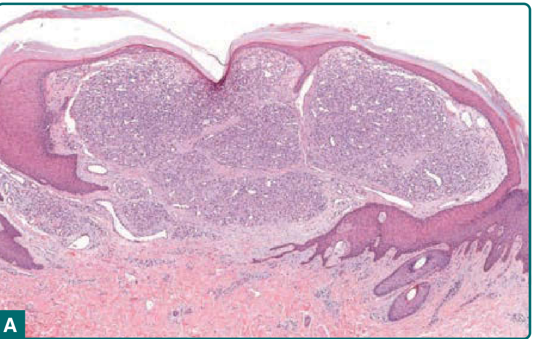
- Lobular capillary hemangioma: lobules of small capillaries lined by bland endothelial cells
- Lobules separated by connective tissue septa
- Set within a loose fibroblastic stroma (fibromucinous)
- Epidermal collarette surrounds the dermal vascular proliferation (characteristic architecture)
- With time: epidermis thins then erodes
- Heavy secondary staphylococcal colonization is common
- Intravascular PG: a rare variant appearing as lobular capillary proliferation within a vein
- Immunostaining: endothelial cells positive for CD31, CD34
Differential Diagnosis
| Condition | Distinguishing Features |
|---|---|
| Amelanotic melanoma | Irregular borders, histology |
| Cherry angioma | Smaller, does not bleed as readily, stable |
| Kaposi sarcoma | Multiple lesions, immunocompromised host |
| Squamous cell carcinoma | Keratotic, histology |
| Infantile hemangioma | Earlier onset, GLUT1 positive |
| Vascular malformation | Present at birth, grows proportionally |
Course & Behavior
- Does not spontaneously involute (unlike infantile hemangiomas)
- After treatment, can recur and form satellite lesions surrounding the original site
- Drug-induced lesions regress after dose reduction or drug discontinuation
- Fitzpatrick's Dermatology, p. 2093
Treatment
| Modality | Notes |
|---|---|
| Curettage + electrocautery/fulguration | First-line; usually curative |
| Shave excision + fulguration/aluminum chloride | Effective, allows histologic assessment |
| Surgical excision | For recalcitrant or larger lesions |
| Laser (CO2, pulsed-dye, Nd:YAG, Er:YAG) | Effective; minimal scarring |
| Cryotherapy | Alternative option |
| Silver nitrate | Small lesions only; no histology possible; risk of silver tattoo |
| Topical timolol | Emerging option (beta-blocker) |
| Imiquimod (under occlusion) | Topical immunomodulator |
| Sclerotherapy (monoethanolamine oleate, sodium tetradecyl sulfate) | Used for oral PG |
| Systemic corticosteroids | Recurrent giant PG |
Special Situations
- Granuloma gravidarum: PG on the gingiva during pregnancy; often regresses postpartum, but surgical excision may be needed
- Nail bed PG: May mimic melanoma - always biopsy
- Recurrent PG with satellites: May require wider excision or laser ablation
- Drug-induced PG (isotretinoin, vemurafenib): Reduce dose or discontinue first; systemic corticosteroids for giant recurrent cases