
Haemophagocytic langerha cell histiocutosis
haemophagocytic lymphohistiocytosis Langerhans cell histiocytosis
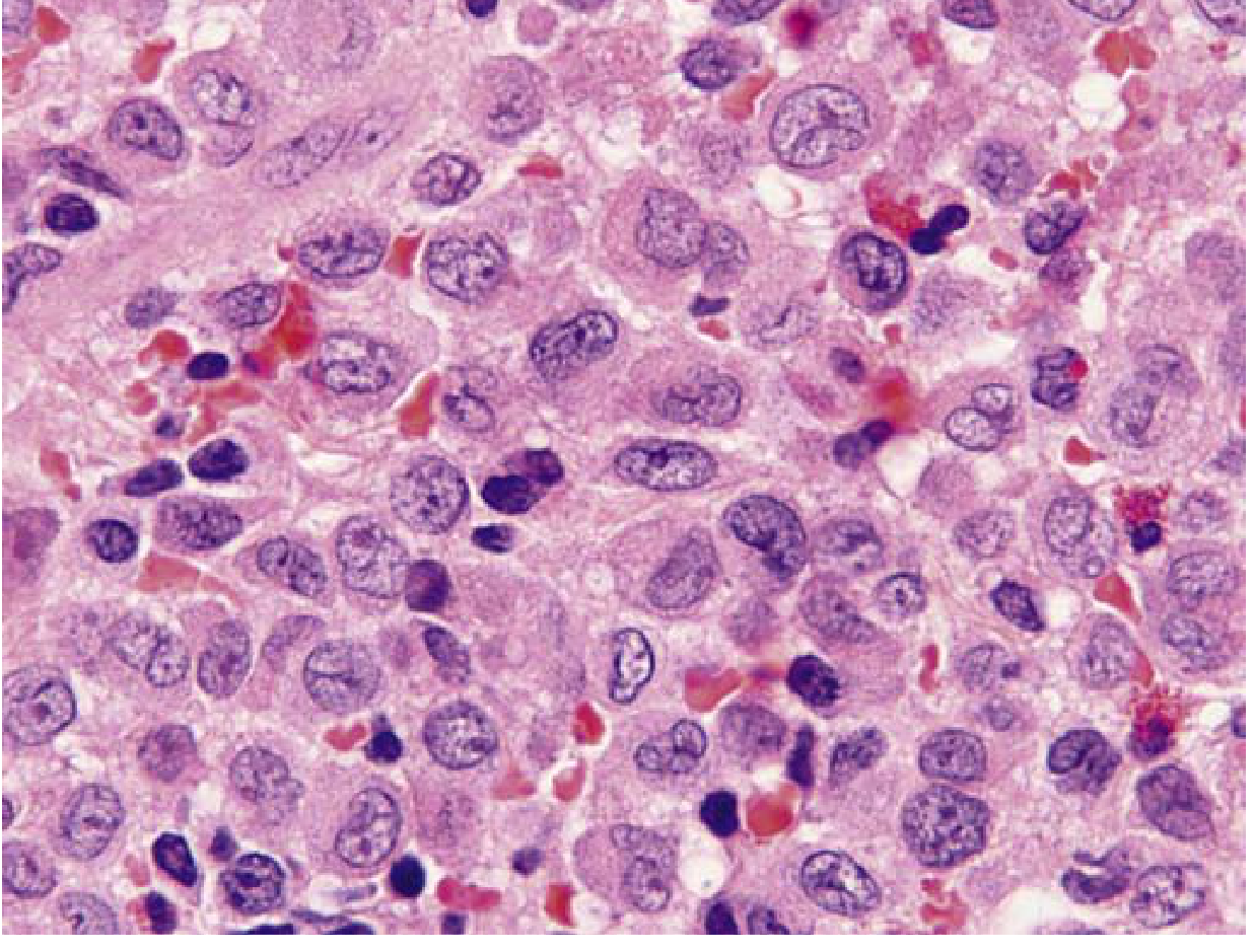
hemophagocytic lymphohistiocytosis bone marrow macrophage phagocytosis
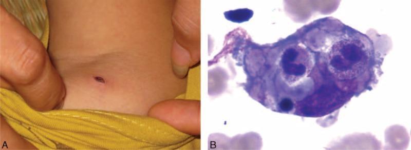
This composite image consists of two panels illustrating clinical and pathological findings associated with scrub typhus and secondary hemophagocytic lymphohistiocytosis (HLH). Panel A is a clinical photograph of a child's shoulder region showing a pathognomonic eschar: a small, oval-shaped, necrotic cutaneous lesion with a dark, crusty center and minimal surrounding erythema. Panel B is a high-power light microscopy image of a bone marrow aspirate stained with Wright-Giemsa. It demonstrates hemophagocytosis, characterized by a large activated macrophage (histiocyte) containing engulfed hematopoietic elements, including nucleated red blood cells or leukocytes, within its cytoplasm. The image serves as an educational tool for identifying the primary diagnostic cutaneous sign of rickettsial infection (Orientia tsutsugamushi) alongside its potential systemic complication, hemophagocytic syndrome.
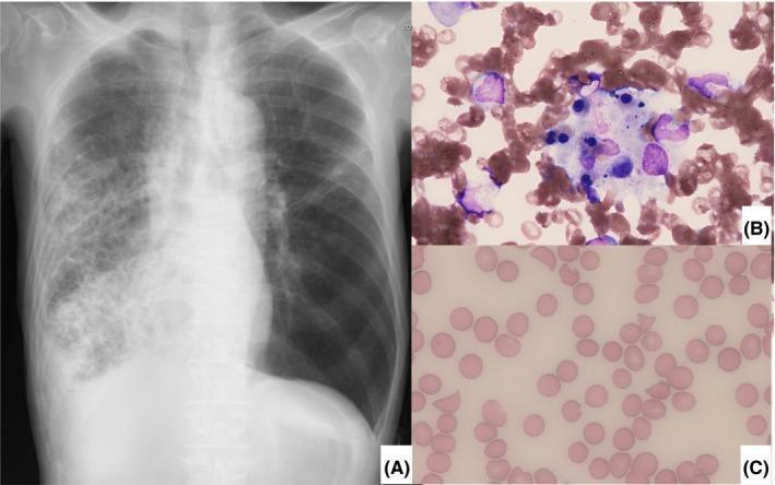
This composite medical image consists of three panels demonstrating clinical findings in a case of severe infection and systemic complications. (A) Anterior-posterior chest X-ray showing extensive, ill-defined heterogeneous opacification and consolidation in the middle and lower zones of the right lung field, consistent with severe pneumonia. (B) High-magnification bone marrow aspirate (Wright-Giemsa stain) revealing active hemophagocytosis, characterized by a large macrophage engulfing multiple hematopoietic cells, including erythrocytes and precursors, indicative of hemophagocytic lymphohistiocytosis (HLH). (C) Peripheral blood smear (Wright-Giemsa stain) showing numerous schistocytes (fragmented red blood cells) including helmet cells and triangular forms, alongside a loss of central pallor in some cells. These findings are diagnostic of microangiopathic hemolytic anemia (MAHA) secondary to thrombotic microangiopathy (TMA). Together, these panels illustrate the progression from a primary pulmonary infection to secondary multisystem hematologic pathologies including septic shock-induced HLH and ADAMTS-13 associated TMA.
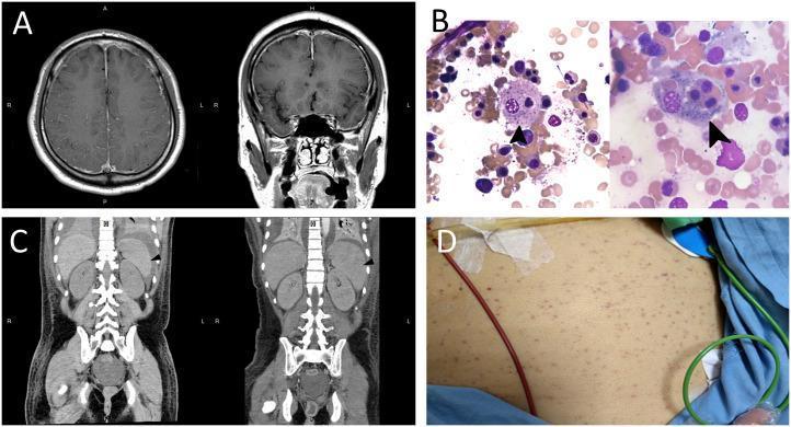
A multi-panel medical figure displaying diagnostic findings for Hemophagocytic Lymphohistiocytosis (HLH). Panel A presents T1-weighted brain MRI scans with gadolinium enhancement in axial (left) and coronal (right) views, showing increased vessel enhancement in the cerebral sulci and bilateral dural thickening, indicative of an inflammatory process. Panel B contains high-power magnification (×1000) Wright-Giemsa stained bone marrow smears; arrows highlight activated macrophages exhibiting hemophagocytosis, specifically engulfing a nucleated cell (left) and platelets (right). Panel C shows sequential coronal abdominal CT scans demonstrating the progression from normal spleen size (left, day 12) to splenomegaly (right, day 18, 12.7 cm). Panel D is a clinical photograph of the torso showing diffuse petechiae. The composite illustrates the neurological, hematological, and systemic manifestations of secondary HLH, including aseptic encephalitis, cytopenias, and organomegaly.
Langerhans cell histiocytosis Birbeck granules CD1a histology
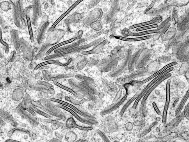
Imaging modality and technique: Transmission Electron Microscopy (TEM); ultrastructural visualization of Birbeck granules within Langerhans cells. Specimen: routine biopsy tissue containing dendritic histiocytes from a suspected Langerhans cell histiocytosis lesion. Location: cutaneous/epidermal/dermal tissue; observed in situ cytoplasmic inclusions. Features: Birbeck granules are zipper-like, pentalaminar rods with a dilated terminal end producing a tennis racket shaped appearance. These cytoplasmic organelles measure approximately 200-400 nm in length and about 33 nm in width. The dilated distal end contains langerin (CD207) and is a hallmark ultrastructural feature of Langerhans cells. Comparative context: Birbeck granules distinguish Langerhans cells from other mononuclear phagocytes on TEM; however, modern diagnostics rely on immunohistochemical markers such as CD1a and CD207 (langerin), making TEM less routinely used today. Pathological significance: the presence of Birbeck granules supports a diagnosis of Langerhans cell histiocytosis when correlated with histology and IHC. Clinical correlation: LCH may present with bone lesions, skin rash, or organ involvement; TEM was historically the gold standard but is now largely supplanted by CD1a and langerin IHC, with ultrastructural confirmation reserved for academic or unresolved cases. Educational utility: illustrates classic ultrastructure of dendritic histiocytes and their diagnostic utility in pathology. This image emphasizes ultrastructural differentiation and context for learners.
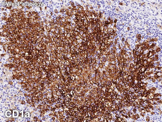
Immunohistochemistry image of pulmonary tissue stained for CD1a demonstrating Langerhans cell histiocytosis. Modality: Immunohistochemistry (IHC) using CD1a primary antibody with DAB chromogen and hematoxylin counterstain. The specimen is lung parenchyma showing a dense cluster of mononuclear dendritic cells with characteristic irregular, reniform (coffee-bean) nuclei surrounded by a background of inflammatory cells. Strong, paranuclear and cytoplasmic CD1a positivity is evident as uniform brown membrane/cytoplasmic labeling of most lesional cells. Langerin (CD207) and S-100 would be co-expressed in the Langerhans cells, though not all panels may be shown in this image. The staining pattern and cellular morphology support the diagnosis of LCH in the pulmonary setting, where Langerhans cells form focal infiltrates and can be associated with smoking-related lung disease. Birbeck granules, while historically demonstrated by electron microscopy, are now replaced by the immunophenotype. This image emphasizes diagnostic specificity of CD1a positivity for identifying Langerhans cells within the alveolar/interstitial compartment. Clinically, pulmonary LCH is frequently linked to adult smokers and may present with nodules or cystic lesions on radiology; histology for CD1a/CD207/S-100 confirms the diagnosis and guides management, including smoking cessation, surveillance, and potential targeted therapies in refractory cases. This panel is essential for diagnostic confirmation in ambiguous clinical-pathological scenarios.
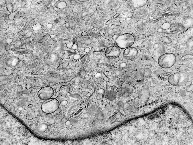
Transmission Electron Microscopy (TEM) image of an epidermal Langerhans cell illustrating Birbeck granules. Birbeck granules are zipper‑like, pentalaminar tubules with a dilated terminus that yields a tennis‑racket appearance; their canonical morphology is highly specific for Langerhans cells. In this specimen, the granules measure approximately 200–400 nanometers in length and about 33 nanometers in width, with a characteristic periodic, curvilinear lattice within the tubule walls. The dilated end often contains structural proteins such as langerin (CD207) and participates in antigen capture and processing. TEM historically served as a gold standard for diagnosing Langerhans cell histiocytosis (LCH) by demonstrating these ultrastructural features; however, modern diagnostic practice relies more on immunohistochemical markers (CD1a and CD207) and molecular tests, making TEM less commonly used. The surrounding cytoplasm shows abundant organelles consistent with active endocytic dendritic cells; cells may exhibit vesicular inclusions and moderate cytoplasmic granularity. This image highlights ultrastructural detail essential for educational purposes and differential diagnosis, particularly in ambiguous cases where immunophenotype is equivocal. Clinically relevant scenarios include suspected LCH in pediatric or adult patients, evaluating skin, bone, or organ involvement, and correlating ultrastructural findings with histology and flow immunophenotyping to confirm diagnosis and guide therapy planning. This image is valuable for teaching.
Histiocytic Disorders: HLH and Langerhans Cell Histiocytosis
PART 1: Haemophagocytic Lymphohistiocytosis (HLH)
Definition and Overview
- Harrison's 22E, p.1348
Pathophysiology
- Cytotoxic lymphocytes cannot kill targets effectively (due to defects in perforin, granzyme secretion, or granule fusion machinery)
- They remain engaged with target cells for prolonged periods
- This drives excessive IFN-gamma release
- IFN-gamma causes unbridled macrophage activation
- Macrophages release toxic levels of TNF, IL-6, and other cytokines
- The result is a cytokine storm resembling sepsis/SIRS, with multi-organ failure
- Robbins Basic Pathology, p.401
Classification
| Category | Gene | Protein |
|---|---|---|
| FHL2 | PRF1 | Perforin (30% of FHL cases) |
| FHL3 | UNC13D | Munc13-4 (30%) |
| FHL4 | STX11 | Syntaxin 11 (10%) |
| FHL5 | STXBP2 | Munc18-2 (20%) |
| XLP1 | SH2D1A | SAP adaptor protein |
| XLP2 | BIRC4/XIAP | XIAP (antiapoptotic) |
| Griscelli syndrome type 2 | RAB27A | Silver-colored hair + immune defects |
| Chediak-Higashi syndrome | LYST | Giant lysosomes in leukocytes |
-
Infection-associated: EBV (most frequent trigger), viral, bacterial, parasitic, fungal
-
Malignancy-associated: especially peripheral T-cell lymphomas
-
Autoimmune-associated (MAS-HLH): systemic JIA, adult-onset Still's disease, SLE, vasculitis
-
Transplant-associated
-
Harrison's 22E, Table 66-1
Epidemiology
- Primary HLH (FHL): incidence ~1 in 50,000 live births; autosomal recessive; median onset 3-6 months
- More common where consanguinity is frequent
- Untreated, median survival is only 1-2 months
- Secondary HLH is far more common in adults
Clinical Features
| System | Features |
|---|---|
| Constitutional | Fever (often high, persistent), sepsis-like state |
| Haematological | Thrombocytopenia (often severe), anaemia, neutropenia - bicytopenia/pancytopenia |
| Hepatic | Hepatosplenomegaly, elevated transaminases, conjugated hyperbilirubinaemia, elevated GGT |
| Coagulation | DIC, hypofibrinogenaemia, severe bleeding |
| CNS | Seizures, decreased consciousness, meningism, cranial nerve palsies, ataxia (~1/3 at diagnosis) |
| Skin | Petechiae, purpura, transient maculopapular rash |
| Lymph nodes | Lymphadenopathy (~50%) |
Bone Marrow Findings

Laboratory Findings (HLH-2004 Diagnostic Criteria)
- Fever
- Splenomegaly
- Cytopenias in ≥2 cell lines
- Hypertriglyceridaemia and/or hypofibrinogenaemia
- Haemophagocytosis in BM, spleen, or lymph nodes
- Low/absent NK cell activity
- Ferritin >500 ng/mL (very high >10,000 µg/L is more specific)
- Elevated soluble CD25 (sIL-2R)
- Robbins Basic Pathology, p.401-402
Additional Investigations
- Perforin expression by flow cytometry - reduced in FHL2
- CD107a degranulation assay (assesses cytotoxic granule exocytosis) - functional screen for genetic HLH
- Genetic testing for FHL mutations
- CSF examination (elevated cells/protein in ~50% of FHL)
- MRI brain (nonspecific findings)
Treatment
- Etoposide + dexamethasone for 8 weeks
- Cyclosporine A added in HLH-2004 protocol
- Intrathecal methotrexate for CNS involvement
- Emapalumab (anti-IFN-gamma monoclonal antibody) - approved for refractory primary HLH
- Ruxolitinib (JAK1/2 inhibitor) - targets the IFN-gamma/JAK pathway
- BRAF inhibitors in specific contexts
- Haematopoietic stem cell transplantation (HSCT) - the only curative treatment for primary HLH; must be performed after achieving remission
- Harrison's 22E, p.137
PART 2: Langerhans Cell Histiocytosis (LCH)
Definition
- Robbins Basic Pathology, p.423-424
Cell of Origin and Immunophenotype
- CD1a (key diagnostic marker)
- Langerin (CD207) - transmembrane protein associated with Birbeck granules
- S-100 protein
- MHC class II antigens
Ultrastructural Hallmark: Birbeck Granules
BRAF Mutation - Key Molecular Pathogenesis
- Robbins Basic Pathology, p.424-425
Clinical Forms
1. Multisystem LCH (Letterer-Siwe Disease) - Most Aggressive
- Children <2 years of age
- Multifocal cutaneous lesions resembling seborrheic dermatitis
- Hepatosplenomegaly, lymphadenopathy
- Pulmonary lesions
- Destructive osteolytic bone lesions (late course)
- Bone marrow infiltration → pancytopenia → recurrent bacterial infections
- Rapidly fatal if untreated; with intensive chemotherapy, ~50% survive 5 years
2. Unisystem LCH (Eosinophilic Granuloma) - Least Aggressive
- Unifocal or multifocal
- Expanding accumulations in medullary cavities of bones (most common: calvaria, ribs, femur)
- Langerhans cells admixed with eosinophils, lymphocytes, plasma cells, neutrophils
- Unifocal: often asymptomatic or causes bone pain; may heal spontaneously or be cured by local excision/irradiation
- Multifocal unisystem disease: multiple erosive bony masses; ~50% have involvement of the posterior pituitary stalk → diabetes insipidus
- Classic triad: Hand-Schuller-Christian triad = calvarial bone defects + diabetes insipidus + exophthalmos
Summary Comparison
| Feature | HLH | LCH |
|---|---|---|
| Cell type | Macrophages (activated) | Langerhans cells (dendritic cell lineage) |
| Nature | Reactive hyperinflammation (primary: genetic; secondary: acquired) | Clonal neoplastic proliferation |
| Marker | Haemophagocytosis on BM biopsy | CD1a, CD207, S-100; Birbeck granules |
| Key mutation | PRF1, UNC13D, STX11, STXBP2 (primary) | BRAF V600E |
| Age | Primary: infants; Secondary: any age | Letterer-Siwe: <2 yrs; eosinophilic granuloma: older children/adults |
| Cytokine | IFN-gamma driven cytokine storm | RAS-MAPK proliferation signal |
| Key lab | Ferritin >10,000, sIL-2R, hypertriglyceridaemia | Bone lytic lesions, diabetes insipidus |
| Treatment | Etoposide + dexamethasone → HSCT | Observation, excision, chemo, BRAF inhibitors |
| Prognosis | Rapidly fatal if untreated (primary) | Variable: unifocal (excellent) to Letterer-Siwe (poor) |
Relationship Between HLH and LCH
- Harrison's Principles of Internal Medicine 22E (2025), Chapter 66-68 (HLH section)
- Robbins & Kumar Basic Pathology, pp. 401-402 (HLH), pp. 423-425 (LCH)
- Galluzzo Mutti & Picarsic, Virchows Arch 2025 - recent review on LCH and histiocytoses in children (PMID 39794638)
- McKinney & Wang, Head Neck Pathol 2025 - recent review on LCH and other histiocytic lesions (PMID 39998733)