
Histology of brain
Reading File
This is a Visual/Broad question - histology involves images and wide coverage. I'll run parallel searches across the library.
Finding Sources
Finding Sources
Finding Sources
Finding Sources
Finding Sources
The Histology textbook has a dedicated neuron section. Let me now read the key histology content and search for more specific brain histology topics in parallel.
Reading File
Finding Sources
Finding Sources
Reading File
Finding Sources
Reading File
Excellent content found. Let me now read the full neuron histology content and the cerebral cortex layers section simultaneously.
Reading File
Reading File
Excellent! I have rich content. Let me now fetch key histology images and read the glial cell content.
Reading File
Loading Image
Loading Image
Now let me get the glial cells section and cortical layers images.
Reading File
Loading Image
Loading Image
Let me now fetch the glial cells content from the histology textbook.
Reading File
Loading Image
I now have enough rich content to deliver a comprehensive, well-illustrated answer on brain histology. Let me compile this.
Histology of the Brain
The brain is composed of two major cell populations: neurons (nerve cells) and neuroglia (supporting cells). The cerebral cortex has a distinctive laminated organization. Here is a systematic overview.
1. Neurons - The Structural and Functional Unit
The neuron is the structural and functional unit of the nervous system. The human brain contains more than 10 billion neurons.
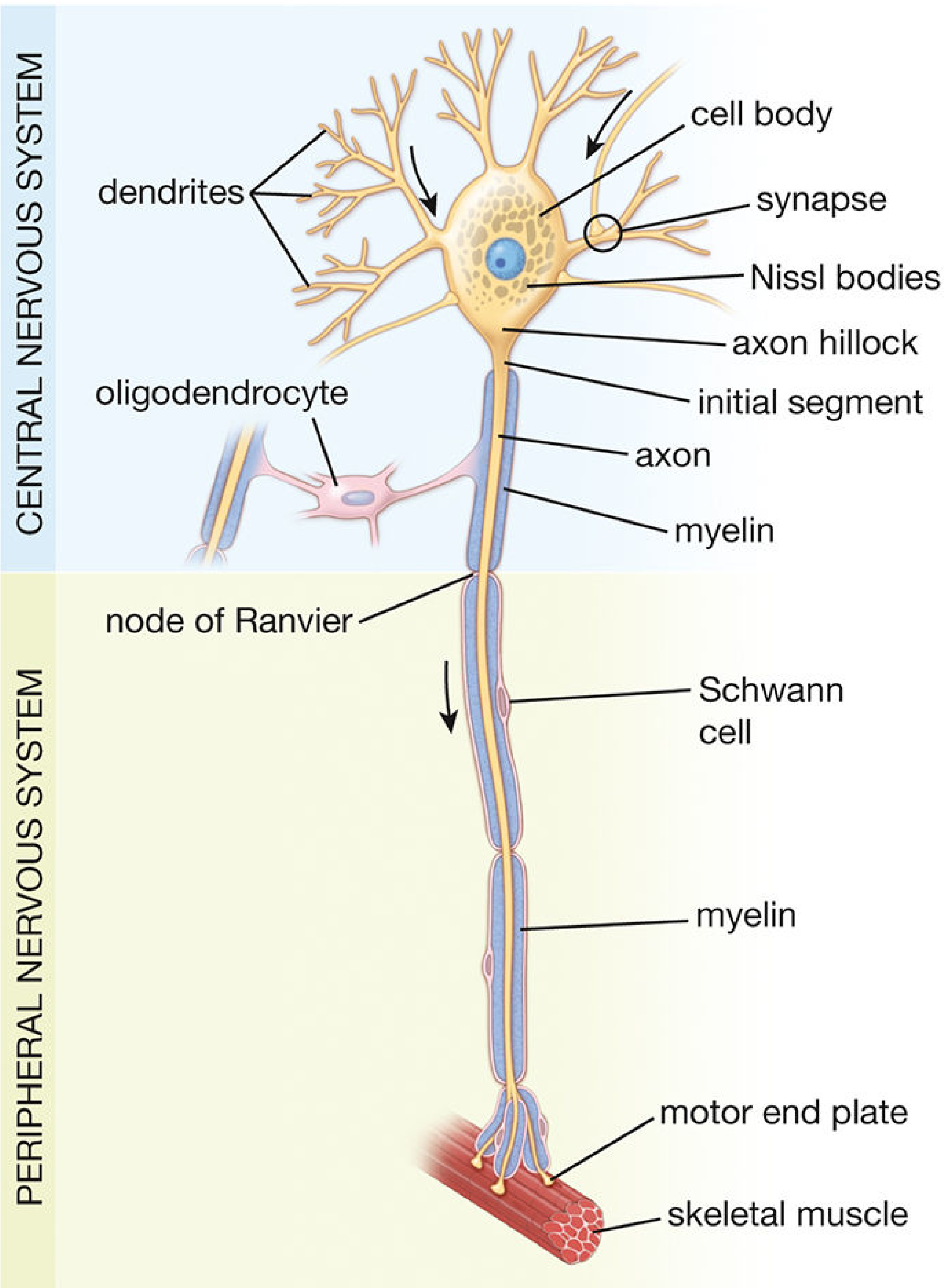
Fig 12.1 - Motor neuron structure (CNS and PNS components) - Histology: A Text and Atlas
Classification of Neurons
| Type | Processes | Location/Function |
|---|---|---|
| Multipolar | 1 axon + 2+ dendrites | Motor neurons, interneurons - most CNS neurons |
| Bipolar | 1 axon + 1 dendrite | Retina, vestibulo-cochlear ganglion, olfactory epithelium |
| Pseudounipolar | Single process (splits into 2) | Dorsal root ganglia; sensory neurons |
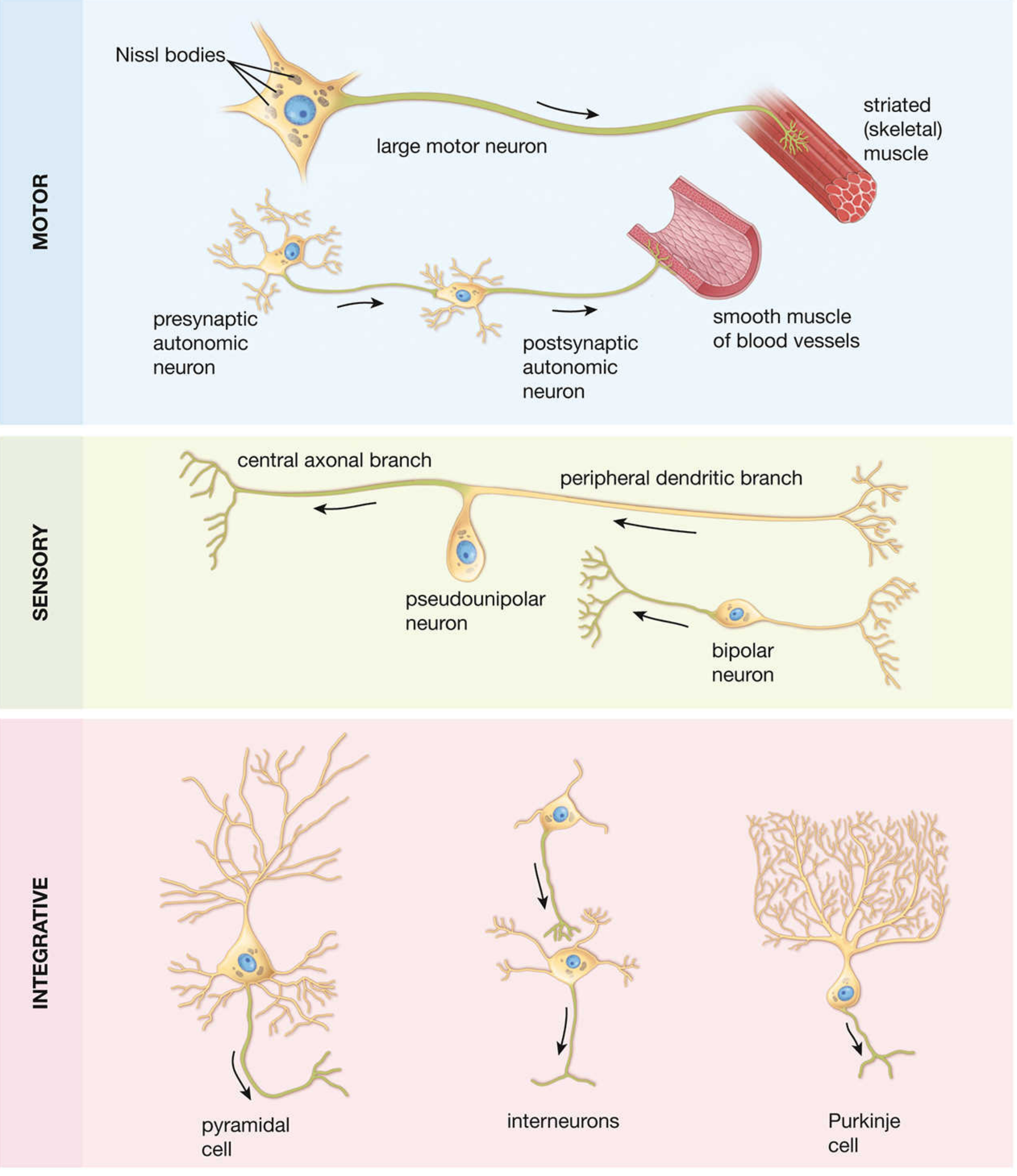
Fig 12.2 - Types of neurons - Histology: A Text and Atlas
2. The Neuron Cell Body (Perikaryon)
The cell body is the protein-producing hub of the neuron.
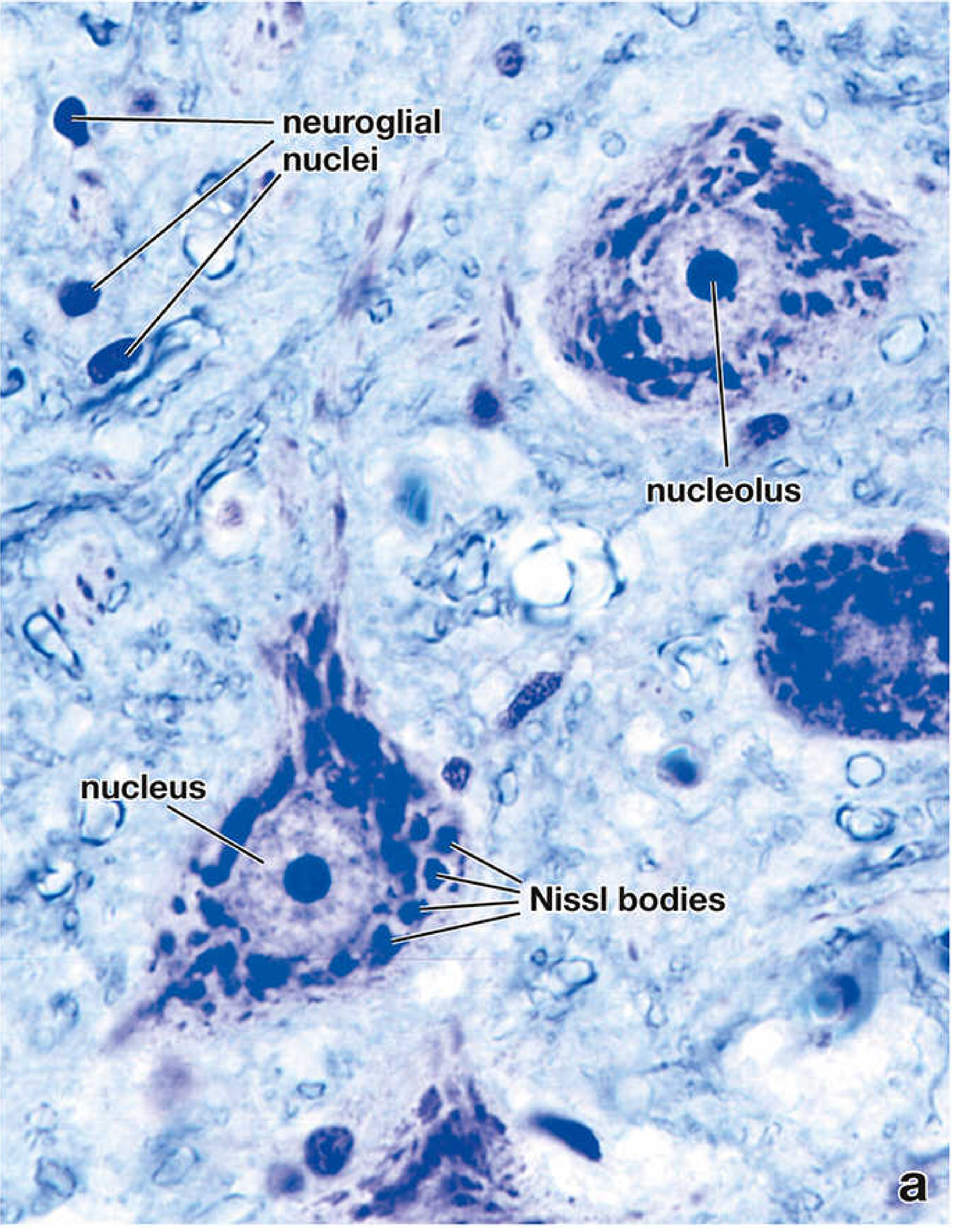
Fig 12.4a - Nerve cell bodies from ventral horn (toluidine blue stain, x640) - Histology: A Text and Atlas
Key histological features of the neuron cell body:
- Large euchromatic nucleus with a prominent nucleolus (reflects intense protein synthesis)
- Nissl bodies - basophilic granules seen with basic dyes (e.g., cresyl violet, thionine); each Nissl body corresponds to a stack of rough endoplasmic reticulum (rER) + free ribosomes. They extend into dendrites but not into the axon
- Golgi apparatus - large, perinuclear
- Mitochondria, lysosomes, neurofilaments (intermediate filaments), neurotubules (microtubules), and transport vesicles
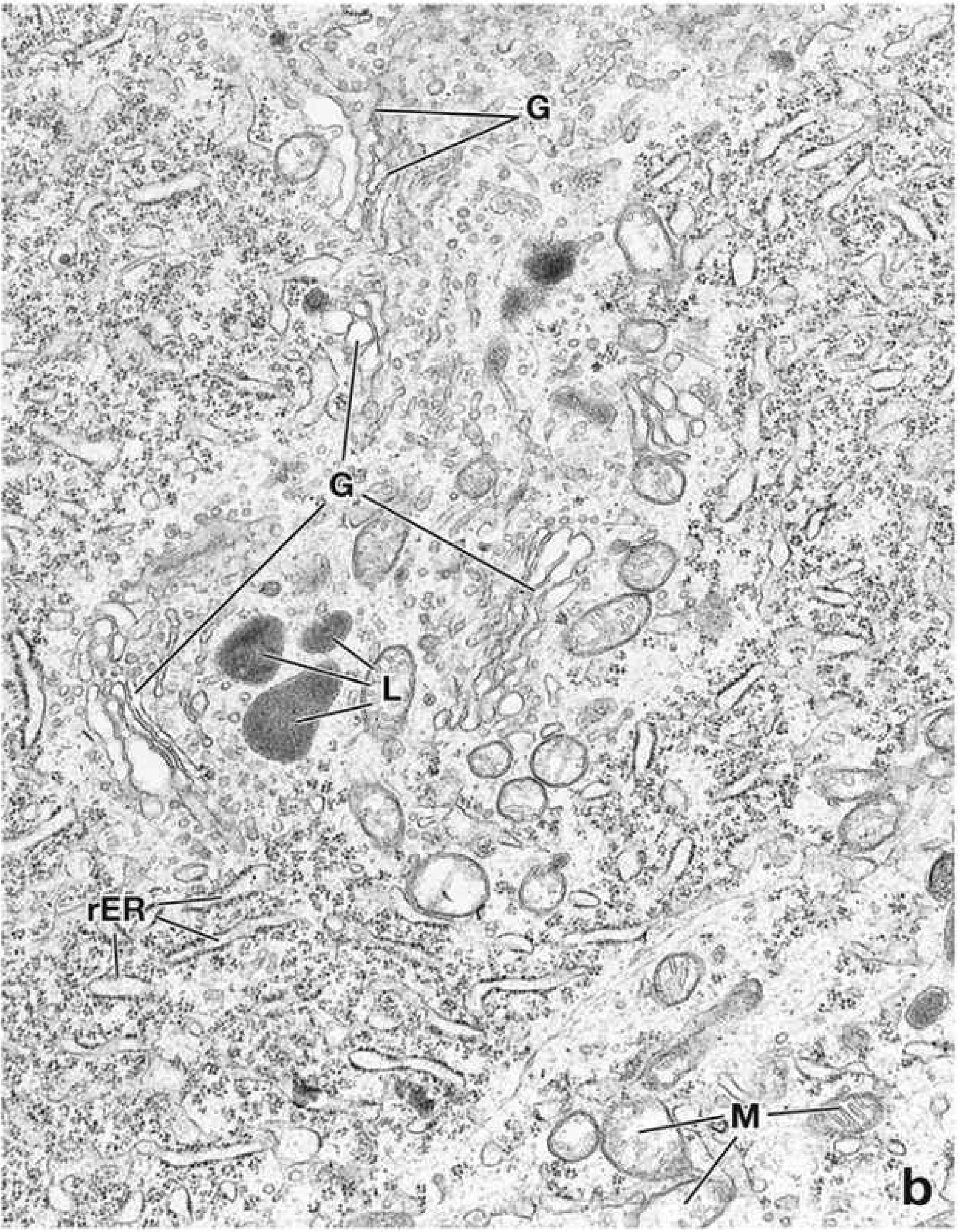
Fig 12.4b - Electron micrograph of nerve cell body (x15,000) - Histology: A Text and Atlas
Axon Hillock
- The region where the axon originates from the cell body
- Lacks Nissl bodies and Golgi cisternae - distinguishes axons from dendrites histologically
- Site of MTOC (centrosome)
- The Axon Initial Segment (AIS) follows immediately after the hillock - this is where action potentials are generated
3. Dendrites and Dendritic Spines
- Dendrites transmit impulses toward the cell body; they contain Nissl bodies, free ribosomes, and Golgi apparatus
- Dendritic spines are short protrusions on dendrites, especially on pyramidal neurons; they are the primary sites of excitatory synapses
- Spines are dynamic structures: their density increases with learning and memory acquisition; stable spines can persist for months
- Loss of dendritic spines on pyramidal neurons in the DLPFC is implicated in schizophrenia
4. Axon
- Conducts impulses away from the cell body
- One axon per neuron; can be over a meter long (e.g., motor neurons)
- Cytoplasm (axoplasm) contains microtubules, neurofilaments, mitochondria, and transport vesicles - but NO Nissl bodies
- Anterograde transport (cell body → terminal): kinesin-driven, fast (200-400 mm/day) or slow (0.5-5 mm/day)
- Retrograde transport (terminal → cell body): dynein-driven; used by viruses (herpes, rabies) and toxins to enter the CNS
5. Synapses
Neurons communicate via specialized junctions called synapses. Types by morphology:
| Synapse Type | Contact |
|---|---|
| Axodendritic | Axon → dendrite (most common; includes spine synapses) |
| Axosomatic | Axon → cell body |
| Axoaxonic | Axon → axon (modulates other synapses) |
Synapse structure:
- Presynaptic knob - contains synaptic vesicles loaded with neurotransmitter
- Synaptic cleft - 20-30 nm gap
- Postsynaptic density - complex of interlinked proteins anchoring receptors and signal transduction machinery
Synapses are not visible on H&E stain but can be demonstrated with the Golgi silver method (appear as oval boutons on the postsynaptic neuron surface).
6. Neuroglia (Supporting Cells of the CNS)
Four main types:
| Cell | LM Appearance | Function |
|---|---|---|
| Astrocytes | Star-shaped; large pale nucleus; GFAP+ | BBB maintenance, synaptic support, scar formation (gliosis), K+ buffering |
| Oligodendrocytes | Small, round dark nucleus; few processes | Produce myelin for CNS axons (one cell myelinates multiple axons) |
| Microglia | Small, elongated, irregular dark nucleus | CNS immune cells (phagocytosis, antigen presentation); derived from monocytes |
| Ependymal cells | Columnar/cuboidal ciliated epithelium | Line ventricles and central canal; produce and circulate CSF |
Key contrast: In the PNS, myelin is produced by Schwann cells (each cell myelinates only one axon segment). In the CNS, myelin is produced by oligodendrocytes.
On routine H&E sections, neuroglial cells appear as small, dark nuclei scattered between neurons - their cell processes are not visible without special stains.
7. Cerebral Cortex - Layered Architecture
The cerebral cortex is a laminated sheet of neurons several millimeters thick covering the cerebral hemispheres. It contains approximately 22 billion neurons connected by ~165 trillion synapses.
Over 90% of the cortex is neocortex, which has a 6-layer (laminar) structure:
| Layer | Name | Main Content |
|---|---|---|
| I | Molecular (plexiform) layer | Few neurons; mainly axons and dendrites of deeper cells; Cajal-Retzius cells |
| II | External granular layer | Small pyramidal and granule cells |
| III | External pyramidal layer | Medium pyramidal cells; association and commissural fibers |
| IV | Internal granular layer | Stellate cells; prominent in sensory cortex (receives thalamic input) |
| V | Internal pyramidal (ganglionic) layer | Large pyramidal cells (Betz cells in motor cortex); project to spinal cord |
| VI | Multiform (fusiform) layer | Mixed cell types; projects to thalamus |
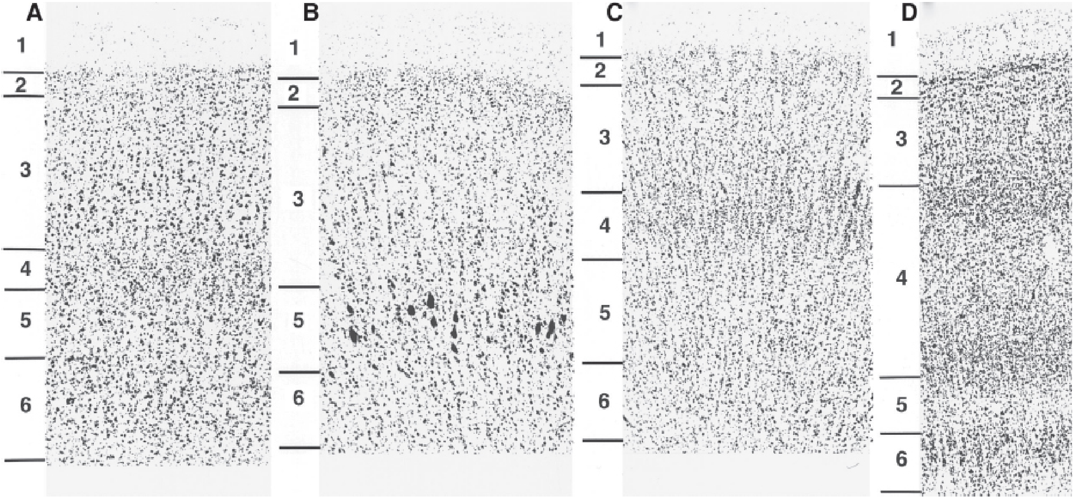
Fig 1.2-19 - Nissl-stained cortical sections comparing 4 regions (DLPFC, Motor, Auditory, Visual cortex). Note the prominent Betz cells in layer V of panel B and the marked differences in laminar organization. - Kaplan & Sadock's Comprehensive Textbook of Psychiatry
Two Major Neuron Types in Neocortex
Pyramidal neurons (~75% of all neocortical neurons):
- Triangular cell body with a single apical dendrite ascending to the cortical surface
- Basal dendrites spread laterally
- Dendritic spines carry most excitatory synapses
- Predominantly excitatory (glutamate); projection neurons
Stellate (granule/nonpyramidal) neurons:
- Small local circuit neurons (interneurons)
- Primarily inhibitory (GABA)
- Multiple subtypes distinguished by neuropeptide content and morphology: chandelier cells, basket cells, Martinotti cells, double-bouquet cells
- Axons arborize locally within gray matter
8. Gray Matter vs. White Matter
| Feature | Gray Matter | White Matter |
|---|---|---|
| Gross appearance | Dark/gray | White (due to myelin) |
| Content | Neuron cell bodies, dendrites, synapses, glial cells | Myelinated axons, oligodendrocytes |
| Location | Cortex (surface), basal ganglia, deep nuclei | Subcortical (below cortex) |
9. Key Histological Stains for Brain Tissue
| Stain | What It Shows |
|---|---|
| H&E | General structure; neurons with pale nucleus and nucleolus; small glial nuclei |
| Nissl (cresyl violet / thionine) | Nissl bodies; neuron cell bodies; glial nuclei |
| Golgi silver | Full neuron morphology including dendrites, spines, synapses |
| Luxol fast blue | Myelin (blue) |
| Bielschowsky silver | Axons and neurofibrils |
| GFAP immunostain | Astrocytes |
| CD68 / Iba-1 | Microglia |
Sources:
- Histology: A Text and Atlas with Correlated Cell and Molecular Biology (9781975181512), Chapter 12 - The Neuron, pp. 908-940
- Kaplan & Sadock's Comprehensive Textbook of Psychiatry (9781975175733), pp. 210-218 - Cerebral Cortex
Spinal cord and lessons
The spinal cord is a cylindrical structure with distinct gray and white matter organization. Gray matter forms an "H" shape centrally (anterior/posterior horns) containing neuron cell bodies, while white matter surrounds it with myelinated tracts. Spinal cord lesions produce predictable syndromes depending on which tracts/horns are damaged - complete transection, hemisection (Brown-Séquard), anterior cord, central cord, etc.
Searching sources...
Finding Sources
Finding Sources
Finding Sources
Finding Sources
Finding Sources
Excellent - the Histology textbook has a dedicated spinal cord section. Let me read it along with the clinical lesions content in parallel.
Reading File
Reading File
Finding Sources
Reading File
Reading File
Loading Image
Reading File
Reading File
Finding Sources
Reading File
Loading Image
Loading Image
I now have all the key content and images needed for a comprehensive answer. Let me compile everything.
Spinal Cord: Histology and Clinical Lesion Syndromes
Part 1 - Histology of the Spinal Cord
Gross Organization
In cross section, the spinal cord shows two distinct compartments:
- Inner butterfly/H-shaped gray matter - contains neuron cell bodies, dendrites, synapses, and neuroglia
- Outer white matter - contains ascending and descending myelinated axon tracts
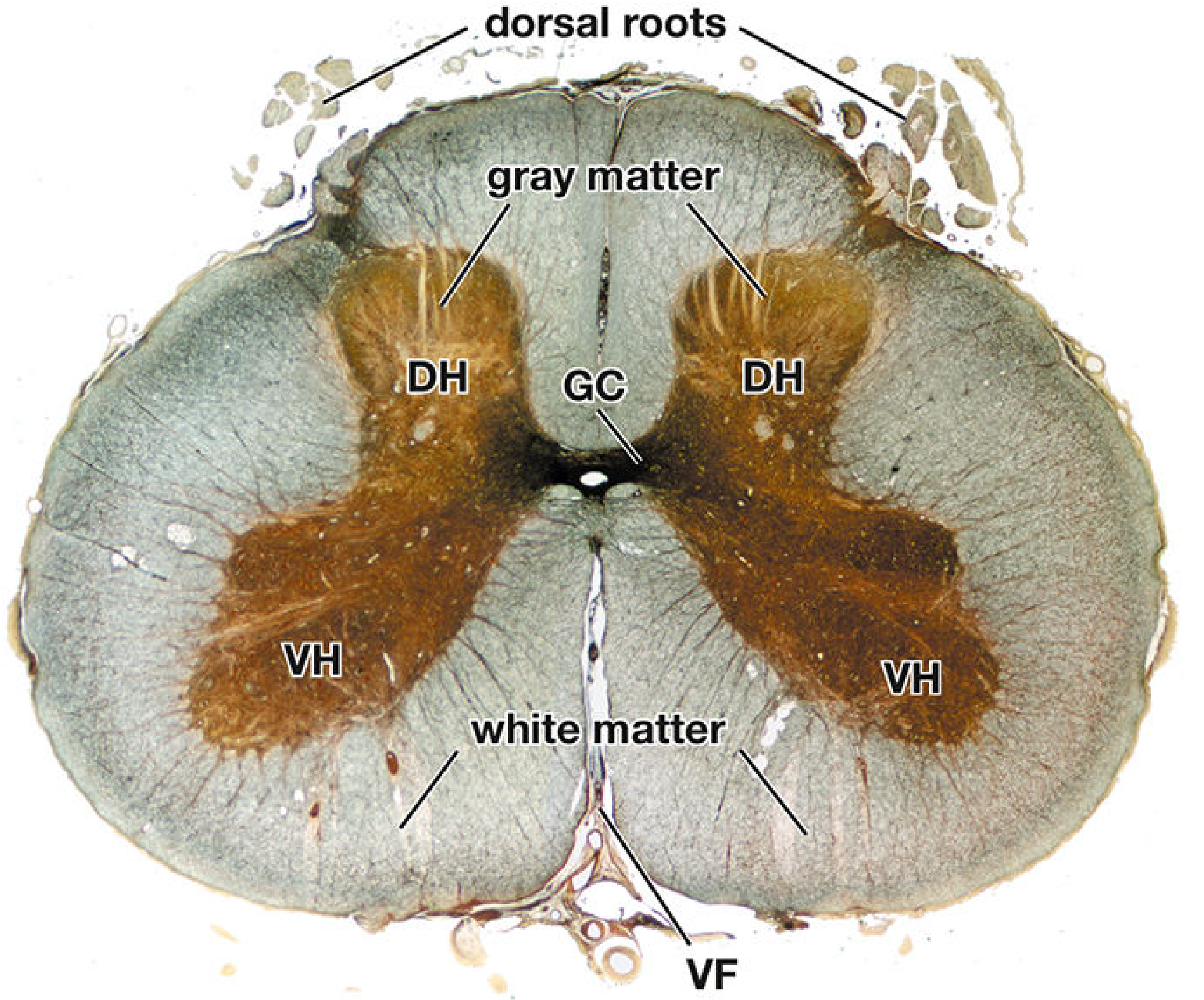
Fig 12.34 - Cross section of human spinal cord, Bielschowsky silver stain (x5) - Histology: A Text and Atlas
Gray Matter
The gray matter is organized into columns (horns) and further sub-divided into Rexed's laminae (I-X):
| Horn | Location | Cell Types | Function |
|---|---|---|---|
| Dorsal (posterior) horn | Dorsal prongs | Sensory interneurons | Receive afferent sensory input from periphery |
| Ventral (anterior) horn | Ventral prongs | Large motor neurons (anterior horn cells) | Efferent - innervate skeletal muscle |
| Lateral horn | T1-L2 only | Preganglionic sympathetic neurons | Sympathetic output |
| Gray commissure | Bridges both halves | Interneurons | Connects the two sides; contains central canal |
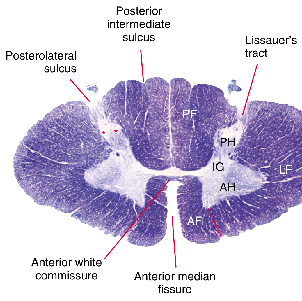
eFig. 9.39 - Spinal cord cross-section at C8 segment - Gray's Anatomy for Students
Anterior Horn Cells (Motor Neurons)
- Large, basophilic multipolar neurons - easily recognized on H&E
- Prominent Nissl bodies in cytoplasm
- Large euchromatic nucleus with prominent nucleolus
- Axon exits via the ventral root → spinal nerve → neuromuscular junction
Dorsal Horn
- Receives pseudounipolar sensory neurons whose cell bodies lie in dorsal root ganglia (DRG)
- Contains substantia gelatinosa (Rexed lamina II) - small neurons processing pain/temperature
- Central segment of DRG neurons enters posterior horn via dorsal root
Rexed's Laminae (I-X)
| Lamina | Region | Significance |
|---|---|---|
| I (Marginal zone) | Tip of dorsal horn | Pain/temperature (superficial) |
| II (Substantia gelatinosa) | Dorsal horn | Pain modulation; opiate receptors |
| III-IV | Dorsal horn | Touch/pressure |
| V | Neck of dorsal horn | Spinothalamic tract origin; wide dynamic range neurons |
| VI | Base of dorsal horn | Proprioception input |
| VII | Intermediate zone | Autonomic (lateral horn T1-L2); Clarke's column (C8-L2) |
| VIII-IX | Ventral horn | Motor interneurons (VIII); alpha and gamma motor neurons (IX) |
| X | Central canal zone | Commissural interneurons |
White Matter - Funiculi and Tracts
The white matter is divided into three funiculi on each side:
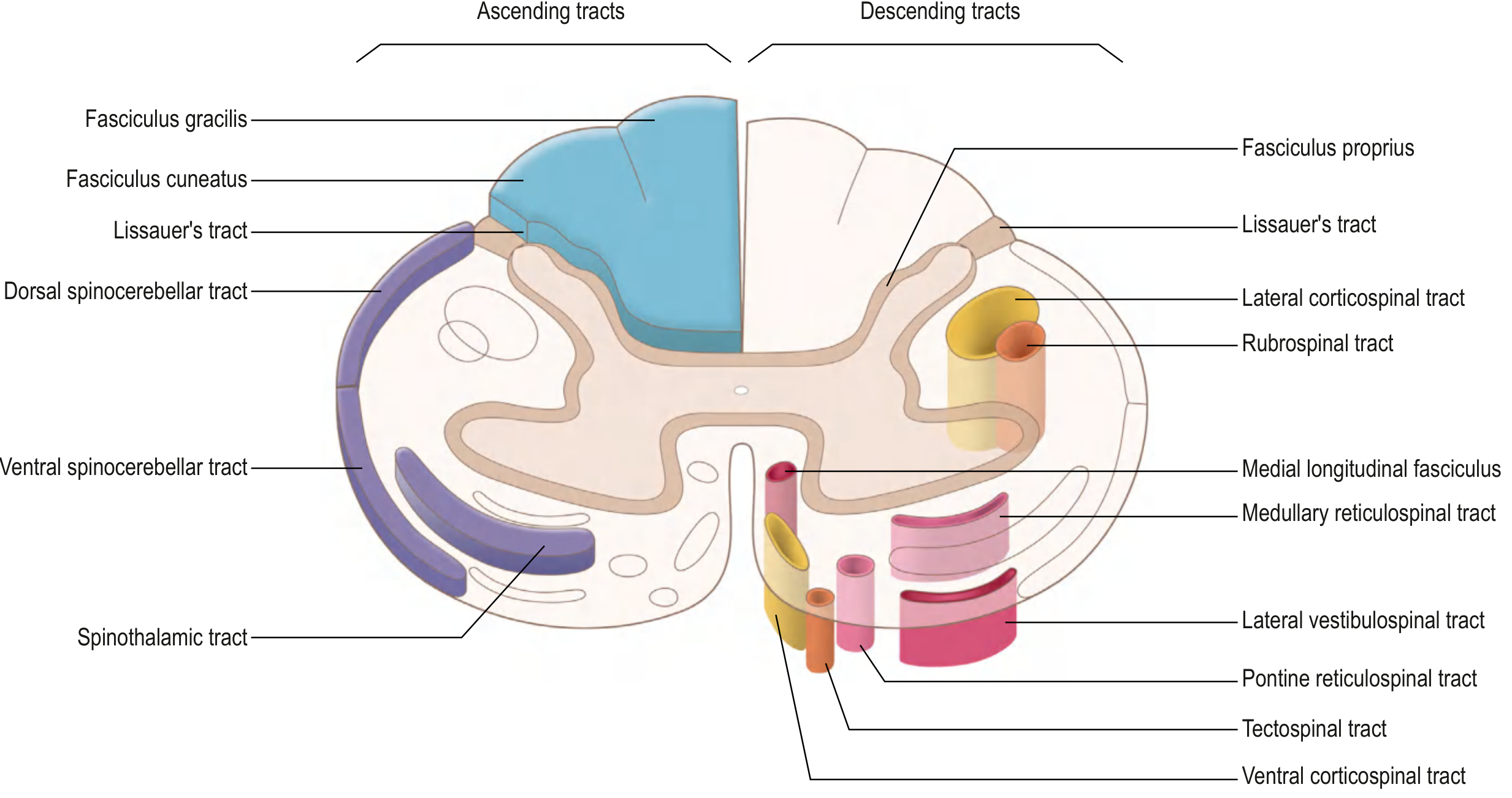
eFig. 9.42 - Ascending and descending tracts in the spinal cord - Gray's Anatomy for Students
Ascending (Sensory) Tracts
1. Posterior Column - Medial Lemniscal Pathway
- Location: Posterior funiculus
- Tracts: Fasciculus gracilis (lower limb/trunk) + Fasciculus cuneatus (upper limb/neck)
- Modalities: Fine touch, vibration, conscious proprioception, 2-point discrimination
- Course: 1st order neuron enters ipsilateral posterior column → ascends to medulla → crosses (internal arcuate fibers) → medial lemniscus → VPL thalamus → primary somatosensory cortex
- Ipsilateral until medulla - lesions below medulla cause ipsilateral loss
2. Anterolateral (Spinothalamic) Pathway
- Location: Anterior and lateral funiculus
- Modalities: Pain, temperature, crude touch
- Course: 1st order neuron enters posterior horn (laminae I, IV, V) → crosses in anterior white commissure (2-3 segments above entry) → ascends contralateral anterolateral column → VPL thalamus → somatosensory cortex
- Crosses within spinal cord - lesions cause contralateral loss below injury level
3. Spinocerebellar Tracts
- Dorsal (posterior) spinocerebellar: Ipsilateral; unconscious proprioception from lower limb
- Ventral (anterior) spinocerebellar: Crossed then re-crossed; unconscious proprioception
Descending (Motor) Tracts
1. Lateral Corticospinal Tract (most important)
- Location: Lateral funiculus (lateral white matter)
- Origin: Primary motor cortex (Betz cells, layer V)
- Course: Motor cortex → corona radiata → posterior limb internal capsule → crus cerebri → pons → crosses in pyramidal decussation at medulla-cord junction → descends ipsilateral lateral column → synapse on anterior horn cells
- Function: Voluntary movement of limbs (especially distal fine movements)
- Lesion above decussation → contralateral UMN signs; lesion in cord → ipsilateral UMN signs
2. Ventral Corticospinal Tract
- Uncrossed; descends in anterior funiculus; controls axial/trunk muscles
3. Rubrospinal Tract - lateral column; from red nucleus; limb flexion
4. Vestibulospinal, Reticulospinal, Tectospinal - anterior/medial column; postural control, axial movements
Meninges
Three connective tissue membranes cover the spinal cord:
| Layer | Composition | Notes |
|---|---|---|
| Dura mater | Dense irregular connective tissue (outermost) | Epidural space between dura and vertebral periosteum contains fat and veins |
| Arachnoid | Delicate leptomeningeal cells; connected to pia by trabeculae | Subarachnoid space contains CSF |
| Pia mater | Thin, closely adheres to cord surface | Denticulate ligaments anchor cord to dural wall |
Part 2 - Spinal Cord Lesion Syndromes
Key Principle: Tract Localization
Understanding which tract is damaged (and where it crosses) predicts the clinical picture:
| Tract/Structure | Crosses | Deficit is... |
|---|---|---|
| Lateral corticospinal | Medullary decussation | Ipsilateral below cord lesion |
| Spinothalamic | Anterior white commissure (spinal cord) | Contralateral, 1-2 levels below lesion |
| Posterior columns | Medulla | Ipsilateral below cord lesion |
1. Complete Cord Transection
Cause: Severe trauma, MS, transverse myelitis
Features below level of lesion:
- Complete bilateral motor loss (UMN if above conus; LMN at level of lesion)
- Complete bilateral loss of all sensory modalities
- Autonomic dysfunction: neurogenic bladder/bowel, loss of sweating
- Acute: Spinal shock - flaccid paralysis, areflexia, urinary retention
- Chronic: Spasticity, hyperreflexia, Babinski sign
2. Brown-Séquard Syndrome (Hemisection)
Cause: Penetrating trauma (stab wounds), unilateral disc herniation, epidural hematoma, MS
Features:
| Deficit | Side | Explanation |
|---|---|---|
| Ipsilateral UMN motor loss (weakness, spasticity) | Same side as lesion | Lateral corticospinal tract - ipsilateral |
| Ipsilateral loss of fine touch, vibration, proprioception | Same side as lesion | Posterior columns - ipsilateral |
| Contralateral loss of pain and temperature | Opposite side | Spinothalamic tract - already crossed |
| Ipsilateral LMN signs at level of lesion | Same side | Anterior horn cell damage at that level |
| Ipsilateral Horner syndrome (if cervical) | Same side | Descending sympathetic fibers |
Prognosis is good - best recovery of all incomplete cord syndromes.
3. Central Cord Syndrome
Cause: Hyperextension injury in elderly with cervical spondylosis (most common incomplete SCI - ~70% of incomplete injuries); cord is pinched between osteophyte/disc anteriorly and buckled ligamentum flavum posteriorly
Features:
- Upper limbs > lower limbs weakness (central fibers of corticospinal tract serving arms are most affected)
- Greater distal > proximal dysfunction in arms
- Variable sensory loss
- Sacral pinprick sensation often preserved (sacral fibers are peripheral in cord)
- Bladder dysfunction common
Most common incomplete cord syndrome. Prognosis is moderate - >50% regain ambulation.
4. Anterior Cord Syndrome
Cause: Anterior spinal artery occlusion or compression (hyperflexion with disc/bone fragment compressing anterior cord)
Structure damaged: Anterior 2/3 of cord (corticospinal tracts + spinothalamic tracts); posterior columns spared
Features:
- Complete motor loss below lesion (bilateral)
- Loss of pain and temperature below lesion (bilateral)
- Preserved: Vibration, proprioception, fine touch (posterior columns intact)
Worst prognosis of all incomplete cord syndromes.
5. Posterior Cord Syndrome
Cause: Rare; tumors, hyperextension injury, subacute combined degeneration (B12 deficiency affecting posterior columns)
Structure damaged: Posterior columns
Features:
- Loss of proprioception and vibration (bilateral)
- Preserved: Motor function, pain, temperature
- Presents as sensory ataxia (positive Romberg test)
6. Conus Medullaris Syndrome
Level: Injury to sacral cord segments (S2-S4) at T11-L2 vertebral level
Features:
- Mixed UMN + LMN picture (sacral LMN + lumbar UMN)
- Areflexic bladder (urinary retention → overflow incontinence)
- Areflexic bowel (fecal incontinence)
- Saddle anesthesia (perineum, genitalia)
- Erectile dysfunction
- Loss of bulbocavernosus reflex and anal wink = poor prognosis
7. Cauda Equina Syndrome
Level: Injury to lumbosacral nerve roots below conus (L1-S5 roots within spinal canal)
Important: This is a peripheral nerve (LMN) injury, not a spinal cord injury
Features:
- Flaccid, areflexic paralysis of lower limbs
- Saddle anesthesia
- Areflexic bladder and bowel
- Absent lower limb reflexes
- Often asymmetric (roots affected unevenly)
- Surgical emergency - early decompression needed
Cauda equina vs conus: Both cause saddle anesthesia and bladder/bowel dysfunction. Cauda equina is pure LMN (flaccid, areflexic). Conus has mixed UMN+LMN features.
Quick Reference: Lesion Pattern Summary
| Syndrome | Motor | Sensory Lost | Sensory Spared | Reflexes |
|---|---|---|---|---|
| Complete transection | Bilateral below | All modalities | None | UMN (chronic) |
| Brown-Séquard | Ipsilateral UMN | Ipsi: posterior columns; Contra: pain/temp | Variable | Ipsi UMN |
| Central cord | UE > LE, bilateral | Variable, sacral sparing | Sacral | UMN |
| Anterior cord | Bilateral below | Pain/temp bilateral | Proprioception/vibration | UMN |
| Posterior cord | None | Proprioception/vibration | Pain/temp/motor | Normal |
| Conus medullaris | LE weak (mixed) | Saddle area | Upper legs variable | Mixed, no bulbocavernosus |
| Cauda equina | Flaccid LE | Saddle area | Upper legs | Absent (LMN) |
Sources:
- Histology: A Text and Atlas (9781975181512), Chapter 12, pp. 998-1010
- Gray's Anatomy for Students (9780323934237), Chapter 9, pp. 1325-1330
- Campbell's Operative Orthopaedics 15th Ed (9780443117633), Chapter 48, pp. 2468-2472
- Bradley and Daroff's Neurology in Clinical Practice (9780323642613), Spinal Cord Injury Syndromes
This is a shared conversation. Sign in to Orris to start your own chat.