
Aniridia
I'll read the medical library skill first, then research aniridia comprehensively.
Reading File
Now let me search the medical library and PubMed in parallel for aniridia.
Finding Sources
Finding Sources
Searching PubMed
aniridia PAX6 genetics management
Good results. Now let me read the key ophthalmology textbook sections and the embryology section in parallel.
Reading File
Reading File
Reading File
Excellent content. Let me read more of the Kanski section (treatment/management) and fetch the clinical images in parallel.
Reading File
Loading Image
Loading Image
I now have comprehensive information from the textbooks. Let me also fetch the gonioscopy image and check the 2026 European guidelines abstract.
Loading Image
Reading Article
PMID: 40892309
I now have comprehensive information from multiple authoritative sources. Here is the full structured review:
Aniridia
Definition
Aniridia is a rare, bilateral panocular developmental disorder characterized by partial or complete absence of the iris. Despite the name, truly total absence of iris tissue is uncommon - gonioscopy almost always reveals a hypoplastic or rudimentary iris frill at the angle. It is best understood as a disorder of the master eye developmental gene PAX6, causing widespread ocular dysgenesis well beyond the iris alone.
- Kanski's Clinical Ophthalmology, 10th ed., p. 420
- The Wills Eye Manual, p. 537
Genetics
| Type | Frequency | Inheritance | PAX6 | Systemic Risk |
|---|---|---|---|---|
| Autosomal dominant (familial) | ~2/3 of cases | AD | PAX6 mutation | None |
| Sporadic (including WAGR) | ~1/3 of cases | New mutation | PAX6 deletion | Wilms tumor ~30% |
| Gillespie syndrome | ~1% | AR | NOT PAX6 | Cerebellar ataxia, learning disability |
Key molecular point: PAX6 lies adjacent to the WT1 gene on chromosome 11p13. When a larger chromosomal deletion encompasses both, the result is WAGR syndrome (Wilms tumor, Aniridia, Genital abnormalities, intellectual Retardation - previously called Miller syndrome). Autosomal dominant aniridia shows complete penetrance but variable expressivity (severity differs between affected family members).
- Kanski's Clinical Ophthalmology, 10th ed., p. 420
- Emery's Elements of Medical Genetics and Genomics
- The Developing Human: Clinically Oriented Embryology, p. 1134
Embryology
The defect results from an arrest of optic cup rim development during approximately the 8th week of gestation. PAX6 acts as a master transcription factor driving differentiation of multiple ocular structures; its dysfunction causes widespread multi-structure dysgenesis rather than isolated iris agenesis.
- The Developing Human: Clinically Oriented Embryology, p. 1134
Clinical Features
Presentation: Typically at birth, with nystagmus and photophobia. Parents often notice apparently large pupils or absence of the colored iris.
Ocular Manifestations (Pan-ocular Disease)
Iris
- Variable severity - from minimal iris hypoplasia (detectable only on retroillumination) to near-total absence
- Gonioscopy typically shows a rudimentary iris frill even in apparently total cases
Cornea (Aniridia-Associated Keratopathy - AAK)
- Tear film instability, dry eye, and epithelial defects
- Limbal stem cell deficiency (LSCD) leads to conjunctivalization of the peripheral cornea (pannus formation)
- End-stage: total central corneal stromal scarring and vascularization
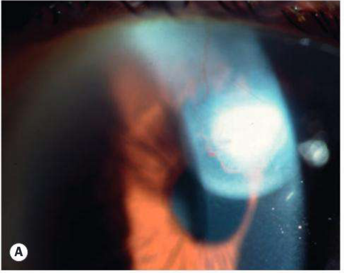
Fig. 11.62A - Advanced aniridia showing corneal vascularization and absent iris (Kanski's)
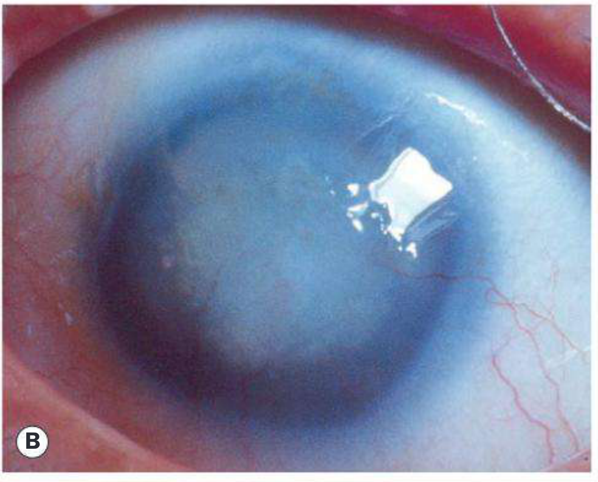
Fig. 11.62B - Near-total aniridia showing dark pupil occupying almost the entire visible eye (Kanski's)
Glaucoma
- Occurs in ~75% of patients
- Usually presents in late childhood or adolescence
- Mechanism: synechial angle closure from contraction of rudimentary iris tissue
- Difficult to manage; prognosis guarded
Foveal and Optic Nerve Hypoplasia
- Foveal hypoplasia is actually a more frequent and reliable clinical sign than visible iris absence (per 2026 European guidelines - Romano et al., Acta Ophthalmol 2026)
- Leads to reduced visual acuity and nystagmus
Lens
- Cataract and lens subluxation may occur
Lids
- Meibomian gland dysfunction is common
WAGR Syndrome
Children with sporadic aniridia have approximately a 30% lifetime risk of Wilms tumor and require systematic surveillance:
- Abdominal ultrasound every 3 months until age 5
- Every 6 months until age 10
- Annually until age 16
- Screening can be stopped if molecular genetic testing confirms absence of WT1 mutation
By contrast, only 1 in 50 Wilms tumor patients has aniridia. The inverse association is important: aniridia is the flag that triggers surveillance; Wilms tumor is not a flag that routinely triggers eye examination.
- Kanski's Clinical Ophthalmology, 10th ed., p. 420
- Henry's Clinical Diagnosis and Management by Laboratory Methods
- Robbins, Cotran & Kumar Pathologic Basis of Disease
Management
Glaucoma
- Medical therapy is usually inadequate long-term
- Trabeculectomy with mitomycin C or combined trabeculectomy-trabeculotomy have been tried but usually fail
- Glaucoma drainage devices offer the best chance of long-term IOP control
- Diode laser cycloablation if other modalities fail
Cornea / Ocular Surface
- Frequent lubricants for associated keratopathy
- Limbal stem cell transplantation (with or without keratoplasty) for severe LSCD/corneal failure
Lens
- Cataract surgery often required
- Tinted IOL implant may reduce photophobia
- Limbal trauma must be minimized during any ocular surgery to preserve remaining stem cell function
Vision Rehabilitation
- Painted (tinted/iris-print) contact lenses or simple tinted lenses to create an artificial pupil - can also reduce nystagmus
- Prosthetic iris implantation has been described in pseudophakic eyes, but may worsen glaucoma (use with caution)
Amblyopia / Refractive
- Refractive errors, amblyopia, and strabismus should be managed aggressively
Systemic
-
Gillespie syndrome: cerebellar rehabilitation, learning support
-
WAGR: Wilms tumor surveillance protocol (above); also screen for genitourinary anomalies and monitor intellectual development
-
Kanski's Clinical Ophthalmology, 10th ed., pp. 420-421
Differential Diagnosis
| Condition | Key Distinguishing Feature |
|---|---|
| Axenfeld-Rieger syndrome | Posterior embryotoxon + iris strands; PITX2/FOXC1 mutations; dental/skeletal anomalies |
| Peters anomaly | Central corneal opacity + iris strands to posterior corneal defect |
| Traumatic mydriasis/iridodialysis | History of trauma; unilateral |
| Congenital idriodialysis | Sector defect, not bilateral absence |
Recent Evidence (2026)
The European COST Action ANIRIDIA-NET published the first multinational clinical guidelines in 2026, formalizing that:
- Foveal hypoplasia - not iris absence - is the most frequent and reliable clinical sign
- PAX6 is the main but not the only causative gene
- Management is challenging due to lack of targeted therapies
- Both medical and non-medical approaches (low vision aids, tinted lenses, psychological support) are formalized components of care
Romano et al., Acta Ophthalmol 2026 (PMID 40892309) - European ANIRIDIA-NET Guidelines
Summary Points
- Aniridia = PAX6 mutation causing pan-ocular disorder (iris + fovea + cornea + angle + lens + optic nerve)
- Autosomal dominant (2/3) has no systemic risk; sporadic (1/3) carries ~30% Wilms tumor risk
- WAGR = Wilms tumor + Aniridia + Genital abnormalities + Retardation (chr 11p13 deletion)
- Gillespie syndrome = AR, non-PAX6, + cerebellar ataxia (rare, 1%)
- Glaucoma occurs in 75% - drainage devices are treatment of choice
- Aniridia-associated keratopathy from limbal stem cell deficiency is a major cause of visual loss
- Sporadic cases need renal ultrasound surveillance every 3 months until age 5
This is a shared conversation. Sign in to Orris to start your own chat.