
Covering of kedney
Reading File
The kidney is covered by several layers: the renal capsule (innermost fibrous layer), perirenal fat (perinephric fat), the renal fascia (Gerota's fascia), and pararenal fat (paranephric fat) on the outside.
Searching sources...
Finding Sources
Finding Sources
Reading File
Reading File
Reading File
Loading Image
Loading Image
Coverings of the Kidney
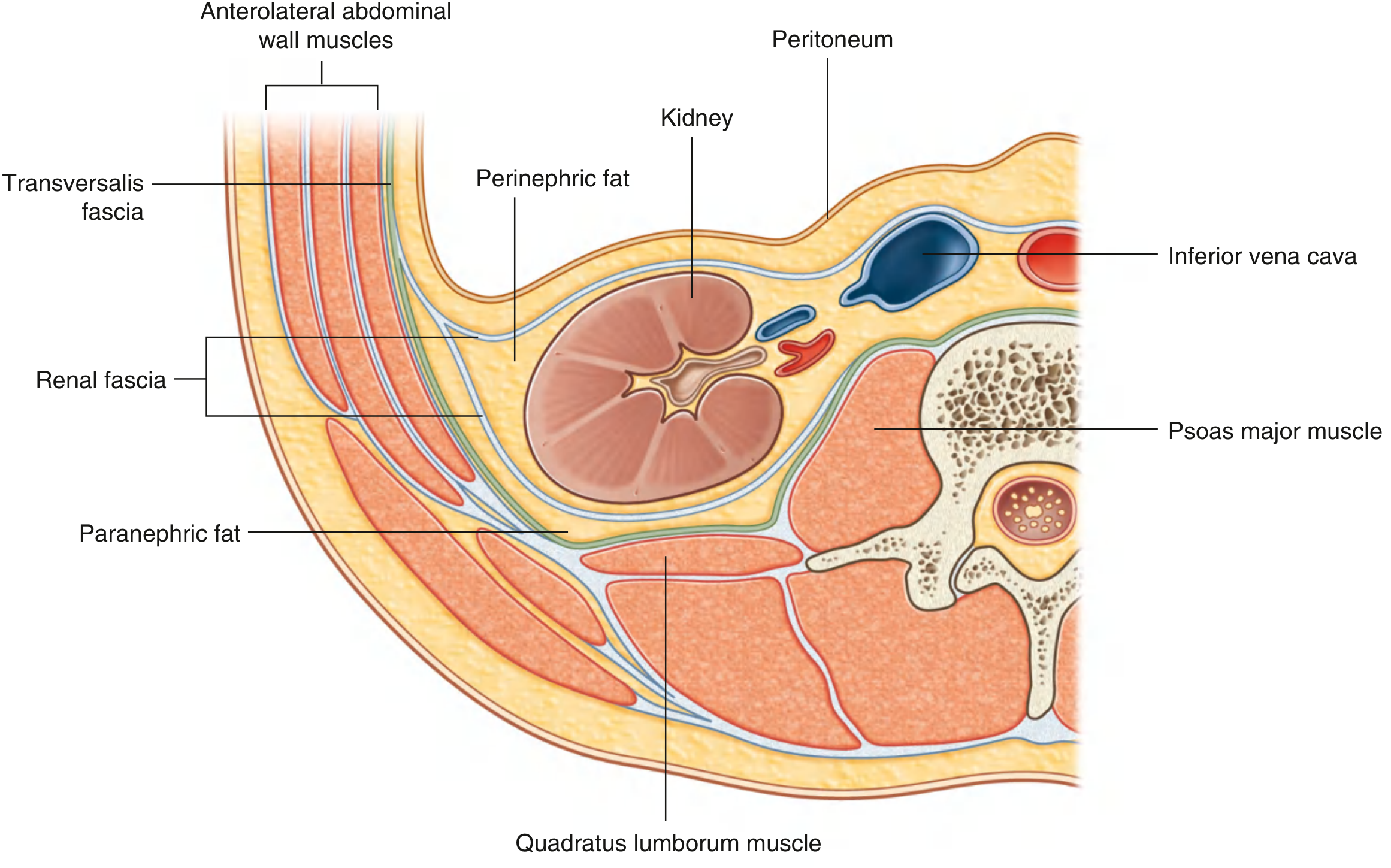
The kidney is a retroperitoneal organ surrounded by three distinct layers of covering, from inside out:
Layer 1 - Renal Capsule (Fibrous Capsule)
The innermost layer. It is a tough, fibrous covering that directly wraps the kidney substance. It is connected to the outer renal fascia by fine connective tissue fiber bundles. This layer must be carefully preserved during surgical dissection to avoid bleeding. The capsule also extends inward into the renal sinuses alongside the vessels.
Layer 2 - Perirenal Fat (Perinephric Fat / Fat Capsule)
Located between the renal capsule and the renal fascia. This adipose layer completely surrounds the kidney and extends into the renal sinuses. It acts as a protective cushion for the kidney. Its thickness varies with the individual's nutritional status - in severe emaciation, it may be absent entirely. When this fat layer is lost, the kidney may descend toward the pelvis, a condition called a floating kidney (nephroptosis).
Layer 3 - Renal Fascia (Gerota's Fascia / Perirenal Fascia)
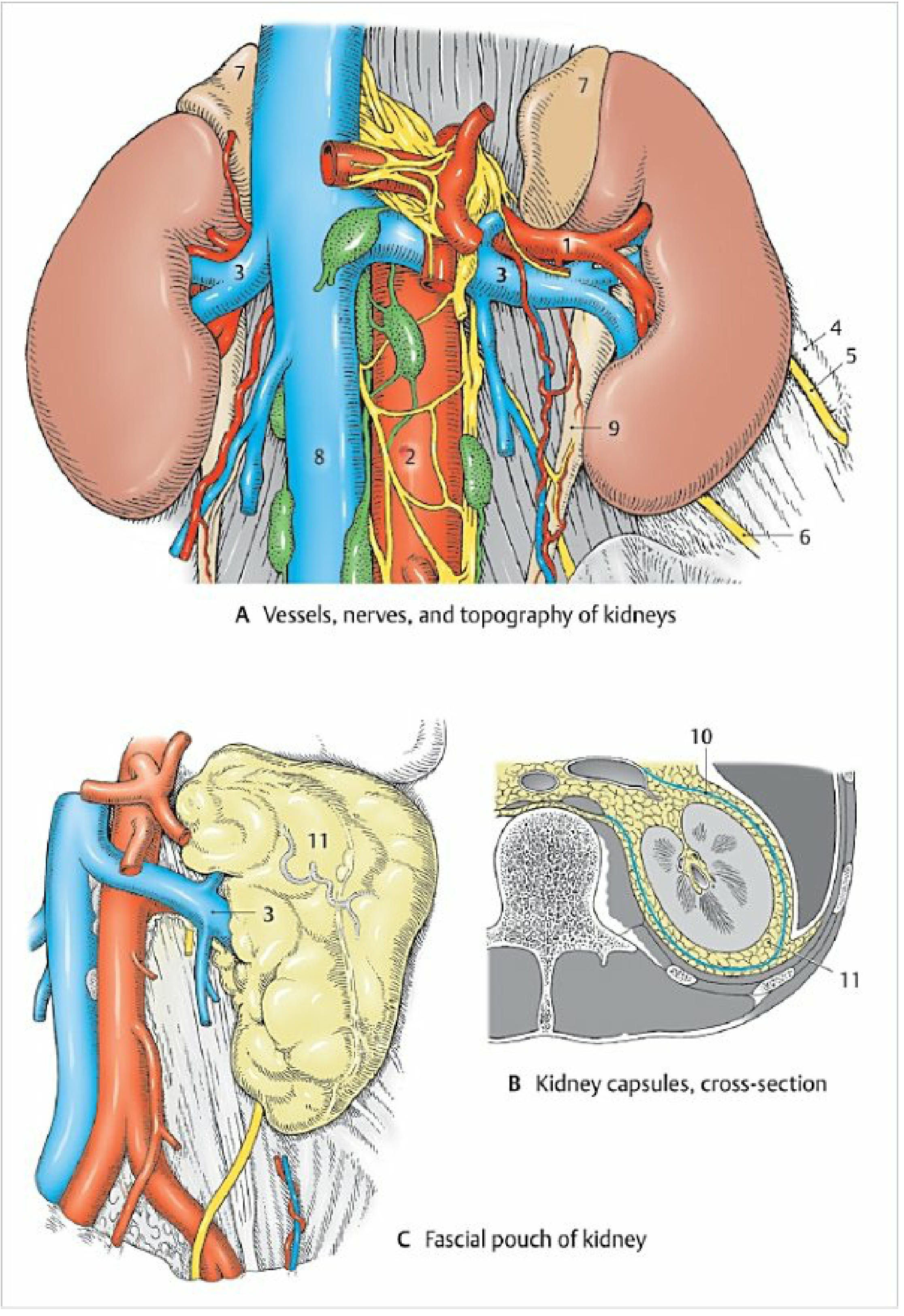
The outermost membranous layer. It is a condensation of extraperitoneal fascia that encloses the perirenal fat, the kidney, and the suprarenal (adrenal) gland (usually separated from the kidney by a thin septum within the fascia). It has:
- An anterior layer - continues over the renal hilum vessels and fuses with the connective tissue of the aorta and inferior vena cava; in some cases crosses midline
- A posterior layer - passes medially between the kidney and the quadratus lumborum to fuse with fascia over psoas major
Attachments and closure of the fascial pouch:
- Superiorly - the two layers fuse above the suprarenal gland and blend with diaphragmatic fascia
- Laterally - the layers fuse at the lateral kidney margin and may connect to the transversalis fascia
- Medially - open (fascial layers diverge toward the midline vessels)
- Inferiorly - open / closed only loosely by fat; the layers continue down around the ureter as periureteric fascia
Layer 4 - Paranephric Fat (Pararenal Fat)
A final external layer of fat located outside the renal fascia - posteriorly between the renal fascia and the quadratus lumborum fascia, and anteriorly between the renal fascia and the parietal peritoneum. It is most prominent on the posterolateral abdominal wall. The renal fascia sends collagen fiber bundles through this layer.
Summary Table
| Layer | Also Called | Location | Key Feature |
|---|---|---|---|
| Renal capsule | Fibrous capsule | Innermost, on kidney surface | Tough, fibrous; connected to renal fascia |
| Perirenal fat | Perinephric fat | Between capsule and renal fascia | Protective cushion; absent in emaciation |
| Renal fascia | Gerota's fascia | Encloses perirenal fat + kidney | Open medially and inferiorly |
| Paranephric fat | Pararenal fat | Outside renal fascia | Most prominent posterolaterally |
Diagrams
Cross-section showing all layers (Gray's Anatomy for Students):
Fascial pouch of kidney and kidney capsules cross-section (Color Atlas of Human Anatomy):
Clinical Significance
- The renal fascia must be incised in any surgical approach to the kidney (e.g., nephrectomy)
- Loss of perinephric fat leads to floating kidney (nephroptosis) - the kidney descends toward the pelvis
- Infections/abscesses can spread within the perirenal space, but the fascia helps contain them; inflammation can involve nearby nerves (subcostal, iliohypogastric, ilioinguinal) causing pain radiating to the groin and genitalia
- The inferior opening of Gerota's fascia has relevance in spread of retroperitoneal fluid or blood - it can track down around the ureter
Sources: Gray's Anatomy for Students, p. 439-440 | Color Atlas of Human Anatomy Vol. 2, p. 398-400 | Fischer's Mastery of Surgery 8th ed., p. 7624
This is a shared conversation. Sign in to Orris to start your own chat.