
Types of sweat glands
The human body has two main types of sweat glands: eccrine and apocrine. Eccrine glands are the primary sweat glands found all over the body, producing watery sweat for thermoregulation. Apocrine glands are larger, found in specific areas (axilla, groin), and produce a thicker secretion that becomes odorous when broken down by bacteria. A third type, apoeccrine, exists in axillary skin and has features of both.
Searching sources...
Reading File
Finding Sources
Finding Sources
Good sources found. Let me read the most relevant sections in parallel - Fitzpatrick's dermatology, Junqueira's histology, and Histology: A Text and Atlas.
Reading File
Reading File
Reading File
Excellent content from all three sources. Let me now fetch the histology images and read a bit more from Fitzpatrick's (apocrine/apoeccrine sections) and Histology text.
Loading Image
Loading Image
Reading File
Reading File
Let me get the apocrine section images and the apoeccrine gland content from Fitzpatrick's.
Reading File
Reading File
Now let me fetch the apocrine histology image and get the apoeccrine section from Fitzpatrick's.
Loading Image
Reading File
Now let me get the apocrine and apoeccrine sections from Fitzpatrick's:
Finding Sources
Reading File
Reading File
Now let me search for the apoeccrine content in Fitzpatrick's:
Finding Sources
I now have comprehensive content from multiple authoritative sources. Let me compile the full answer.
Types of Sweat Glands
The human body has 2 to 4 million sweat glands (200-400/cm²). They are broadly classified into two main types - eccrine and apocrine - with a third, intermediate type called apoeccrine.
1. Eccrine Sweat Glands
Distribution
Distributed over nearly the entire body surface, with highest density on the:
- Palms of hands (~620/cm²)
- Soles of feet
- Forehead, axillae, and trunk
- Absent from: vermilion border of the lips, nail beds, glans penis, clitoris, and labia minora
Structure
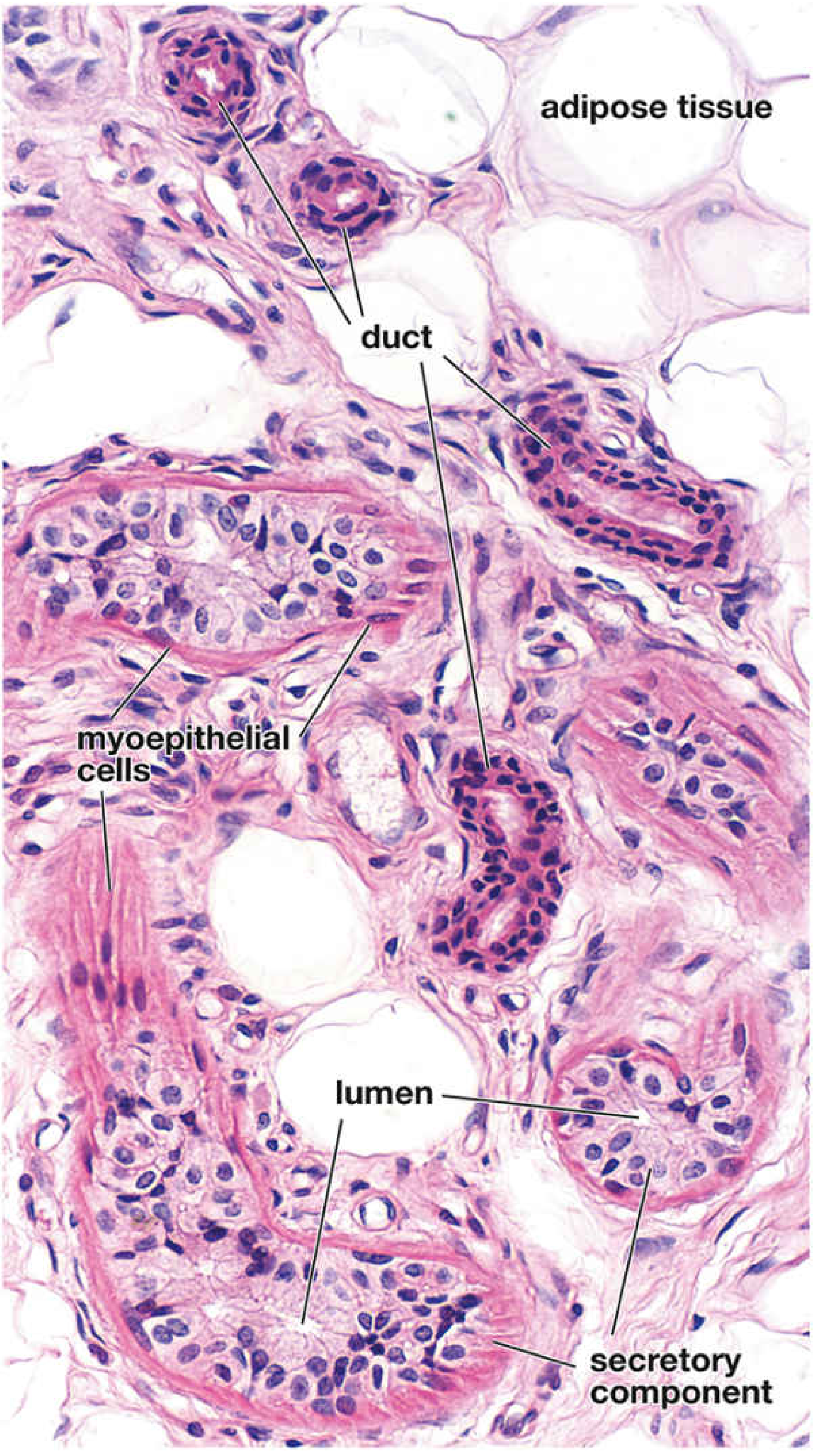
Each eccrine gland is a simple coiled tubular gland in two segments:
- Secretory coil: located deep in the dermis or upper hypodermis; contains three cell types:
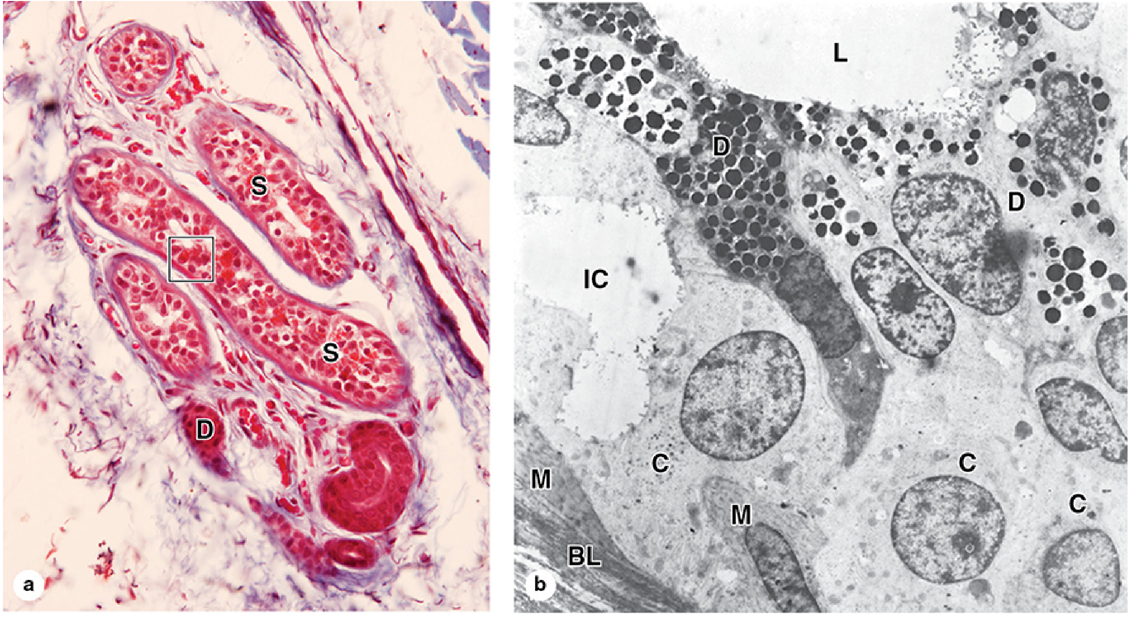
- Clear cells - on the basal lamina, produce the primary watery sweat; rich in mitochondria with extensive basolateral infoldings
- Dark cells - line the lumen; filled with eosinophilic secretory granules; release glycoproteins with bactericidal activity (e.g., dermcidin, β-defensin)
- Myoepithelial cells - on the basal lamina; contract to propel secretion into the duct
- Duct: double layer of cuboidal cells rich in Na⁺/K⁺-ATPase; absorbs Na⁺ from the primary secretion, producing hypotonic sweat; opens independently to the epidermal surface (NOT via a hair follicle)
Histology - eccrine sweat gland (H&E, ×320):
Eccrine gland ultrastructure - LM and TEM:
Secretion
- Primary secretion is isotonic NaCl (similar to blood plasma ultrafiltrate), also contains K⁺, HCO₃⁻, glucose, lactate, urea, uric acid, and antimicrobial peptides
- Ductal reabsorption of Na⁺ and Cl⁻ produces a hypotonic final sweat
- Can produce up to 10 L/day in acclimatized individuals
- Also serves as an auxiliary excretory organ for nitrogenous waste and excess salts
Innervation
- Innervated by sympathetic cholinergic fibers (an exception to the rule - sympathetic system normally uses norepinephrine)
- Acetylcholine (ACh) is the primary stimulus, acting via muscarinic receptors and intracellular Ca²⁺
- Also respond to adrenergic agents (beta-adrenergic, mediated via cAMP)
- Stimulated by: thermal stress (hypothalamic thermoregulatory center), emotional stress (palms, soles, axillae first)
- Botulinum toxin blocks sweating by inhibiting ACh release
Function
Primary: Thermoregulation through evaporative cooling
Secondary: Increased gripping strength (palms/soles), minor excretory function
Development
Anlagen first appear in the 3.5-month fetus on palms and soles, extending over the whole body by the sixth fetal month.
2. Apocrine Sweat Glands
Distribution
Confined to specific body regions:
- Axillae (armpits) - most prominent
- Perineum and anogenital area
- Areolae and nipples of the breast
- External genitalia
- Modified apocrine glands: ceruminous glands (external auditory canal), glands of Moll (eyelash follicles)
- Not functional until puberty; development depends on sex hormones
Structure
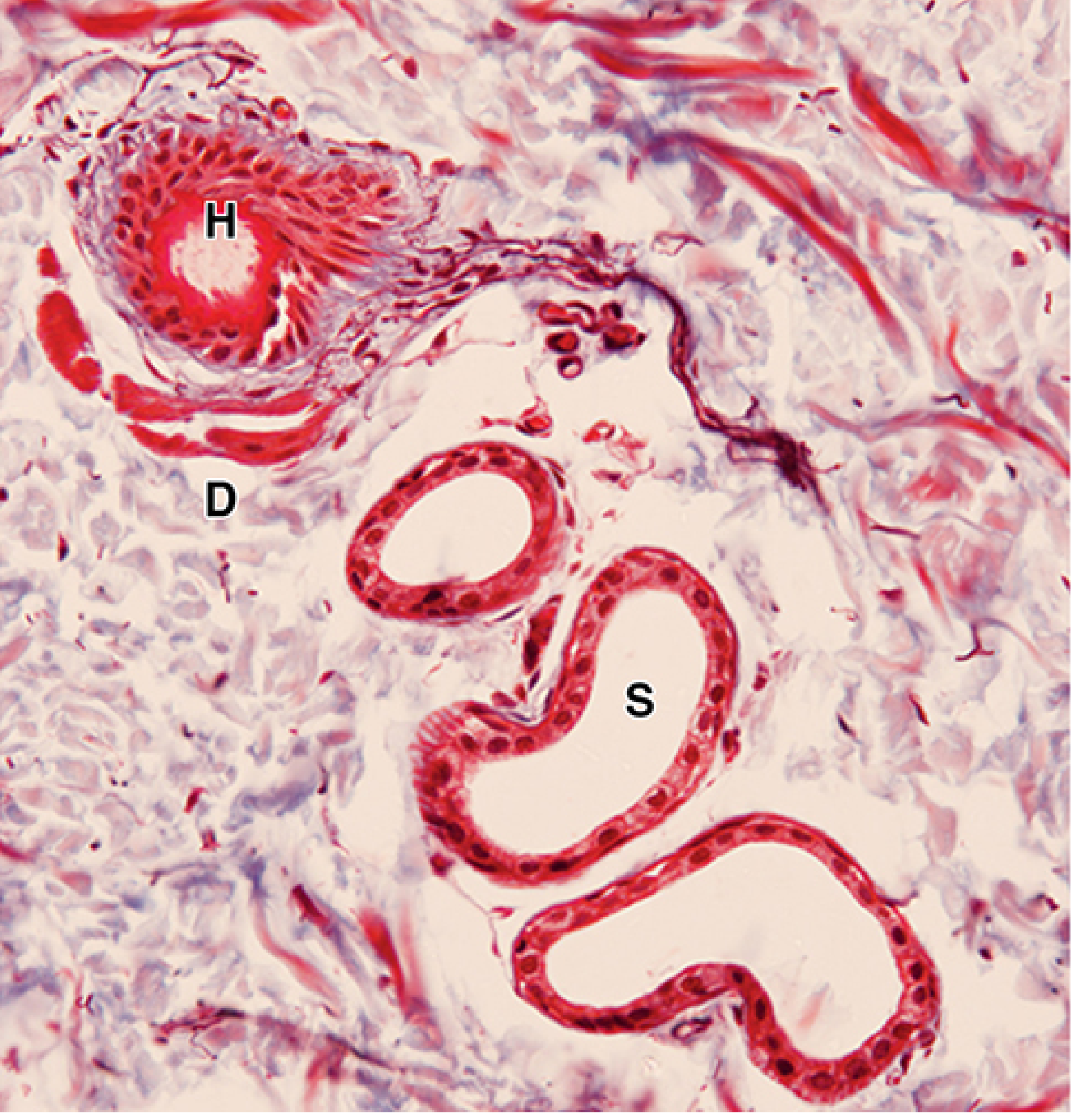
- Coiled tubular glands located in subcutaneous fat near the dermis
- Secretory portion: simple epithelium of cuboidal or columnar cells, with a much larger lumen than eccrine glands; surrounded by myoepithelial cells
- Duct: stratified cuboidal epithelium (2-3 layers), NO myoepithelial cells; opens into the hair follicle infundibulum (not directly to skin surface)
- Ducts follow a relatively straight path - unlike the coiled eccrine duct
- No ductal reabsorption occurs (unlike eccrine)
Histology - apocrine sweat gland (Mallory trichrome, ×200):
Secretion
- Secretion is milky, cloudy, slightly viscous, protein-rich
- Contains proteins, carbohydrates, ammonia, lipids, and pheromone precursors
- Initially odorless - odor develops when skin bacteria (especially Corynebacterium spp.) break down the secretion
- pH: 6 to 7.5
- Secretion mode: mainly merocrine (exocytosis), but cells show apical blebs suggesting some apocrine mechanism (the name is now considered controversial)
- Secretion mixes with sebum in the hair follicle canal
Innervation
- Sympathetic adrenergic innervation (norepinephrine, epinephrine)
- Beta-adrenergic and purinergic receptors identified
- No muscarinic receptors detected - any cholinergic effect is thought to be humoral
- Stimulated by emotional stress, fear, pain, sexual stimulation - but NOT by heat
- Menstrual cycle influences secretion in females
Function
- Pheromone production (well established in mammals; likely vestigial in humans)
- Male pheromones (androstenol, androstenone) influence the female menstrual cycle
- Female pheromones (copulins) influence male perception and hormonal status
- Axillary hair increases odor by providing a surface for bacterial colonization
3. Apoeccrine Sweat Glands
A third type, described primarily in axillary skin, with structural and functional features intermediate between eccrine and apocrine glands:
- Develop from eccrine-like precursors in the axilla during puberty
- Larger than eccrine but smaller than apocrine glands
- Duct opens directly to the skin surface (like eccrine), not into hair follicles
- Respond to both cholinergic and adrenergic stimuli
- Can produce large volumes of secretion
- Clinically significant in axillary hyperhidrosis - apoeccrine glands may account for a major portion of axillary sweat volume
Comparison Table
| Feature | Eccrine | Apocrine | Apoeccrine |
|---|---|---|---|
| Distribution | Whole body | Axilla, groin, areola | Axilla only |
| Lumen size | Small | Large | Intermediate |
| Duct opens into | Skin surface | Hair follicle | Skin surface |
| Secretion type | Watery, hypotonic | Viscous, protein-rich | Watery (large volume) |
| Innervation | Cholinergic (sympathetic) | Adrenergic | Both |
| Stimulated by | Heat, emotion | Emotion only | Both |
| Develops at | Fetal (3.5 mo) | Puberty | Puberty |
| Primary function | Thermoregulation | Pheromones/odor | Axillary sweating |
| Odor | None (directly) | Yes (bacterial action) | Minimal |
Sources:
- Fitzpatrick's Dermatology, Vol. 1-2, pp. 109-114 (Ch. 6: Sweat Glands)
- Junqueira's Basic Histology, 17e, pp. 936-939
- Histology: A Text and Atlas with Correlated Cell and Molecular Biology, pp. 1362-1373
This is a shared conversation. Sign in to Orris to start your own chat.