Here are your comprehensive, exam-ready notes on Upper Limb Arteries for First Year MBBS:
🩸 Upper Limb Arteries — First Year MBBS Notes
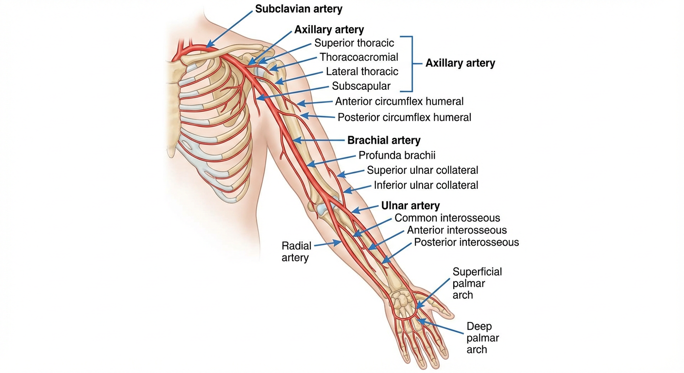
OVERVIEW — Arterial Tree of the Upper Limb
Subclavian Artery
     ↓ (at outer border of 1st rib)
Axillary Artery
     ↓ (at lower border of teres major)
Brachial Artery
     ↓ (bifurcates at neck of radius / cubital fossa)
Radial Artery        Ulnar Artery
     ↓                    ↓
        Deep Palmar Arch (mainly radial)
        Superficial Palmar Arch (mainly ulnar)
DIAGRAM 1 — Complete Upper Limb Arterial Tree
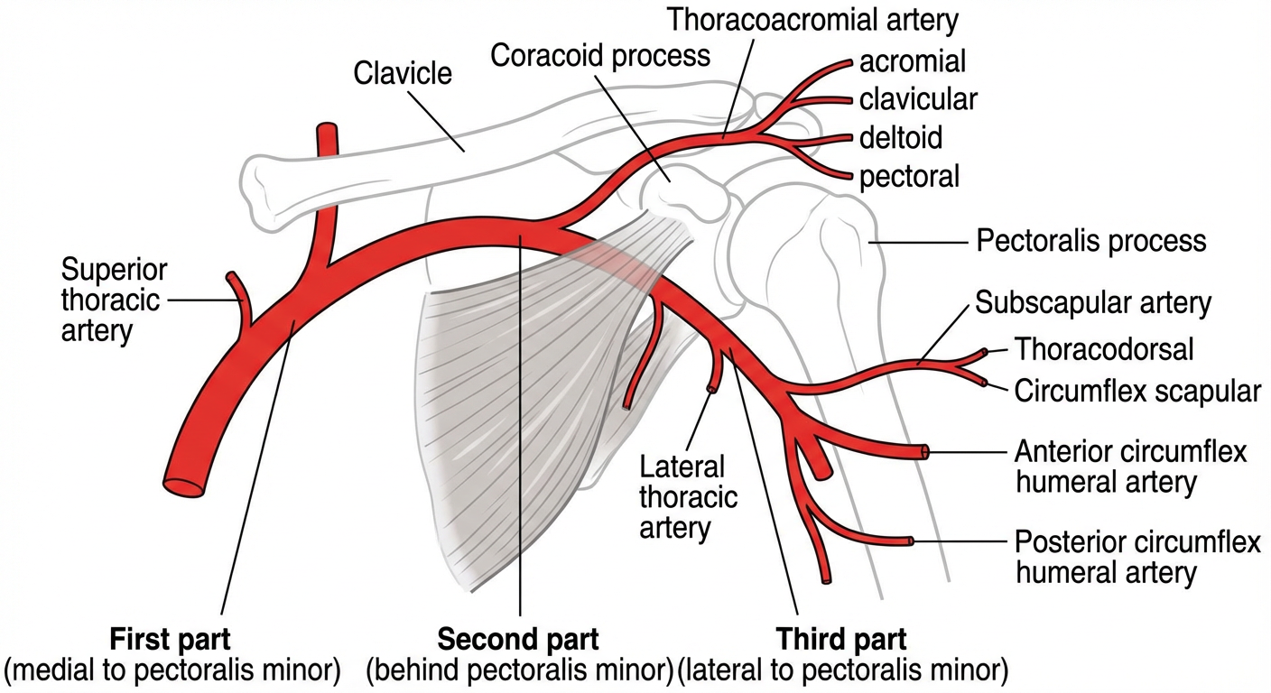
DIAGRAM 2 — Axillary Artery (3 Parts & Branches)
1. SUBCLAVIAN ARTERY
| Feature | Right | Left |
|---|
| Origin | Brachiocephalic trunk | Arch of aorta |
| Course | Arches over 1st rib, enters axilla | Same, slightly longer |
Key Branches (mnemonic: VIT C D)
| Branch | Supply |
|---|
| Vertebral artery | Brain, spinal cord |
| Internal thoracic artery | Anterior chest wall, breast |
| Thyrocervical trunk | Thyroid, neck muscles |
| Costocervical trunk | Deep neck, upper intercostals |
| Dorsal scapular artery | Rhomboids, levator scapulae |
Exam tip: The subclavian becomes the axillary artery at the outer border of the 1st rib.
2. AXILLARY ARTERY
Extent
- From: Outer border of 1st rib
- To: Lower border of teres major (where it becomes brachial artery)
Three Parts (relative to Pectoralis Minor)
| Part | Position | No. of Branches | Branches |
|---|
| 1st part | Medial to pectoralis minor | 1 | Superior thoracic artery |
| 2nd part | Behind pectoralis minor | 2 | Thoracoacromial artery, Lateral thoracic artery |
| 3rd part | Lateral to pectoralis minor | 3 | Subscapular, Anterior circumflex humeral, Posterior circumflex humeral |
Mnemonic for parts: "1, 2, 3 branches" → 1st part = 1, 2nd = 2, 3rd = 3
Branches in Detail
2nd Part — Thoracoacromial Artery (4 branches — mnemonic: CAPS)
- Clavicular branch → sternoclavicular joint
- Acromial branch → acromioclavicular joint
- Pectoral branch → pectoralis major & minor
- Shoulder/Deltoid branch → deltoid muscle
3rd Part
| Branch | Accompanies | Passes through | Supply |
|---|
| Subscapular artery (largest branch) | — | Divides into thoracodorsal & circumflex scapular | Subscapularis, latissimus dorsi, scapular anastomosis |
| Anterior circumflex humeral | Musculocutaneous nerve | Anterior to surgical neck | Shoulder joint (minor supply) |
| Posterior circumflex humeral | Axillary nerve | Quadrangular space | Deltoid, shoulder joint (major supply) |
Exam tip: Posterior circumflex humeral artery and axillary nerve both pass through the quadrangular space. Surgical neck fracture of humerus can damage both!
3. BRACHIAL ARTERY
Extent
- From: Lower border of teres major
- To: Neck of radius (bifurcates in cubital fossa)
Surface Marking
- Medial bicipital groove → midpoint of cubital fossa
- Pulsation felt medial to biceps tendon in antecubital fossa
Relations in Arm
| Medial | Lateral | Anterior | Posterior |
|---|
| Ulnar nerve (upper) → moves anterior (lower) | Biceps brachii | Skin, fascia, median nerve crosses it | Triceps, coracobrachialis |
Note: Median nerve crosses anterior to brachial artery (lateral → medial) at midarm.
Branches of Brachial Artery
| Branch | Origin | Accompanies | Anastomosis |
|---|
| Profunda brachii (Deep artery of arm) | Upper, posterior | Radial nerve in spiral groove | Radial collateral & radial recurrent → around elbow |
| Superior ulnar collateral | Middle of arm | Ulnar nerve | Posterior ulnar recurrent |
| Inferior ulnar collateral | Lower arm | — | Anterior ulnar recurrent |
| Nutrient artery to humerus | Mid-brachial | — | — |
| Muscular branches | Throughout | — | — |
Exam tip: Profunda brachii accompanies the radial nerve in the spiral groove. Fracture of the shaft of humerus (midshaft) = radial nerve + profunda brachii injury → wrist drop.
Elbow Anastomosis
Profunda brachii → Radial collateral ─────────┐
                → Middle collateral ───────────┤
Superior ulnar collateral ─────────────────────┤→ Elbow joint network
Inferior ulnar collateral ─────────────────────┤
                                               │
Radial recurrent ──────────────────────────────┤
Anterior/Posterior ulnar recurrent ────────────┘
4. RADIAL ARTERY
Extent
- From: Bifurcation of brachial artery (neck of radius)
- To: Joins deep branch of ulnar in palm → Deep palmar arch
Course
- Forearm: under brachioradialis → lies on flexor pollicis longus → on pronator teres
- Wrist: at radial styloid (where pulse is felt — "radial pulse")
- Snuffbox: crosses anatomical snuffbox (floor = scaphoid + trapezium)
- Palm: passes between 1st & 2nd metacarpal heads → deep palm
Branches
| Region | Branch | Supply |
|---|
| Forearm | Radial recurrent artery | Elbow anastomosis |
| Forearm | Muscular branches | Forearm muscles |
| Wrist | Palmar carpal branch | Palmar carpal arch |
| Wrist | Superficial palmar branch | Thenar eminence |
| Dorsal wrist | Dorsal carpal branch | Dorsal carpal arch |
| Dorsal wrist | 1st dorsal metacarpal artery | Dorsum of thumb, index |
| Palm | Princeps pollicis | Thumb (both sides) |
| Palm | Radialis indicis | Radial side of index finger |
| Palm | Deep palmar arch | Main contribution to deep arch |
Clinically important: Radial artery is used for ABG sampling, coronary angiography (radial approach), and radial artery flap surgery.
5. ULNAR ARTERY
Extent
- From: Bifurcation of brachial artery (neck of radius)
- To: Enters palm superficial to flexor retinaculum → Superficial palmar arch
Course
- Passes deep to pronator teres (not between its two heads — unlike median nerve)
- Lies on flexor digitorum profundus
- Enters wrist superficial to flexor retinaculum, lateral to pisiform
- Enters Guyon's canal (ulnar canal) with the ulnar nerve
Branches
| Region | Branch | Supply |
|---|
| Elbow | Anterior ulnar recurrent | Front of elbow anastomosis |
| Elbow | Posterior ulnar recurrent | Back of elbow anastomosis |
| Forearm | Common interosseous artery | Divides into anterior & posterior |
| Forearm | Muscular branches | Forearm muscles |
| Wrist | Palmar carpal branch | Palmar carpal arch |
| Wrist | Dorsal carpal branch | Dorsal carpal arch |
| Palm | Superficial palmar arch | Main contribution |
| Palm | Deep branch | Contributes to deep arch |
Common Interosseous Artery (important!)
Common Interosseous Artery (from ulnar)
         ↓
    ─────┴─────
    │           │
Anterior      Posterior
Interosseous  Interosseous
(runs on      (pierces
interosseous  interosseous
membrane)     membrane)
- Anterior interosseous: Supplies deep flexors of forearm, pronator quadratus
- Posterior interosseous: Supplies extensor compartment of forearm
6. PALMAR ARCHES
Superficial Palmar Arch
| Feature | Detail |
|---|
| Main formation | Ulnar artery (terminal) |
| Completion | Superficial palmar branch of radial artery (sometimes median or princeps pollicis) |
| Level | Corresponds to level of fully extended thumb tip |
| Branches | 4 common palmar digital arteries → 7 proper palmar digital arteries |
| Supplies | Medial 3½ digits (ulnar nerve territory fingers) |
Deep Palmar Arch
| Feature | Detail |
|---|
| Main formation | Radial artery (terminal) |
| Completion | Deep branch of ulnar artery |
| Level | ~1 cm proximal to superficial arch (level of metacarpal bases) |
| Branches | 3 palmar metacarpal arteries, recurrent branches to carpal bones |
| Supplies | Metacarpals, intrinsic muscles |
Exam tip — Allen's Test: Compress both radial and ulnar arteries at the wrist → release one at a time → tests integrity of palmar arches and collateral circulation.
7. ANASTOMOSES — Summary Table
| Site | Arteries Participating | Clinical Importance |
|---|
| Scapular anastomosis | Subscapular (from axillary) ↔ Suprascapular & dorsal scapular (from subclavian) | Axillary artery ligation above subscapular = safe |
| Elbow anastomosis | Radial/Ulnar collaterals ↔ Radial/Ulnar recurrents | Allows brachial artery ligation above elbow |
| Palmar arches | Radial ↔ Ulnar | Allen's test |
| Dorsal carpal arch | Radial ↔ Ulnar (dorsal carpal branches) | Hand perfusion |
8. CLINICAL CORRELATIONS (High-Yield Exam Points)
| Condition | Artery Involved | Mechanism | Clinical Feature |
|---|
| Wrist drop | Profunda brachii + Radial nerve | Midshaft humeral fracture / Saturday night palsy | Wrist drop, loss of finger extension |
| Deltoid paralysis | Post. circumflex humeral + Axillary nerve | Surgical neck fracture / shoulder dislocation | Flat (flattened) shoulder, loss of abduction |
| Volkmann's ischemic contracture | Brachial artery | Supracondylar fracture → brachial artery kinking | Flexion deformity of forearm and fingers |
| Thoracic outlet syndrome | Subclavian/axillary artery | Compression at thoracic outlet (cervical rib) | Arm pain, pulseless limb, Adson's test +ve |
| Radial pulse absent | Radial artery | Trauma, thrombosis | Use ulnar pulse / Allen's test to assess |
| Ulnar artery aneurysm | Ulnar artery | Hypothenar hammer syndrome (repetitive trauma) | Mass over hypothenar, ulnar nerve symptoms |
| Aneurysm of axillary artery | Axillary artery | Crutch palsy / trauma | Brachial plexus compression signs |
9. SURFACE MARKING — Quick Reference
| Artery | Surface Marking |
|---|
| Axillary artery | Arm abducted 90° — from midpoint of clavicle to lower border of teres major |
| Brachial artery | Medial bicipital groove (groove between biceps & medial intermuscular septum) |
| Radial pulse | 1 cm medial to radial styloid process, lateral to FCR tendon |
| Ulnar pulse | Medial to FCU tendon at the wrist |
| Radial artery in snuffbox | Anatomical snuffbox (between EPL and EPB/APL) |
FCR = Flexor Carpi Radialis | FCU = Flexor Carpi Ulnaris | EPL = Extensor Pollicis Longus | EPB = Extensor Pollicis Brevis | APL = Abductor Pollicis Longus
10. QUICK REVISION MNEMONICS
| Mnemonic | Remembers |
|---|
| "Screw The Lawyer, Save A Patient" | Subclavian → Axillary (at 1st rib), Axillary → Brachial (at teres major) |
| "1-2-3" | Axillary artery parts have 1, 2, 3 branches respectively |
| "CAPS" | Branches of thoracoacromial trunk: Clavicular, Acromial, Pectoral, Shoulder/Deltoid |
| "VIT C D" | Branches of subclavian artery: Vertebral, Internal thoracic, Thyrocervical, Costocervical, Dorsal scapular |
| "Radial is DEEP, Ulnar is SUPER" | Deep palmar arch = mainly Radial; Superficial palmar arch = mainly Ulnar |
11. FLOWCHART — Pulse Points of the Upper Limb
Subclavian ─── posterior triangle of neck (above clavicle)
     ↓
Axillary ────── axilla (compressible against humerus)
     ↓
Brachial ────── medial bicipital groove / antecubital fossa
     ↓
Radial ─────── anterior wrist (lateral to FCR) ← Most commonly used
Ulnar ──────── anterior wrist (medial to FCU)
References: BD Chaurasia's Human Anatomy Vol. 1 (Upper Limb & Thorax), Gray's Anatomy 41st ed., Snell's Clinical Anatomy by Regions 10th ed.
These notes cover all major exam topics: origins, courses, branches, relations, anastomoses, surface markings, and clinical applications. Focus especially on the brachial artery (Volkmann's, wrist pulse), axillary artery (3 parts & branches), and palmar arches (Allen's test) as these are the most frequently examined in First Year MBBS anatomy practicals and theory papers.