Here are comprehensive, exam-ready notes on Upper Limb Arteries for your First Year MBBS:
🩸 UPPER LIMB ARTERIES — First Year MBBS Notes
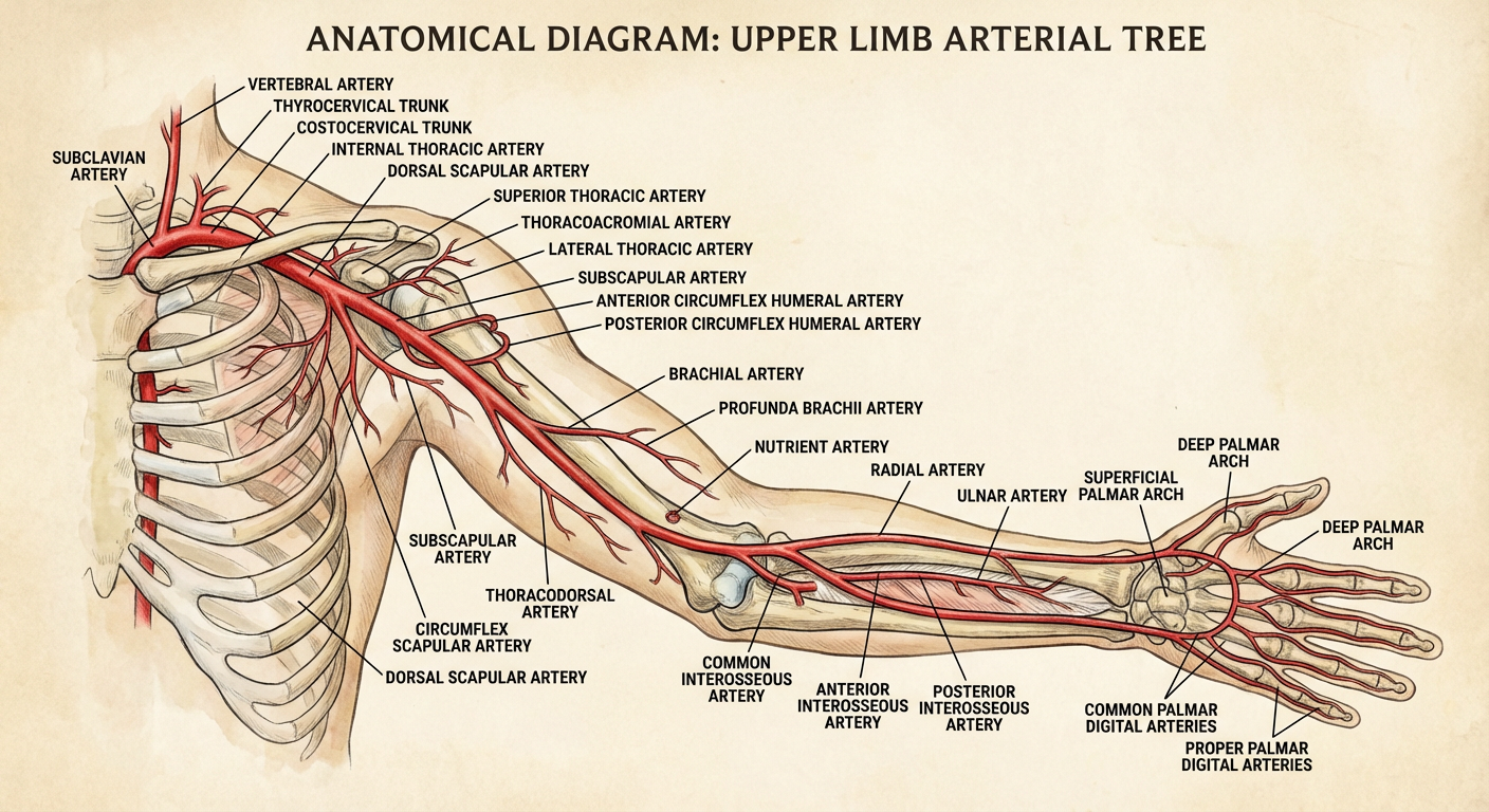
OVERVIEW: Arterial Tree of the Upper Limb
Subclavian Artery
      ↓ (at outer border of 1st rib)
Axillary Artery
      ↓ (at lower border of teres major)
Brachial Artery
      ↓ (bifurcates at neck of radius / cubital fossa)
Radial Artery          Ulnar Artery
      ↓                      ↓
   Deep Palmar Arch ←→ Superficial Palmar Arch
              ↓
        Digital Arteries
📌 OVERALL LABELED DIAGRAM
1. SUBCLAVIAN ARTERY
Origin
| Side | Origin |
|---|
| Right | Brachiocephalic trunk (behind right sternoclavicular joint) |
| Left | Arch of aorta (directly) — longer intrathoracic course |
Parts (relative to Scalenus Anterior)
| Part | Position |
|---|
| 1st | Medial to scalenus anterior |
| 2nd | Behind scalenus anterior |
| 3rd | Lateral to scalenus anterior → becomes axillary at outer border of 1st rib |
Branches (mnemonic: VIT C D)
| Branch | Part | Supply |
|---|
| Vertebral artery | 1st | Brain, spinal cord |
| Internal thoracic artery | 1st | Anterior thoracic wall, breast |
| Thyrocervical trunk | 1st | Thyroid, scapular region |
| Costocervical trunk | 2nd | Deep neck muscles, upper 2 intercostal spaces |
| Dorsal scapular artery | 3rd (or thyrocervical) | Rhomboids, levator scapulae |
Thyrocervical trunk gives: Inferior thyroid, Suprascapular, Transverse cervical arteries
Costocervical trunk gives: Superior intercostal, Deep cervical arteries
2. AXILLARY ARTERY
Extent
- Begins: at outer border of 1st rib (continuation of subclavian)
- Ends: at lower border of teres major (continues as brachial)
Parts (defined by Pectoralis Minor)
| Part | Position | Number of Branches |
|---|
| 1st | Medial to pectoralis minor | 1 branch |
| 2nd | Posterior to pectoralis minor | 2 branches |
| 3rd | Lateral to pectoralis minor | 3 branches |
Mnemonic: 1-2-3 (medial-posterior-lateral = 1-2-3 branches)
Branches
1st Part (1 branch):
- Superior thoracic artery → upper 2 intercostal spaces, serratus anterior
2nd Part (2 branches):
- Thoracoacromial artery → Pectoral, acromial, deltoid, clavicular branches (mnemonic: PADC)
- Lateral thoracic artery → Serratus anterior, breast, pectoral muscles
3rd Part (3 branches):
- Subscapular artery (largest branch) → divides into:
- Circumflex scapular artery (anastomoses around scapula)
- Thoracodorsal artery (supplies latissimus dorsi)
- Anterior circumflex humeral artery → small, encircles surgical neck anteriorly
- Posterior circumflex humeral artery → larger, passes through quadrangular space with axillary nerve; supplies deltoid, shoulder joint
Scapular Anastomosis (Important for Exam!)
After subclavian/axillary ligation, collateral flow via:
- Suprascapular ↔ Circumflex scapular (from subscapular)
- Transverse cervical ↔ Subscapular
3. BRACHIAL ARTERY
Extent
- Begins: lower border of teres major
- Ends: neck of radius (in cubital fossa) — bifurcates into radial & ulnar
Course
- Runs medial to biceps brachii → enters cubital fossa → lies medial to biceps tendon, lateral to median nerve
Important relation in cubital fossa (lateral to medial): Biceps Tendon → Brachial Artery → Median Nerve
Mnemonic: BAM (from lateral to medial: Biceps, Artery, Median nerve) — or "Be Absolutely Meticulous"
Branches
| Branch | Supply |
|---|
| Profunda brachii (Deep brachial) | Posterior compartment of arm; accompanies radial nerve in radial groove; gives radial collateral & middle collateral branches |
| Nutrient artery of humerus | Humerus |
| Superior ulnar collateral | Elbow anastomosis (with posterior ulnar recurrent) |
| Inferior ulnar collateral | Elbow anastomosis (with anterior ulnar recurrent) |
| Muscular branches | Arm muscles |
Cubital Anastomosis (Periarticular Anastomosis of Elbow)
| Descending vessels | Anastomose with |
|---|
| Radial collateral (from profunda brachii) | Radial recurrent (from radial artery) |
| Middle collateral (from profunda brachii) | Posterior interosseous recurrent |
| Superior ulnar collateral | Posterior ulnar recurrent |
| Inferior ulnar collateral | Anterior ulnar recurrent |
4. RADIAL ARTERY
Extent
- Origin: bifurcation of brachial (cubital fossa, neck of radius)
- Ends: anastomoses with deep branch of ulnar artery → forms deep palmar arch
Course
- Forearm: Descends under brachioradialis; becomes superficial in lower 1/3 (palpable radial pulse here, between FCR tendon and brachioradialis)
- Wrist: Winds around lateral aspect of carpus → through anatomical snuffbox → enters palm between heads of 1st dorsal interosseous
Branches
| Segment | Branch | Supply |
|---|
| Forearm | Radial recurrent artery | Elbow anastomosis |
| Muscular branches | Lateral forearm muscles |
| Palmar carpal branch | Palmar carpal arch |
| Superficial palmar branch | Thenar muscles / joins superficial palmar arch |
| Wrist/Hand | Dorsal carpal branch | Dorsal carpal arch → dorsal metacarpal arteries |
| Princeps pollicis | Thumb (both sides) |
| Radialis indicis | Lateral side of index finger |
| Deep palmar arch | (main termination) |
5. ULNAR ARTERY
Extent
- Origin: bifurcation of brachial (cubital fossa)
- Ends: anastomoses with superficial palmar branch of radial → forms superficial palmar arch
Course
- Passes deep to pronator teres → descends between FDS and FDP → enters hand through Guyon's canal (lateral to pisiform, medial to hook of hamate)
Branches
| Segment | Branch | Supply |
|---|
| Forearm | Anterior ulnar recurrent | Elbow anastomosis |
| Posterior ulnar recurrent | Elbow anastomosis |
| Common interosseous artery | Divides into anterior & posterior interosseous |
| Muscular branches | Medial forearm muscles |
| Palmar carpal branch | Palmar carpal arch |
| Dorsal carpal branch | Dorsal carpal arch |
| Hand | Deep branch | Deep palmar arch |
| Superficial palmar arch | Main termination |
Common Interosseous Artery (Key Branch)
- Short trunk from upper ulnar artery
- Divides at upper border of interosseous membrane into:
- Anterior interosseous → anterior compartment of forearm, nutrient artery to radius & ulna; perforates membrane → joins dorsal carpal arch
- Posterior interosseous → posterior compartment of forearm (with posterior interosseous nerve); gives posterior interosseous recurrent artery (elbow anastomosis)
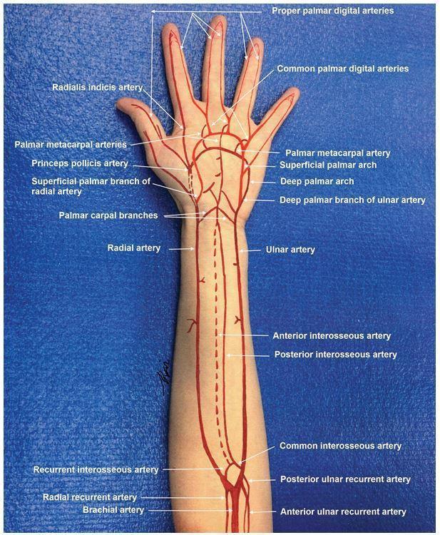
6. PALMAR ARCHES
Superficial Palmar Arch
| Feature | Detail |
|---|
| Main contribution | Ulnar artery (direct continuation) |
| Completing vessel | Superficial palmar branch of radial |
| Level | Level of fully extended thumb (distal to deep arch) |
| Branches | 3–4 common palmar digital arteries → bifurcate into proper palmar digital arteries supplying adjacent sides of fingers |
Deep Palmar Arch
| Feature | Detail |
|---|
| Main contribution | Radial artery |
| Completing vessel | Deep branch of ulnar artery |
| Level | 1 cm proximal to superficial arch |
| Branches | 3 palmar metacarpal arteries → join common palmar digital arteries |
Mnemonic: "Radial = Deep, Ulnar = Superficial" → R-D, U-S
Allen's Test
Used to assess dual blood supply to the hand before arterial cannulation:
- Compress both radial and ulnar arteries
- Patient clenches fist → opens (hand blanches)
- Release one artery → hand should flush pink within 5–7 seconds
- Repeat for the other artery
- Positive Allen's test = flushing does NOT occur → dominant supply from the tested artery is absent → do NOT cannulate that artery
7. DIGITAL ARTERIES
| Artery | Supply |
|---|
| Princeps pollicis (from radial) | Both sides of thumb + dorsum |
| Radialis indicis (from radial) | Radial side of index finger |
| Proper palmar digital arteries (from common palmar digital) | Adjacent sides of fingers 2–5 |
8. CLINICAL CORRELATIONS (High-Yield for Exams)
| Condition | Artery Involved | Clinical Feature |
|---|
| Radial pulse | Radial artery | Felt at wrist between FCR and brachioradialis; used for pulse rate, BP |
| Brachial BP measurement | Brachial artery | Auscultated in cubital fossa |
| Profunda brachii injury | Radial groove fracture (humerus) | Damages radial nerve + profunda → wrist drop |
| Anterior interosseous nerve syndrome | Anterior interosseous artery area | Pinch weakness (FPL, FDP to index) |
| Posterior circumflex humeral injury | Quadrangular space | Axillary nerve damage → deltoid paralysis |
| Volkmann's ischemic contracture | Brachial artery occlusion | Supracondylar fracture of humerus in children |
| Raynaud's phenomenon | Digital arteries | Vasospasm → pallor-cyanosis-redness sequence |
| Thoracic outlet syndrome | Subclavian artery (3rd part) | Compression between clavicle & 1st rib or scalenus anterior |
9. QUICK REVISION TABLE: All Arteries at a Glance
| Artery | Origin | Termination | Key Branch |
|---|
| Subclavian | Brachiocephalic (R) / Aorta (L) | Outer border of 1st rib | Vertebral, Internal thoracic, Thyrocervical trunk |
| Axillary | Outer border of 1st rib | Lower border of teres major | Subscapular (largest), Post. circumflex humeral |
| Brachial | Lower border of teres major | Neck of radius | Profunda brachii |
| Radial | Brachial bifurcation | Deep palmar arch | Princeps pollicis |
| Ulnar | Brachial bifurcation | Superficial palmar arch | Common interosseous |
| Anterior interosseous | Common interosseous | Dorsal carpal arch | Nutrient arteries to radius & ulna |
| Posterior interosseous | Common interosseous | Dorsal carpal arch | Post. interosseous recurrent |
10. MNEMONIC SUMMARY
| Topic | Mnemonic |
|---|
| Subclavian branches | VIT C D (Vertebral, Internal thoracic, Thyrocervical, Costocervical, Dorsal scapular) |
| Axillary parts & branches | 1 – 2 – 3 (branches per segment) |
| Cubital fossa contents (lat → med) | BAM (Biceps tendon, Artery, Median nerve) |
| Thoracoacromial branches | PADC (Pectoral, Acromial, Deltoid, Clavicular) |
| Palmar arches | R-D, U-S (Radial = Deep, Ulnar = Superficial) |
These notes cover the full upper limb arterial anatomy as per standard first-year MBBS syllabi (Gray's Anatomy, BD Chaurasia). Focus especially on: axillary artery parts & branches, brachial artery relations in the cubital fossa, common interosseous artery, palmar arches, and the clinical correlations for exam vivas and written questions.