
Cerebrum physiology mbbs 1st year
Reading File
The cerebrum is the largest part of the brain, responsible for higher functions like consciousness, voluntary movement, sensory processing, memory, and language. For MBBS 1st year physiology, the key topics include the cerebral cortex (motor, sensory, association areas), basal ganglia, limbic system, and cortical functions.
Searching sources...
Finding Sources
Finding Sources
Finding Sources
Finding Sources
Finding Sources
Finding Sources
Finding Sources
I found relevant sections in Guyton & Hall (the standard MBBS 1st year physiology textbook) and Ganong's. Let me read those sections now.
Reading File
Finding Sources
Reading File
Finding Sources
Reading File
Reading File
Reading File
Loading Image
Loading Image
Reading File
Loading Image
Reading File
I now have comprehensive information from Guyton & Hall to write a thorough MBBS 1st year cerebrum physiology answer. Let me compile it.
Cerebrum Physiology - MBBS 1st Year
(Source: Guyton and Hall Textbook of Medical Physiology, Guyton & Hall)
1. Overview of the Cerebrum
The cerebrum is the largest part of the brain. Its functional surface - the cerebral cortex - is a thin layer of neurons (2-5 mm thick) covering all convolutions of the cerebrum. Total cortical area is about 25% of a square meter, and it contains over 80 billion neurons.
2. Histological Structure of the Cerebral Cortex
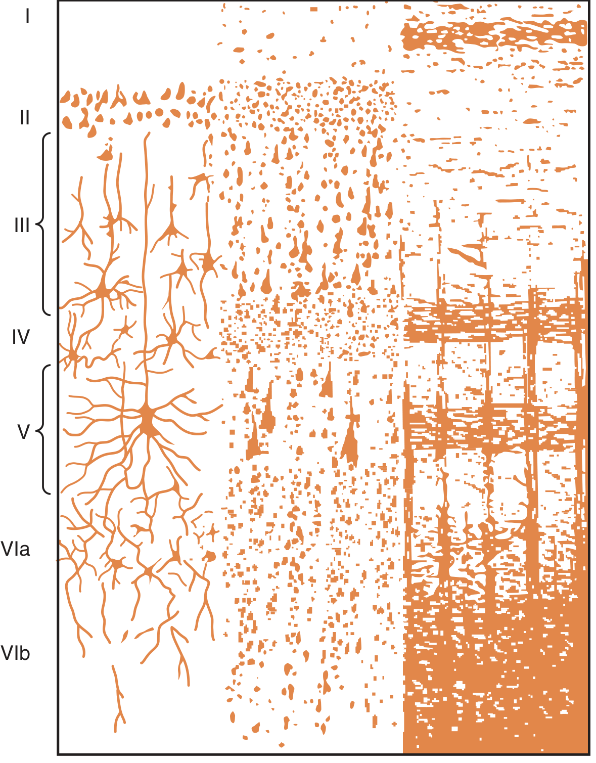
The cortex is arranged in 6 layers (Fig. 58.1):
| Layer | Name | Contents |
|---|---|---|
| I | Molecular layer | Sparse neurons, many fibers |
| II | External granular layer | Small granule (stellate) cells |
| III | External pyramidal layer | Pyramidal cells |
| IV | Internal granular layer | Granule cells - sensory input terminates here |
| V | Large pyramidal cell layer | Large pyramidal cells - major output layer |
| VI | Fusiform (polymorphic) layer | Fusiform cells - output signals |
Three main neuron types:
- Granular (stellate) cells - short axons, function as interneurons; excitatory (release glutamate) or inhibitory (release GABA); concentrated in sensory and association areas
- Pyramidal cells - source of long fibers going all the way to the spinal cord (corticospinal tract); give rise to large subcortical association fiber bundles
- Fusiform cells - contribute to output signals from cortex
3. Thalamocortical System
All cortical areas have extensive two-way connections with the thalamus. When thalamic connections are cut, the functions of the corresponding cortical area are almost entirely lost. The thalamus and cortex together are called the thalamocortical system. Almost all sensory pathways pass through the thalamus before reaching the cortex (exception: olfaction).
4. Functional Areas of the Cerebral Cortex
A. Motor Areas
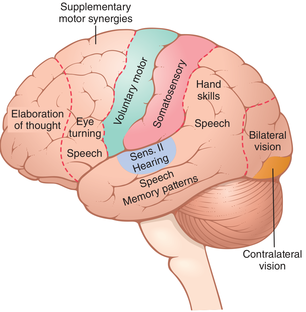
Primary Motor Cortex (Area 4)
- Located in the first convolution of the frontal lobe, anterior to the central sulcus
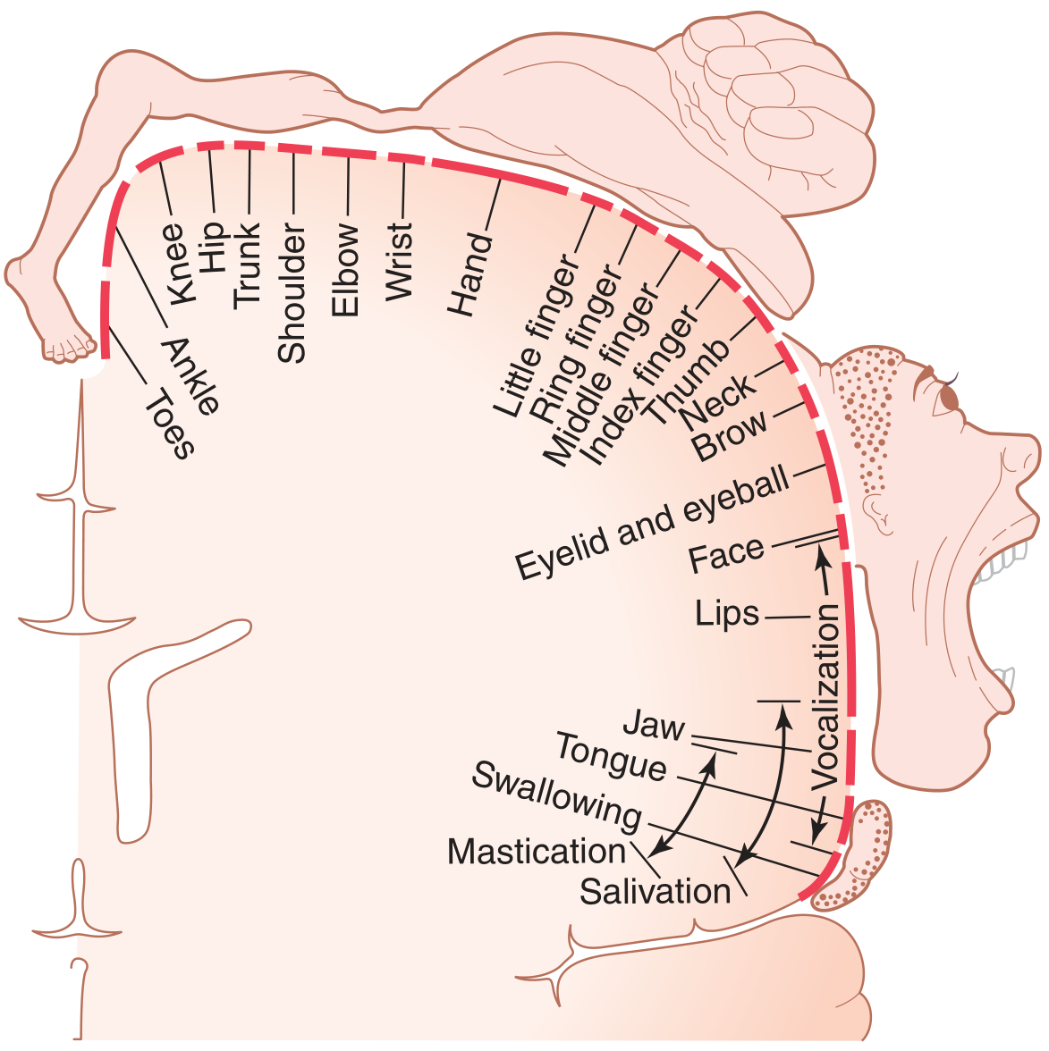
- Has somatotopic (topographical) organization - different body parts are represented in specific regions (motor homunculus)
- Face and mouth: near the sylvian fissure (lateral)
- Hand and arm: mid-portion
- Trunk: near apex
- Leg and foot: dips into longitudinal fissure
- More than half the primary motor cortex is devoted to hands and speech muscles (reflecting their precision)
Premotor Area (Area 6)
- Lies 1-3 cm anterior to the primary motor cortex
- Generates complex "patterns" of movement
- Develops a "motor image" of the total movement to be performed
- Works with basal ganglia to provide patterns of motor activity
Supplementary Motor Area
- Abuts the premotor area superiorly
- Functions similar to the premotor area
- Controls postural and synergistic movements
B. Sensory Areas
Primary Somatosensory Cortex (Areas 3, 1, 2)
- Located in the parietal lobe, posterior to the central sulcus
- Receives somatic sensations from the body (touch, pain, temperature, proprioception)
- Also has somatotopic organization - sensory homunculus
Primary Visual Cortex (Area 17)
- Located in the occipital lobe (calcarine sulcus region)
- Receives visual signals from the retina via lateral geniculate body of thalamus
Primary Auditory Cortex (Areas 41, 42)
- Located in the superior temporal gyrus (Heschl's gyri)
- Processes sound frequency and intensity
C. Association Areas
1. Parieto-Occipitotemporal Association Area
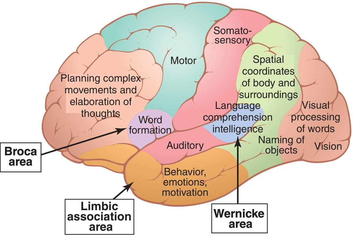
- Provides high-level interpretation of signals from all surrounding sensory areas
- Functions include:
- Spatial coordinate analysis of body and surroundings (posterior parietal cortex)
- Wernicke's area (posterior superior temporal gyrus) - language comprehension and intelligence; most important region for higher intellectual function
- Angular gyrus - processing of visual language (reading); damage = alexia (word blindness)
- Naming of objects
2. Prefrontal Association Area
- Works with premotor area to plan complex bodily movements
- "Elaboration of thought" - planning for action
- Contains working memory (short-term memory for active reasoning)
3. Limbic Association Area
- Located in the anterior pole of temporal lobe, orbital frontal cortex, and cingulate gyrus
- Controls behavior, emotions, and motivation
5. Language Areas (Speech and Communication)
Wernicke's Area
- Located in the posterior part of the superior temporal gyrus (dominant hemisphere)
- Responsible for language comprehension
- Damage → Wernicke's aphasia: can hear words but cannot interpret meaning; speech is fluent but incoherent ("word salad")
Broca's Area (Area 44, 45)
- Located in the lateral frontal lobe (inferior frontal gyrus)
- Responsible for motor speech production (word formation)
- Damage → Broca's aphasia: understands language but cannot speak fluently (non-fluent, effortful speech)
Dominant Hemisphere
- In approximately 95% of people, the left hemisphere is dominant
- The Wernicke area is up to 50% larger in the left hemisphere even at birth
6. Memory
| Type | Duration | Mechanism |
|---|---|---|
| Short-term memory | Seconds to minutes | Reverberating neural circuits; presynaptic facilitation |
| Intermediate long-term | Days to weeks | Temporary chemical/physical changes at synapses |
| Long-term memory | Years to lifetime | Permanent structural changes - new synaptic connections (consolidation) |
Classification by content:
- Declarative memory - memory of facts, events, experiences (hippocampus-dependent)
- Skill (procedural) memory - motor skills, habits (basal ganglia-dependent)
Role of the hippocampus: Bilateral destruction of the hippocampus causes inability to form new long-term declarative memories (anterograde amnesia).
7. Key Brodmann Areas Summary
| Brodmann Area | Location | Function |
|---|---|---|
| Area 4 | Precentral gyrus | Primary motor cortex |
| Area 6 | Premotor cortex | Complex movement patterns |
| Areas 3, 1, 2 | Postcentral gyrus | Primary somatosensory |
| Area 17 | Occipital lobe | Primary visual cortex |
| Areas 41, 42 | Superior temporal gyrus | Primary auditory cortex |
| Area 44, 45 | Inferior frontal gyrus | Broca's area (motor speech) |
| Area 22 | Superior temporal gyrus | Wernicke's area |
| Area 39 | Angular gyrus | Visual language processing |
8. Electroencephalogram (EEG) and Consciousness
The EEG records electrical activity of the cerebral cortex. Four main wave types:
- Alpha waves (8-13 Hz) - awake, relaxed, eyes closed
- Beta waves (14-30 Hz) - active thinking, eyes open
- Theta waves (4-7 Hz) - light sleep, children
- Delta waves (<4 Hz) - deep sleep, severe brain disease when awake
Quick Exam Points
- Cerebral cortex has 6 layers; sensory input arrives at layer IV, motor output leaves via layers V and VI
- Primary motor cortex = Area 4 (precentral gyrus); primary sensory cortex = Areas 3, 1, 2 (postcentral gyrus)
- More than half the motor cortex represents the hands and speech
- Wernicke's area = language comprehension (damage = fluent but meaningless speech)
- Broca's area = motor speech (damage = non-fluent aphasia)
- Left hemisphere is dominant in 95% of people
- Thalamus is essential for cortical function - thalamo-cortical system
- Hippocampus is essential for conversion of short-term to long-term declarative memory
- Guyton and Hall Textbook of Medical Physiology, pp. 687, 716-722
This is a shared conversation. Sign in to Orris to start your own chat.