
Give in detail about walleyes ring and detail about each it’s component
Reading File
The "Waldeyer's ring" (often spelled "Waldeyer's ring" or "Waldeyer ring") is a ring of lymphoid tissue in the pharynx. Let me search the medical library for detailed information.
Searching sources...
Finding Sources
Finding Sources
Searching the Web
Waldeyer's ring anatomy components detail lymphoid tissue pharynx
Now let me read the most detailed textbook sections on Waldeyer's ring components:
Reading File
Reading File
Reading File
Reading File
Excellent data gathered. Let me also fetch the pharynx diagram and get a bit more detail on each tonsil component:
Loading Image
Finding Sources
Finding Sources
Let me now read the Color Atlas section on the tonsils for detailed component descriptions:
Reading File
Reading File
Let me fetch the tonsils diagram from the Color Atlas:
Loading Image
I now have comprehensive information from multiple authoritative textbooks. Here is the full, detailed answer:
Waldeyer's Ring - Detailed Overview
Waldeyer's ring (also called the pharyngeal lymphoid ring or anulus lymphoideus pharyngis) is a discontinuous, roughly circular arrangement of lymphoid tissue encircling the entrance to the naso- and oropharynx. It was described by the German anatomist Heinrich Wilhelm Gottfried von Waldeyer-Hartz in 1884. The ring is positioned at the gateway of both the respiratory and alimentary tracts, placing it at the body's first point of contact with inhaled and ingested antigens.
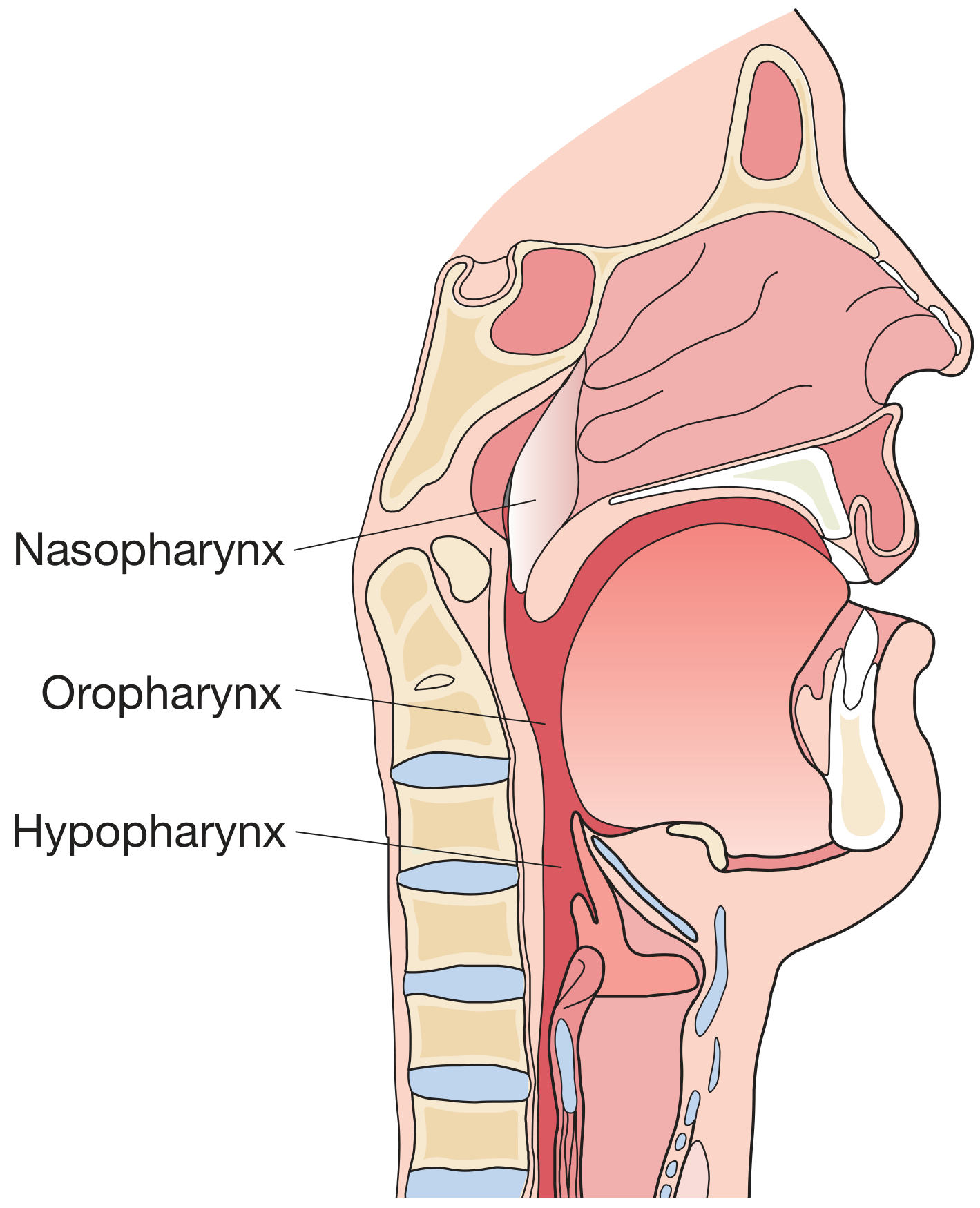
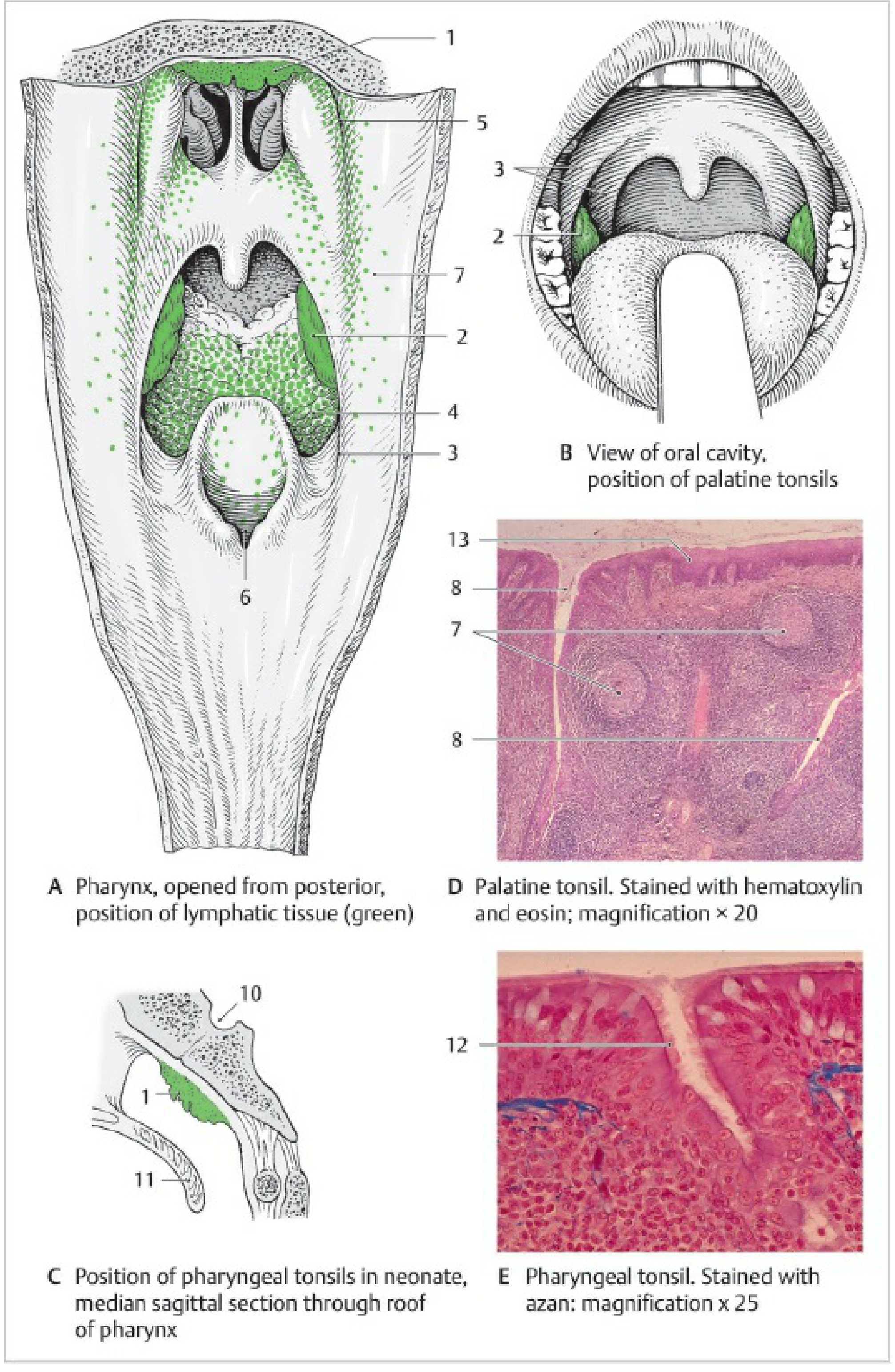
Biological Role
Waldeyer's ring is part of mucosa-associated lymphoid tissue (MALT) - specifically the nasal-associated lymphoid tissue (NALT). Its functions include:
- First-line immune defence - it is the initial immunological barrier against antigens entering via nasal and oral routes, particularly in children.
- B lymphocyte proliferation occurs in germinal centres within the tonsils, generating specific antibody responses.
- Immunoglobulin production - IgG, IgA, IgM, IgD, complement, interferon, lysozymes, and cytokines all accumulate in tonsillar tissue.
- Immune maturation and sampling - the ring samples the antigen environment at the entry to the aerodigestive tract.
- The lymphoid tissue undergoes physiological hypertrophy during early childhood as the child encounters increasing antigenic stimuli, and there is often parallel hypertrophy of cervical lymph nodes. - Bailey and Love's Short Practice of Surgery
Components of Waldeyer's Ring
The ring has two concentric divisions - an inner ring and an outer ring.
Inner Ring (Major Components)
| Component | Location |
|---|---|
| Pharyngeal tonsil (Adenoid) | Roof/posterior wall of nasopharynx |
| Tubal tonsils (Gerlach's tonsils) | Around Eustachian tube openings |
| Palatine tonsils | Lateral oropharynx, between the pillars |
| Lingual tonsil | Posterior one-third of tongue |
Outer Ring (Minor/Accessory Components)
| Component | Location |
|---|---|
| Lateral pharyngeal bands | Behind the posterior tonsillar pillars |
| Intertonsillar lymphoid tissue | Lamina propria between the tonsils |
Detailed Description of Each Component
1. Pharyngeal Tonsil (Adenoid)
- Location: Situated at the junction of the roof and posterior wall of the nasopharynx, inferior to the sphenoid bone. It is the superior (topmost) component of Waldeyer's ring.
- Structure: A single midline mass of lymphoid tissue. Unlike the palatine tonsils it has no crypts, but instead has deep folds or furrows on its surface. In children it can be prominent enough to obstruct the nasopharynx.
- Epithelium: Covered by pseudostratified ciliated (respiratory) columnar epithelium (since it lies in the nasopharynx where ciliated epithelium predominates).
- Size: Enlarges rapidly during childhood (peak size at 5-6 years) and involutes after puberty.
- Function: Samples antigens from inspired air. It processes antigens and presents them to T-helper and B cells, facilitating first-line immune response.
- Clinical significance:
- Adenoid hypertrophy in children can cause nasal obstruction, mouth breathing, snoring, and obstructive sleep apnea.
- Enlarged adenoids can block the Eustachian tube orifice, causing recurrent otitis media with effusion (glue ear) and conductive hearing loss.
- Surgical removal = adenoidectomy.
- Adenoiditis (acute or chronic infection) is common in childhood.
- The fossae of Rosenmüller (pharyngeal recesses) lie adjacent and are a common site for nasopharyngeal carcinoma.
2. Tubal Tonsils (Gerlach's Tonsils)
- Location: Small lymphoid nodules situated at (and around) the pharyngeal openings of the Eustachian (auditory) tubes in the nasopharynx - specifically within the lip of the fossa of Rosenmüller, posterior to the torus tubarius.
- Structure: A collection of smaller secondary lymphoid nodules; they are viewed as a direct continuation of the pharyngeal tonsil. There are two tubal tonsils (one on each side). They are not always present or well-defined in all individuals.
- Epithelium: Covered by respiratory epithelium (pseudostratified ciliated columnar), consistent with the nasopharyngeal location.
- Function: Guards the opening of the Eustachian tube from ascending infection. Part of the general sentinel function of the ring.
- Clinical significance:
- Enlargement can obstruct the pharyngeal opening of the auditory tube, resulting in hearing impairment, nasal speech, and chronic ear infections (recurrent otitis media).
- Source: Color Atlas of Human Anatomy, Vol. 2
3. Palatine Tonsils
- Location: Within the lateral wall of the oropharynx, sitting in the tonsillar fossa (tonsillar bed) between the palatoglossal fold (anterior pillar) and the palatopharyngeal fold (posterior pillar).
- Structure:
- Ovoid masses of lymphoid tissue, one on each side.
- Free medial surface has a pitted appearance with 10-20 pits leading into deep, blind-ending crypts that traverse the full thickness of the tonsil. Crypts allow antigen entry and are responsible for trapping debris (which can lead to tonsilloliths).
- The recessus palatinus (intratonsillar cleft) is a specific cleft-like space within the substance of the tonsil, representing a remnant of the second pharyngeal pouch.
- The tonsil sits in a hemicapsule of connective tissue (not a full capsule), which facilitates tonsillectomy.
- Epithelium: Covered by stratified non-keratinized squamous epithelium - consistent with oral cavity epithelium, allowing direct antigen presentation.
- Size: Enlarges rapidly until ages 5-6, maximal size at puberty, then involutes. Little lymphoid tissue remains in old age.
- Blood supply: Predominantly the tonsillar artery (branch of the facial artery) off the external carotid artery. Multiple anastomosing branches contribute (descending palatine, ascending pharyngeal, dorsal lingual).
- Venous drainage: Via the paratonsillar vein (external palatine vein) within the capsule, and a plexiform system outside it.
- Lymph drainage: To the jugulodigastric (tonsillar) node of the deep cervical lymph chain - the most important node to palpate when assessing tonsil pathology.
- Nerve supply: Tonsillar branch of the glossopharyngeal nerve (CN IX) and the lesser palatine nerve (V2 - maxillary division of CN V).
- Function: Immunological early warning system; site of vigorous B lymphocyte proliferation. Encounters pathogens entering through mouth and nose, activating specific immune responses.
- Clinical significance:
- Tonsillitis (acute or chronic) - commonest cause is Group A beta-haemolytic streptococcus (GABHS).
- Peritonsillar abscess (quinsy) - collection of pus between tonsil capsule and superior pharyngeal constrictor.
- Tonsillectomy - surgical removal for recurrent tonsillitis (Paradise criteria) or obstructive hypertrophy.
- Most commonly affected site in Waldeyer's ring lymphoma (usually diffuse large B-cell NHL).
- Source: Scott-Brown's Otorhinolaryngology Head & Neck Surgery
4. Lingual Tonsil
- Location: At the posterior one-third (base) of the tongue, inferior-anterior component of Waldeyer's ring.
- Structure:
- Has a bumpy/nodular surface with numerous crypt-like infoldings of the oral mucosa.
- Crypts are surrounded by secondary lingual nodules.
- The posterior lingual glands (mucous-secreting) open at the base of the crypts.
- Unlike the palatine tonsil, the lingual tonsil is not encapsulated; it is flat and diffuse.
- Epithelium: Covered by stratified non-keratinized squamous epithelium (oral type).
- Function: Provides immune surveillance over the posterior tongue and oropharyngeal inlet.
- Clinical significance:
- Can undergo hypertrophy, causing dysphagia, snoring, or obstructive sleep apnea (especially post-tonsillectomy when compensatory hypertrophy occurs).
- Rarely removed surgically but can be treated with coblation or laser reduction.
- Can be a site of squamous cell carcinoma (HPV-related oropharyngeal cancer, increasingly recognized).
5. Lateral Pharyngeal Bands
- Location: Aggregates of lymphoid tissue in the mucosa of the lateral and posterior walls of the pharynx, posterior to the posterior tonsillar pillars.
- Structure: The lymphoid tissue can form small nodules on the posterior pharyngeal wall. On examination they appear as small areas of salmon-pink, slightly raised swellings.
- Function: Part of the interstitial lymphoid component of the ring; act as supplementary immune surveillance tissue between the major tonsils.
- Clinical significance:
- Lateral pharyngitis - inflammatory swelling of the pharyngeal mucosa involving the lateral bands, presenting with sore throat and dysphagia.
- These become more prominent after tonsillectomy (compensatory hypertrophy).
- Source: Color Atlas of Human Anatomy, Vol. 2
6. Intertonsillar Lymphoid Tissue (MALT in Lamina Propria)
- Location: Numerous collections of lymphoid tissue in the lamina propria just beneath the epithelium throughout the pharyngeal mucosa, in the intertonsillar intervals.
- Structure: Diffuse MALT (mucosa-associated lymphoid tissue) - not organized into tonsil-like structures but present as scattered lymphocyte collections and solitary lymphoid nodules.
- Function: Provides continuous immunological coverage along the entire mucosal lining of the pharynx, filling in the "gaps" between the major tonsillar components to complete the ring.
Summary Table
| Component | Location | Epithelium | Key Feature | Clinical Condition |
|---|---|---|---|---|
| Pharyngeal tonsil (Adenoid) | Roof/posterior nasopharynx | Respiratory (ciliated) | No crypts; folds/furrows | Adenoid hypertrophy, otitis media |
| Tubal tonsils (Gerlach's) | Around Eustachian tube openings | Respiratory (ciliated) | Small nodule collection | Eustachian tube obstruction |
| Palatine tonsils | Oropharynx, between faucial pillars | Stratified squamous | 10-20 deep crypts; hemicapsule | Tonsillitis, quinsy, lymphoma |
| Lingual tonsil | Posterior 1/3 of tongue | Stratified squamous | Bumpy; crypt-like folds | Compensatory hypertrophy, SCC |
| Lateral pharyngeal bands | Lateral/posterior pharyngeal walls | Stratified squamous | Salmon-pink nodules | Lateral pharyngitis |
| Intertonsillar MALT | Throughout pharyngeal lamina propria | Variable | Diffuse scattered lymphocytes | Chronic pharyngitis |
Clinical Correlates of the Whole Ring
- Lymphoma - Waldeyer's ring is the commonest site of head and neck lymphoma (predominantly non-Hodgkin lymphoma, diffuse large B-cell type). The tonsils are most commonly affected. In contrast to Hodgkin's lymphoma (where the ring is rarely involved), NHL commonly involves Waldeyer's ring. - Grainger & Allison's Diagnostic Radiology
- Immune maturation in children - The ring undergoes hypertrophy as a normal response to increasing antigen exposure in childhood; tonsillectomy/adenoidectomy has no proven negative immunological impact. - K.J. Lee's Essential Otolaryngology
- Nasopharyngeal carcinoma - The adjacent fossae of Rosenmüller (at the tubal tonsil level) are the commonest primary site.
- MALT lymphomas - Up to 20% of Waldeyer's ring NHLs are MALT-type, with a close association with synchronous GI tract involvement (endoscopy is part of staging in many centres).
This is a shared conversation. Sign in to Orris to start your own chat.