
Appendicitis sign Psoas Obturator Rovsing and other
appendicitis clinical signs psoas obturator Rovsing diagram
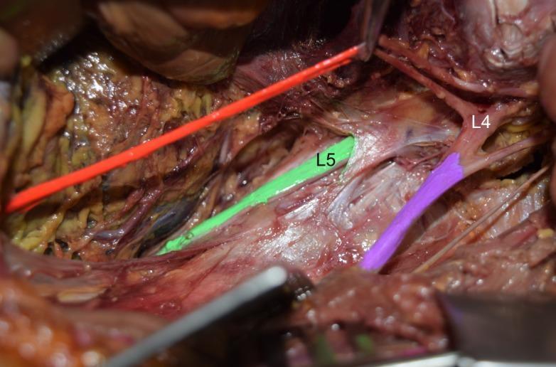
Anatomical Diagram/Clinical Photograph: This image depicts a cadaveric dissection of the left posterior abdominal wall focusing on the lumbar plexus. The psoas major muscle has been removed to expose the neural structures. Key anatomical landmarks are color-coded for clarity: the L4 ventral ramus and femoral nerve are highlighted in purple, the L5 ventral ramus is highlighted in green, and the obturator nerve is shown in orange/red. The primary educational focus is an anatomical variation where the lumbosacral trunk is absent. Visually, there is a clear separation between the L4 and L5 ventral rami, with no evidence of the standard fusion that typically forms the lumbosacral trunk. The femoral nerve (purple) is seen emerging from the L4 level, running lateral and parallel to the L5 ramus (green). This clinical photograph serves as an educational tool for gross anatomy and neurosurgery, illustrating variations in lumbosacral plexus connectivity and its implications for regional anesthesia or surgical approaches to the pelvic and lumbar regions.
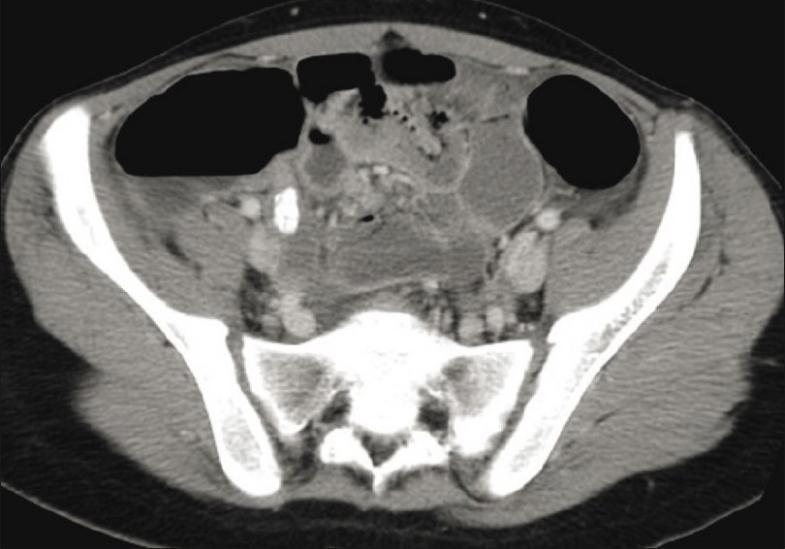
This diagnostic axial CT image of the lower abdomen and pelvis demonstrates findings consistent with acute appendicitis. The scan reveals a distended and wall-thickened appendix in the right iliac fossa. A key visual feature is the presence of a high-attenuation, calcified appendicolith (fecalith) located within the lumen of the inflamed appendix. Associated secondary signs of inflammation are evident, including localized fat stranding and soft tissue haziness in the periappendiceal region. Surrounding anatomical structures include the pelvic bones (ilium and sacrum), psoas muscles, and multiple bowel loops containing air and contrast material. The image serves as a classic educational example of radiological diagnosis for appendicitis in a clinical emergency setting, highlighting the primary obstructed appendix and the secondary inflammatory response in the adjacent peritoneal fat.
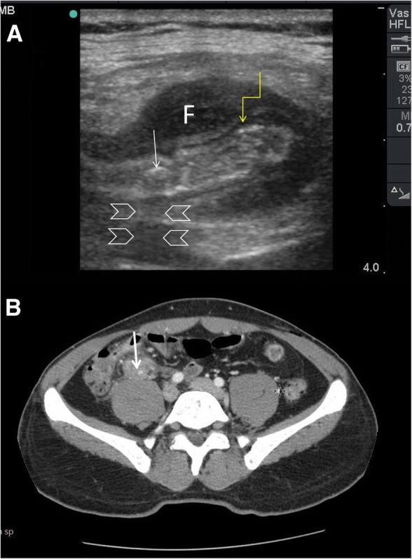
This composite figure demonstrates the diagnostic imaging of acute appendicitis via Point-of-Care Ultrasound (POCUS) and Computed Tomography (CT). Image A is a high-frequency linear probe ultrasound showing a cross-section of a thickened, non-compressible appendix. Key features include a hypoechoic, edematous muscularis propria (indicated by a yellow arrow) and an intraluminal hyperechoic appendicolith/faecolith (white arrow) exhibiting prominent posterior acoustic shadowing (arrowheads). A localized anechoic fluid collection (marked 'F') surrounds the inflamed appendix, consistent with periappendiceal fluid or a phlegmon. Image B is an axial CT scan of the abdomen and pelvis with intravenous contrast, confirming the ultrasound findings in the right iliac fossa. It shows a dilated appendix with wall enhancement and an associated hyperdense faecolith (white arrow) situated anterior to the psoas muscle. This comparison illustrates the utility of POCUS in identifying classic sonographic signs of appendicitis—wall thickening, fecaliths with shadowing, and periappendiceal fluid—and its correlation with definitive CT imaging in a clinical setting of fever and right lower quadrant pain.
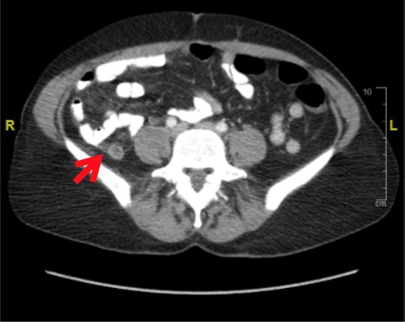
This diagnostic image is an axial computed tomography (CT) scan of the lower abdomen and pelvis, oriented in a cross-sectional view with the patient's right (R) and left (L) sides indicated. A red arrow in the right lower quadrant points to a dilated and thickened appendix, which measures approximately 10 mm in diameter. The visual hallmark of acute appendicitis is demonstrated by the presence of periappendiceal fat stranding—increased density and haziness in the surrounding mesenteric fat—suggesting localized inflammation. Anatomical landmarks include the vertebral body of the spine centrally, the psoas muscles bilaterally, and loops of bowel. The scan illustrates the classic radiological presentation of uncomplicated acute appendicitis, used for diagnostic confirmation in clinical emergency medicine. The imaging provides evidence of inflammation without visual signs of perforation or abscess formation at this level.
Clinical Signs of Appendicitis
1. McBurney's Point Tenderness
- How: Direct palpation at a point one-third of the distance from the right anterior superior iliac spine (ASIS) to the umbilicus.
- Positive: Maximal tenderness at this point.
- Significance: The most classic finding in acute appendicitis. Tenderness here reflects localized peritoneal irritation as the inflammation progresses from the appendix to the overlying peritoneum.
- Goldman-Cecil Medicine; Current Surgical Therapy, 14th ed.
2. Rovsing's Sign
- How: Deep palpation of the left lower quadrant (LLQ).
- Positive: Pain is referred to the right lower quadrant (RLQ).
- Mechanism: Pressure in the LLQ pushes coils of ileum slightly to the right, which then press on the inflamed appendix. It is an indirect assessment for localized peritoneal inflammation in the RLQ.
- S Das Manual of Clinical Surgery, 13th ed.; Maingot's Abdominal Operations; Harrison's Principles, 22nd ed.
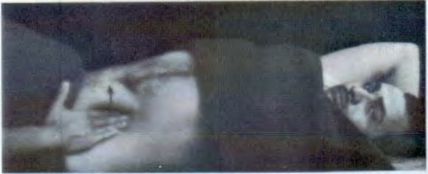
3. Psoas Sign (Cope's Psoas Test / Iliopsoas Sign)
- How (classical): Patient lies in the left lateral decubitus position (turned to the left); the examiner hyperextends the right thigh posteriorly. Alternatively, with the patient supine, they actively flex the right hip against resistance.
- Positive: Pain is produced in the RLQ.
- Mechanism: A retrocecal appendix lies directly on the psoas major muscle. When inflamed, any stretching of this muscle (by hip extension or active flexion against resistance) irritates the adjacent inflamed appendix, producing pain.
- Also: Can be positive in psoas abscess or other retroperitoneal inflammatory processes.
- S Das Manual of Clinical Surgery; Fischer's Mastery of Surgery, 8th ed.; Sleisenger & Fordtran, 10th ed.
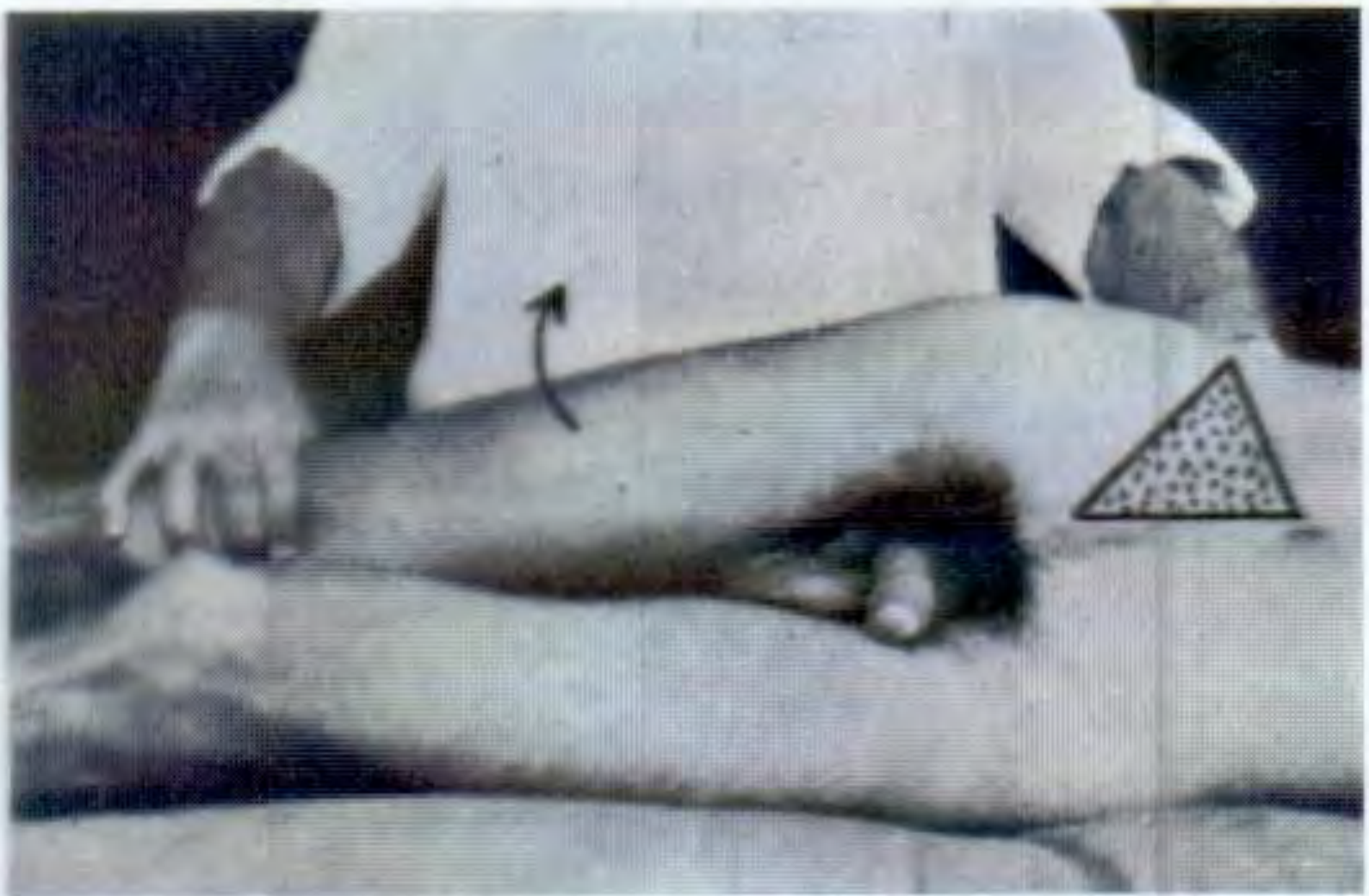
4. Obturator Sign (Cope's Obturator Test)
- How: Patient is supine with the right hip and knee flexed to 90°. The examiner internally rotates the right hip.
- Positive: Pain is reproduced in the RLQ or suprapubic area.
- Mechanism: A pelvic appendix lies on the obturator internus muscle. Internal rotation of the hip stretches this muscle, irritating the adjacent inflamed pelvic appendix.
- Also: Can suggest pelvic inflammatory disease (PID) or other pelvic pathology.
- S Das Manual of Clinical Surgery; Yamada's Textbook of Gastroenterology; Harrison's Principles, 22nd ed.
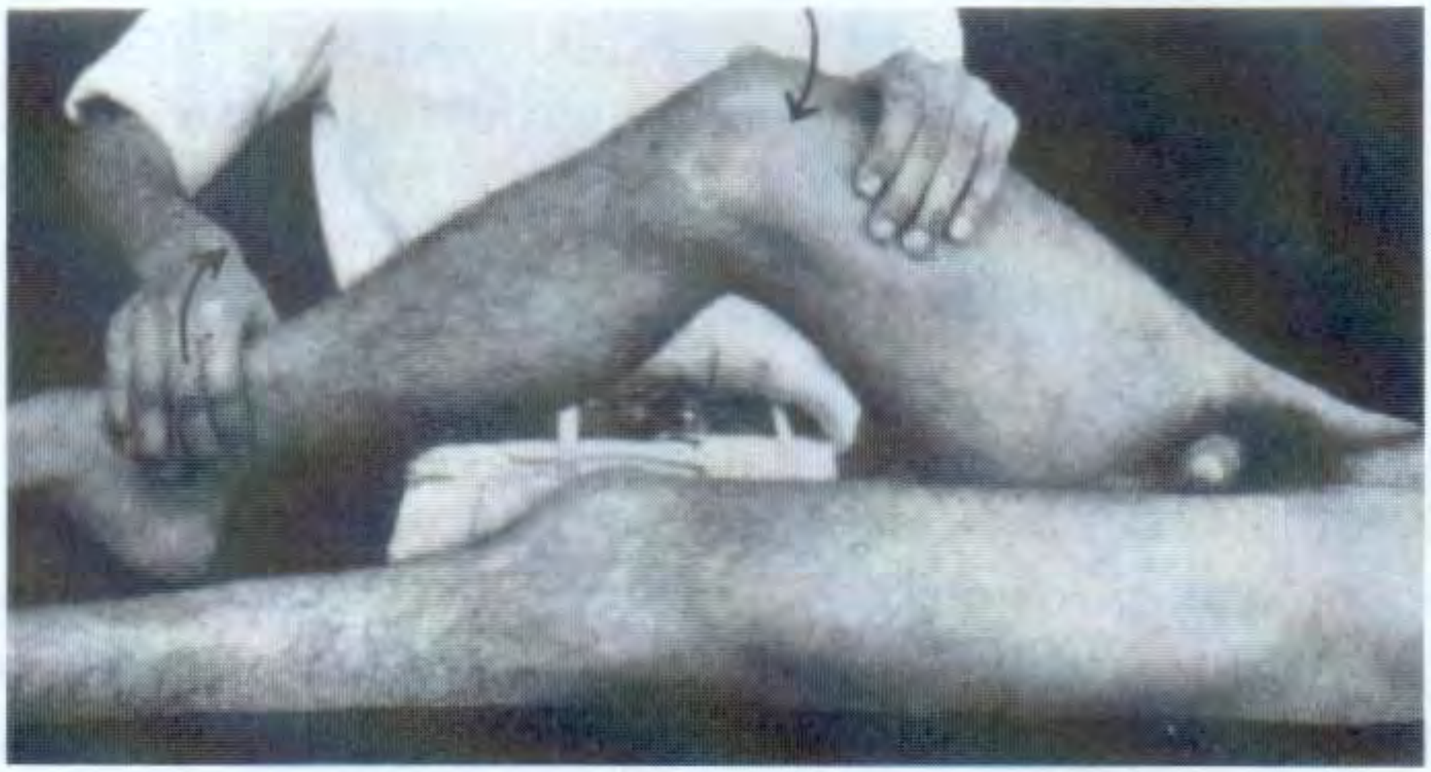
5. Rebound Tenderness (Blumberg's Sign)
- How: Deep pressure is applied over the tender area, then suddenly released.
- Positive: Pain is greater on release than on pressure.
- Mechanism: Sudden movement of inflamed parietal peritoneum causes sharp pain.
- Note: Many modern textbooks (including Yamada's) recommend abandoning this test as it causes unnecessary pain and has limited accuracy compared to gentle percussion tenderness. Percussion tenderness is preferred.
- Yamada's Textbook of Gastroenterology, 7th ed.
6. Dunphy's Sign (Cough Sign)
- How: Ask the patient to cough.
- Positive: Coughing worsens RLQ pain.
- Mechanism: Increased intra-abdominal pressure during coughing jostles the inflamed peritoneum. Patients with appendicitis also report pain on bumpy car rides, sneezing, or Valsalva maneuvers for the same reason.
- Harrison's Principles, 22nd ed.
7. Baldwing's Test
- How: A hand is placed over the patient's right flank; the patient is asked to raise the right lower limb off the bed with the knee extended (straight leg raise).
- Positive: Immediate pain complaint in retrocecal appendicitis.
- Mechanism: Similar to the psoas sign - the retrocecal appendix contacts the psoas major; active hip flexion (SLR) contracts and shortens the psoas, compressing the inflamed appendix.
- S Das Manual of Clinical Surgery, 13th ed.
Summary Table
| Sign | Maneuver | Positive Finding | Appendix Position |
|---|---|---|---|
| McBurney's | Palpate McBurney's point | Maximal tenderness | Typical |
| Rovsing's | Palpate LLQ | Pain referred to RLQ | Typical |
| Psoas (Cope's) | Hyperextend right hip (left lateral) OR active flexion against resistance | RLQ pain | Retrocecal |
| Obturator (Cope's) | Flex right hip + internal rotation | RLQ / pelvic pain | Pelvic |
| Rebound (Blumberg's) | Deep pressure then sudden release | Pain worse on release | Any (peritonitis) |
| Dunphy's / Cough sign | Ask patient to cough | Worsened RLQ pain | Any |
| Baldwing's | Straight leg raise (right) | RLQ pain | Retrocecal |
Key Clinical Points
- Retrocecal appendix (most common variant, ~65%): RLQ tenderness may be less prominent; psoas sign and Baldwing's test are more useful.
- Pelvic appendix: May present with dysuria, frequency, diarrhea, or tenesmus; obturator sign is key; rectal/vaginal examination is important.
- All these signs are highly specific but poorly sensitive - a negative sign does not exclude appendicitis.
- Scoring systems (Alvarado, AIR score for adults; Pediatric Appendicitis Score) incorporate combinations of symptoms, signs, and labs to improve diagnostic accuracy.
- Current Surgical Therapy operative characteristics table: Rovsing's sensitivity 30-68%, specificity 58-91%; Obturator sensitivity 21-34%, specificity 94%; Psoas sensitivity 13-42%, specificity 79-97%. - Current Surgical Therapy, 14th ed.