
Thrombosis & embolus
Reading File
Now I'll search the medical library for comprehensive content on thrombosis and embolism in parallel.
Finding Sources
Finding Sources
Good leads. Now let me pull the core pathology content on thrombosis and embolism from Goldman-Cecil and Harrison's - the most comprehensive sources.
Finding Sources
Finding Sources
Robbins Pathologic Basis of Disease is the best source for pathology of thrombosis and embolism. Let me read those sections plus the embolism heading search.
Reading File
Finding Sources
Excellent. Now let me read the Embolism section from Robbins Cotran (the most authoritative pathology text), and also fetch the Virchow triad diagram.
Reading File
Loading Image
I have rich content. Let me also read the rest of the embolism section and fetch a key thrombus morphology image.
Reading File
I have a comprehensive set of information. Now let me compile a thorough answer from all the Robbins Cotran content I've gathered.
Thrombosis & Embolism
Source: Robbins, Cotran & Kumar - Pathologic Basis of Disease
THROMBOSIS
A thrombus is a solid mass of blood constituents (platelets, fibrin, RBCs, WBCs) formed within the vascular system during life. It is pathological clot formation - distinct from normal hemostasis.
Virchow's Triad
The three primary abnormalities leading to thrombosis form Virchow's triad:
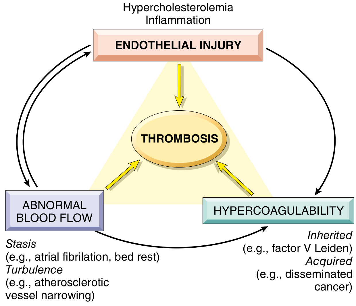
1. Endothelial Injury
The most important factor, especially for arterial and cardiac thrombosis where high flow rates impede clot formation. Platelet adherence and activation is a necessary prerequisite under high shear stress.
Mechanisms of injury:
- Physical injury, infectious agents, abnormal blood flow
- Inflammatory mediators and cytokines
- Metabolic abnormalities (hypercholesterolemia, homocystinemia)
- Toxins from cigarette smoke
Prothrombotic endothelial changes:
- Procoagulant changes: Downregulation of thrombomodulin, protein C, and tissue factor pathway inhibitor (TFPI) - shifts balance toward coagulation
- Antifibrinolytic effects: Secretion of plasminogen activator inhibitors (PAIs), reduced tissue plasminogen activator (t-PA) expression
- Platelet-activating effects: Increased expression of vWF and tissue factor
2. Abnormal Blood Flow (Stasis or Turbulence)
- Turbulence promotes arterial and cardiac thrombosis by causing endothelial injury/dysfunction
- Stasis is the dominant factor in venous thrombosis
Turbulence and stasis promote thrombosis by:
- Disrupting laminar flow, bringing platelets into contact with endothelium
- Preventing dilution of activated clotting factors by fresh blood
- Retarding influx of clotting factor inhibitors
- Promoting endothelial cell activation
Clinical examples:
- Atrial fibrillation - blood stasis in the atrial appendage
- Aneurysms - turbulence and stasis in the sac
- Atherosclerotic plaques - turbulence around stenoses
- Prolonged bed rest / immobility - venous stasis in lower limbs
- Hyperviscosity states (polycythemia, sickle cell disease)
3. Hypercoagulability
Defined as any alteration in the coagulation pathways that predisposes to thrombosis.
| Type | Examples |
|---|---|
| Primary (Inherited) | Factor V Leiden mutation (most common hereditary thrombophilia), Prothrombin gene mutation (G20210A), Antithrombin III deficiency, Protein C or S deficiency |
| Secondary (Acquired) | Prolonged bed rest/immobility, MI, atrial fibrillation, tissue damage (surgery, fracture), cancer (Trousseau syndrome), antiphospholipid antibody syndrome, HIT (heparin-induced thrombocytopenia), pregnancy, OCP use, nephrotic syndrome |
Morphology of Thrombi
Arterial thrombi (white thrombi):
- Rich in platelets and fibrin
- Typically at sites of endothelial injury (atherosclerotic plaques)
- Grow in a retrograde direction from point of attachment
- Show lines of Zahn - pale platelet/fibrin layers alternating with darker RBC-rich layers (pathognomonic of antemortem thrombus)
Venous thrombi (red/stasis thrombi):
- Rich in RBCs (hence "red")
- Occur in slower-flow venous circulation
- Most commonly in deep veins of lower extremity (DVT)
- Lines of Zahn are less distinct
Mural thrombi:
- In heart chambers or aortic lumen
- Post-MI (endocardial injury), atrial fibrillation (stasis), aortic aneurysm
Fate of a Thrombus
Thrombi have four possible fates:
| Fate | Description |
|---|---|
| Propagation | Accumulates more platelets and fibrin, enlarges |
| Embolization | Dislodges and travels to distant sites |
| Dissolution (lysis) | Fibrinolytic activity dissolves the clot (most desirable) |
| Organization and recanalization | Older thrombi become organized by ingrowth of endothelial cells, smooth muscle, fibroblasts; may eventually recanalize to restore flow |
Consequences of Thrombosis
- Local vascular occlusion - ischemia/infarction distal to the clot
- Distal embolization - thrombus fragment travels and blocks a distant vessel
- Venous congestion - in venous thrombosis, backup of blood causing edema, pain, and ulceration
EMBOLISM
An embolus is a detached intravascular solid, liquid, or gaseous mass that is carried by blood from its point of origin to a distant site, where it causes partial or complete vascular occlusion.
Most emboli are dislodged thrombi - hence the collective term thromboembolism.
Types of Emboli
1. Pulmonary Embolism (PE)
- Origin: 70-80% arise from deep vein thrombosis (DVT) of the legs
- Incidence: ~60-120 cases/100,000 per year (US); 60,000-100,000 deaths/year
- ~20% die before or shortly after diagnosis
Pathophysiology by size:
| Embolus Size | Effect |
|---|---|
| Large/massive | Occludes main pulmonary artery or saddle embolus at bifurcation → sudden death, acute cor pulmonale |
| Medium | Occludes a medium pulmonary artery → pulmonary hemorrhage (rarely infarction due to dual lung supply) |
| Small/multiple | Multiple small emboli over time → pulmonary hypertension, right heart failure |
| Paradoxical | Crosses patent foramen ovale → systemic embolization |
Pulmonary infarction occurs in <10% because the lung has a dual blood supply (pulmonary + bronchial arteries). It occurs when bronchial flow is also compromised (left heart failure, pneumonia).
Clinical features:
- Pleuritic chest pain, dyspnea, tachycardia, hemoptysis
- Large emboli: sudden-onset shock, right heart failure
- Multiple small emboli: progressive dyspnea, pulmonary HTN
2. Systemic Thromboembolism
- Most arise from intracardiac mural thrombi (2/3 associated with left ventricular MI; 25% with dilated left atrium in atrial fibrillation)
- Also from aortic aneurysms, ulcerated atherosclerotic plaques, valvular vegetations
- Travel to: legs (most common ~75%), brain, intestines, kidneys, spleen
3. Fat Embolism
- Source: Fat globules from bone marrow after fracture of long bones (femur, tibia) or severe burns
- Enters pulmonary circulation via torn marrow vessels
- Fat embolism syndrome: Occurs 1-3 days after injury
- Pulmonary insufficiency (dyspnea, tachypnea)
- Neurological symptoms (irritability, restlessness, coma)
- Petechial rash (thrombocytopenia + anemia)
- Mortality: ~10%
4. Air Embolism
- Gas bubbles in the circulation obstruct flow
- Can occur from entry of air during surgery (neurosurgical, cardiac), obstetric procedures, IV catheter placement, chest trauma
- Decompression sickness (Caisson disease): Rapid decompression from high-pressure environments causes nitrogen gas bubbles to form in tissues and blood
- Acute: bends (joint pain), chokes (pulmonary symptoms)
- Chronic: aseptic necrosis of bone (especially femoral head)
5. Amniotic Fluid Embolism
- Rare but catastrophic obstetric complication
- Amniotic fluid (with fetal cells, hair, vernix) enters maternal circulation through uterine tears or ruptured cervical veins
- Triggers: pulmonary vascular obstruction, anaphylactoid reaction, DIC
- Mortality: up to 80%
6. Cholesterol (Atheroembolism)
- Showers of cholesterol crystals from ulcerated atherosclerotic plaques
- Affects small arteries of kidneys, brain, extremities
- Often triggered by arterial catheterization
Comparison: Thrombosis vs. Embolus
| Feature | Thrombus | Embolus |
|---|---|---|
| Definition | Clot formed in situ | Detached mass traveling in blood |
| Composition | Platelets, fibrin, RBCs | Usually dislodged thrombus; can be fat, air, tumor |
| Formation | Virchow's triad (injury, stasis, hypercoag) | Usually from thrombosis elsewhere |
| Attachment | Attached to vessel wall | Not attached - free-floating |
| Lines of Zahn | Present (arterial) | Absent |
| Location | Where formed | Distal, at branch points or narrowings |
| Consequence | Local occlusion, embolism | Distal infarction/obstruction |
Key points to remember:
- Virchow's triad (endothelial injury, stasis/turbulence, hypercoagulability) underlies all thrombosis
- Arterial thrombi = platelet-rich, white; venous thrombi = RBC-rich, red
- Lines of Zahn distinguish antemortem thrombus from postmortem clot
- PE is the most common and clinically significant form of embolism
- Fat embolism presents 1-3 days post-fracture with the triad of respiratory failure, neurologic symptoms, and petechiae
- Robbins, Cotran & Kumar Pathologic Basis of Disease, pp. 126-137
This is a shared conversation. Sign in to Orris to start your own chat.