
Triad of stargads disease
Reading File
Searching for the triad of Stargardt disease now.
search_medical_text
Stargardt disease triad features
Clinical Assessment of Patients with Inherited Retinal Degenerations
Page 2

GradeResponse
GradeResponse
Triad of Stargardt Disease
Stargardt disease (STGD1) is the most common inherited macular dystrophy, caused by mutations in the ABCA4 gene. Its classic triad consists of:
1. Central Visual Loss (Macular Atrophy)
- Progressive bilateral loss of central vision, typically presenting in the first or second decade of life
- Results from degeneration of photoreceptors and the retinal pigment epithelium (RPE) in the macula
- The macula takes on a characteristic "beaten-bronze" appearance
2. Pisciform (Fish-Shaped) Flecks
- Yellowish-white, fish-shaped (pisciform) flecks scattered throughout the posterior pole and mid-periphery
- Represent lipofuscin accumulation (bis-retinoid A2E) within RPE cells due to defective ABCA4-mediated clearance
- Pathognomonic for Stargardt disease
3. "Dark Choroid" / Choroidal Silence on Fluorescein Angiography (FA)
- The RPE, laden with lipofuscin, blocks background choroidal fluorescence on FA
- Results in a "dark" or "silent" choroid — seen in ~80% of cases
- This finding is nearly pathognomonic and helps distinguish Stargardt from other flecked retina syndromes
Classic Fundus Appearance
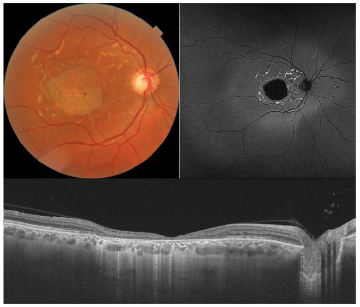
Color fundus (top left): beaten-bronze macular atrophy with pisciform flecks. FAF (top right): central hypoautofluorescence (RPE atrophy) with surrounding hyperautofluorescent flecks. OCT (bottom): complete loss of photoreceptors and RPE over the fovea.
Quick Summary Table
| Feature | Finding |
|---|---|
| Central vision | Progressive loss (central scotoma) |
| Fundus lesions | Yellowish-white pisciform flecks |
| FA finding | Dark/silent choroid (~80%) |
| Gene | ABCA4 (autosomal recessive) |
| Pathology | Lipofuscin (A2E) accumulation in RPE |
The triad is sometimes summarized as: macular atrophy + pisciform flecks + dark choroid on FA.
This is a shared conversation. Sign in to Orris to start your own chat.