

1000013502.jpg
Give me The all topics answer with proper explanation
I can see this is a medical/physiology topic about Basic Concepts in Electrical Stimulation. Let me read the medical library skill first, then research all four topics comprehensively.
Reading File
Now let me search the medical library for all four topics in parallel:
Finding Sources
Finding Sources
Finding Sources
Now let me read the key sections in parallel:
Reading File
Reading File
Reading File
Now let me read propagation of action potential and motor unit sections:
Finding Sources
Reading File
Finding Sources
Reading File
Reading File
Reading File
Reading File
Reading File
Reading File
Reading File
Now let me get the propagation section from Guyton and Ganong:
Reading File
Reading File
I now have comprehensive content from authoritative textbooks (Guyton & Hall, Kandel's Principles of Neural Science, Neuroscience: Exploring the Brain). Let me now fetch the key diagrams to include in the answer:
Loading Image
Loading Image
Loading Image
I now have all the material needed. Let me fetch one more key propagation diagram:
Loading Image
I now have all the information needed for a complete, comprehensive answer. Here is the full explanation of all four topics from Chapter B - Basic Concepts in Electrical Stimulation:
Chapter B: Basic Concepts in Electrical Stimulation
Topic 1: Resting Membrane Potential
Definition
The resting membrane potential (RMP) is the electrical potential difference across the cell membrane of an excitable cell (nerve or muscle) when it is not transmitting a signal. In large nerve fibers, the RMP is approximately -70 mV (the inside is 70 mV more negative than the outside). In skeletal muscle fibers, it is slightly more negative: -80 to -90 mV.
Why the Cell Interior Is Negative
Three key factors establish and maintain the RMP:
1. The Na⁺-K⁺ ATPase Pump (Electrogenic Pump)
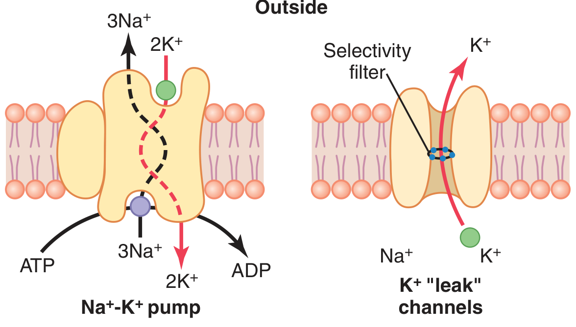
This pump actively transports 3 Na⁺ ions OUT and 2 K⁺ ions IN for every cycle, using ATP energy. Because more positive ions are pumped out than in, it creates a net deficit of positive charge inside the membrane. This contributes about -4 mV directly to the resting potential.
The pump also establishes the following ionic concentration gradients across the resting nerve membrane:
| Ion | Outside (mEq/L) | Inside (mEq/L) |
|---|---|---|
| Na⁺ | 142 | 14 |
| K⁺ | 4 | 140 |
2. Potassium (K⁺) Diffusion Through Leak Channels
The membrane has K⁺ "leak" channels that allow K⁺ to diffuse freely even at rest. Because K⁺ is 35 times more concentrated inside than outside (ratio 140:4), K⁺ tends to diffuse OUT down its concentration gradient. This outward flow of positive ions leaves the inside more negative. The Nernst equilibrium potential for K⁺ alone would be about -94 mV.
3. Sodium (Na⁺) Diffusion - Minor Contribution
The membrane has a very slight permeability to Na⁺. Na⁺ tends to diffuse IN (because concentration outside >> inside), partially offsetting the K⁺ effect. The Nernst potential for Na⁺ alone would be +61 mV. However, because the membrane is ~100 times more permeable to K⁺ than Na⁺, the final resting membrane potential (calculated by the Goldman equation) is approximately -86 mV from diffusion alone. Adding the Na⁺-K⁺ pump contribution of -4 mV gives the observed resting potential of approximately -90 mV (or closer to -70 mV in neurons, depending on the exact ion permeabilities).
Summary of Contributions to RMP:
- K⁺ diffusion alone → -94 mV
- Na⁺ diffusion (slight) → reduces negative value
- Goldman equation result (both ions) → ~ -86 mV
- Electrogenic Na⁺-K⁺ pump → adds ~ -4 mV more
- Final RMP ≈ -70 mV (neuron), -80 to -90 mV (skeletal muscle)
Topic 2: Action Potential
Definition
An action potential (AP) is a rapid, self-amplifying reversal of membrane potential that travels along the membrane of an excitable cell. It begins as a sudden change from the resting negative potential to a positive potential, then quickly returns to negative. It is also called a spike, nerve impulse, or discharge.
Phases of an Action Potential
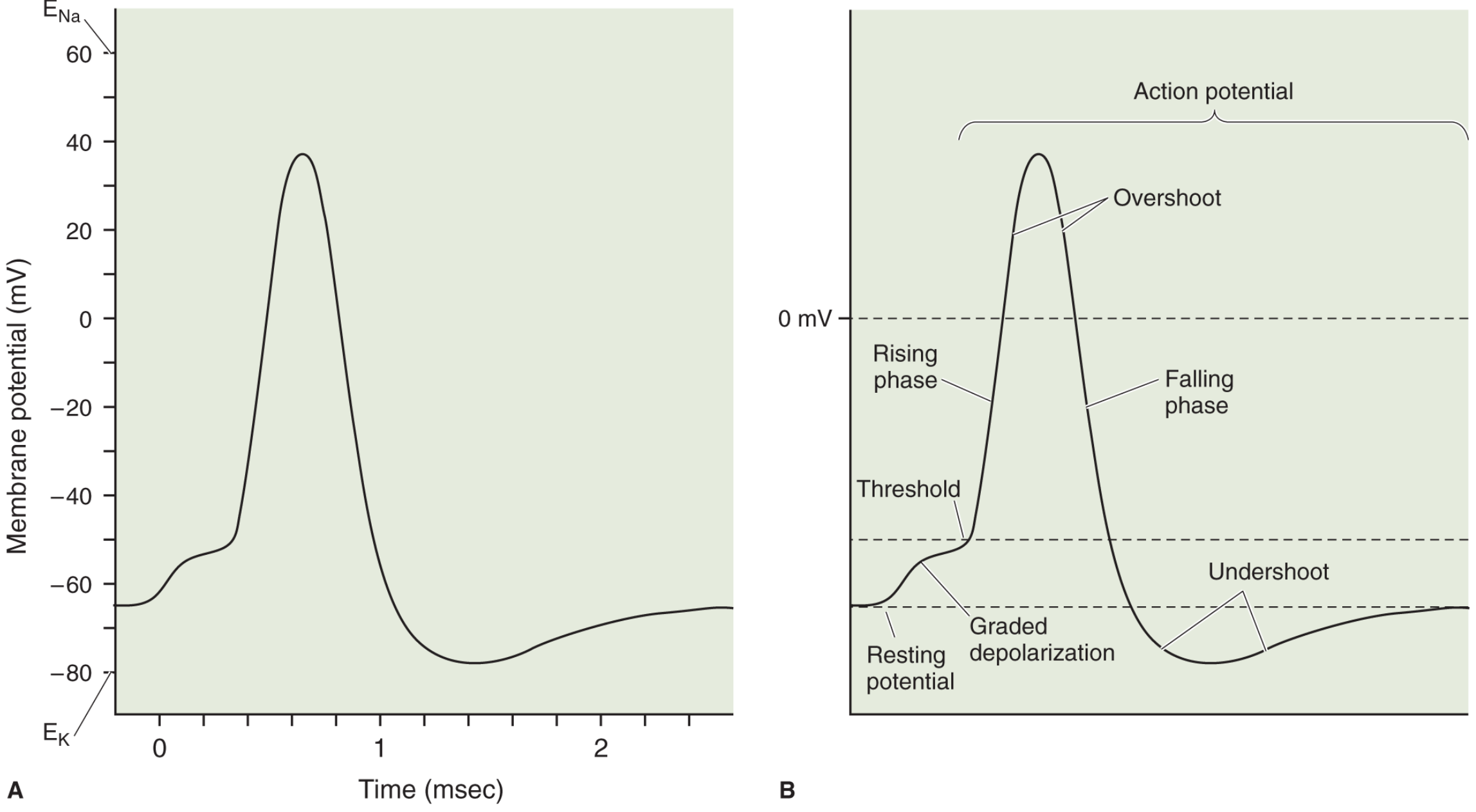
Phase 1 - Resting Stage (-70 mV)
The membrane is polarized. Na⁺ channels are closed (activation gate shut). K⁺ leak channels are open. The fiber is at rest.
Phase 2 - Depolarization (Rising Phase)
A stimulus depolarizes the membrane toward threshold (approximately -55 mV). When threshold is reached, voltage-gated Na⁺ channels open explosively (a positive-feedback cycle: depolarization opens more Na⁺ channels → more Na⁺ rushes in → more depolarization). Membrane potential rapidly rises from -70 mV to +35 to +40 mV (the overshoot) in a fraction of a millisecond. At the peak, sodium conductance increases up to 5,000-fold.
Phase 3 - Repolarization (Falling Phase)
- Within <1 ms, the inactivation gate of Na⁺ channels closes - stopping Na⁺ entry
- Voltage-gated K⁺ channels open (more slowly) - K⁺ rushes OUT
- The rapid efflux of K⁺ restores the negative membrane potential
Phase 4 - Undershoot (After-Hyperpolarization)
K⁺ channels remain open slightly longer than needed, causing the membrane to briefly become more negative than the resting potential (-80 mV). This is the undershoot or after-hyperpolarization. Once the K⁺ channels close, the membrane returns to normal resting potential.
Voltage-Gated Ion Channels
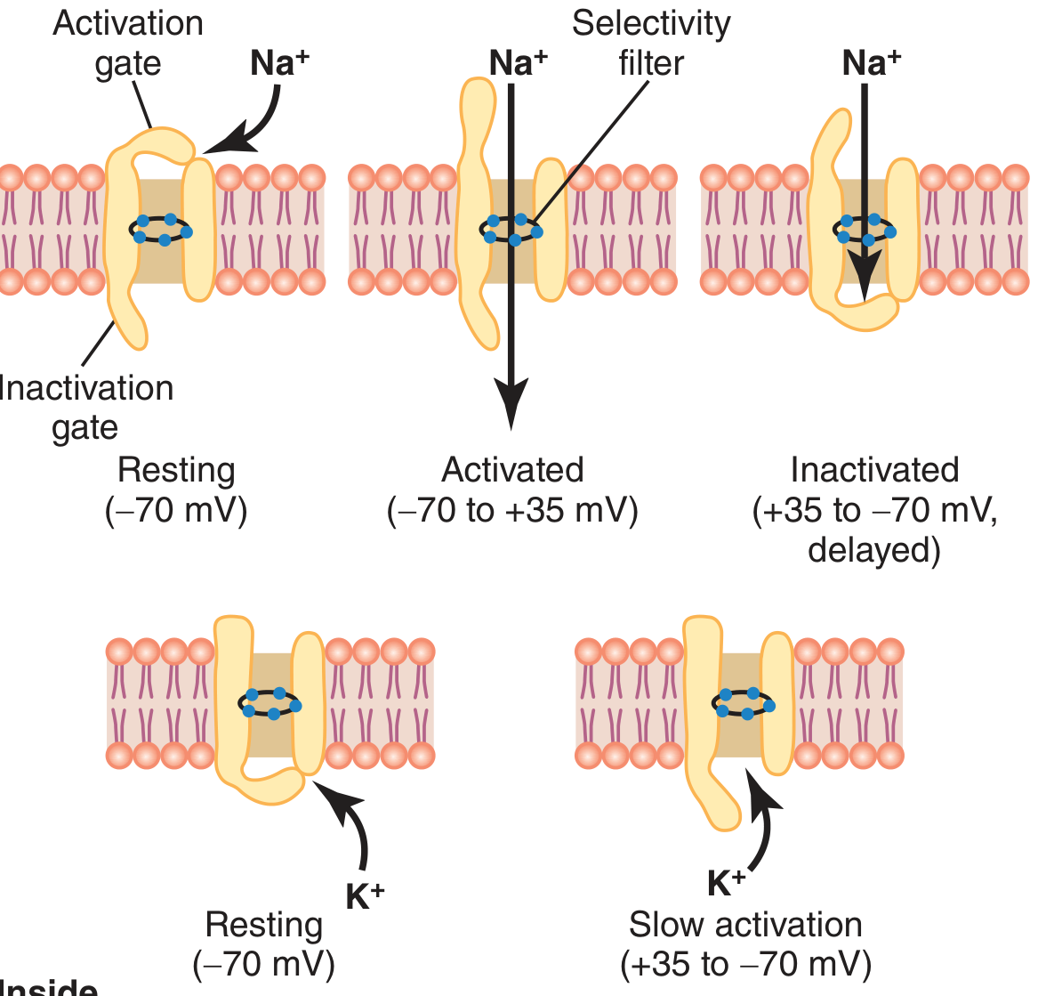
The voltage-gated Na⁺ channel has two gates:
- Activation gate (outside): opens rapidly when membrane reaches threshold
- Inactivation gate (inside): closes more slowly (a few ten-thousandths of a second after the activation gate opens), terminating Na⁺ influx
The voltage-gated K⁺ channel has one gate that opens slowly during depolarization (delayed rectifier) - its opening causes repolarization.
Na⁺ and K⁺ Conductance Changes
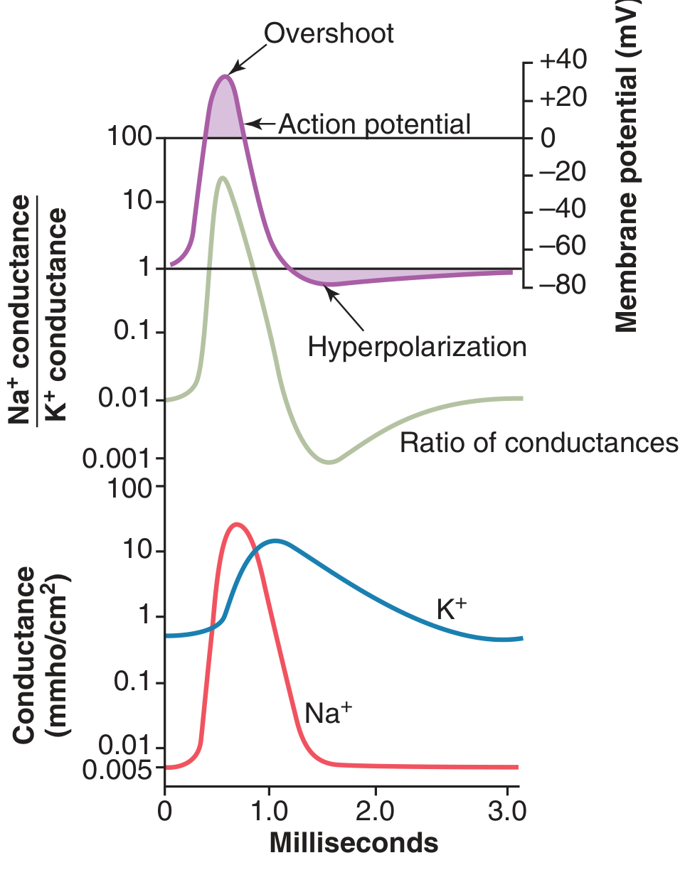
- At the onset of the AP, Na⁺ conductance rises more than 1,000-fold relative to K⁺ conductance - driving depolarization
- As Na⁺ channels inactivate, K⁺ conductance rises ~30-fold - driving repolarization
- The entire AP lasts approximately 1-2 milliseconds in a neuron; 1-5 ms in skeletal muscle
Key Features of Action Potentials
- All-or-none law: An AP either fires fully or not at all - there is no partial AP
- Refractory period: After an AP, the membrane cannot be re-excited (absolute refractory period) or needs a stronger-than-normal stimulus (relative refractory period), because the inactivation gate of Na⁺ channels will not reopen until the membrane repolarizes
- Threshold: Typically ~15-30 mV above the resting potential (approximately -55 mV in neurons)
Topic 3: Propagation of the Action Potential
Definition
Propagation is the process by which an action potential generated at one point on an excitable membrane self-generates and travels along the entire length of the membrane, from the point of stimulation to the fiber's end.
Mechanism of Propagation
When an AP fires at one location on the nerve fiber:
- The depolarized spot becomes positive inside while adjacent resting areas remain negative inside
- This creates a local current flow - positive ions flow intracellularly from the active (depolarized) region toward the adjacent resting region, and extracellularly in the reverse direction
- These local currents depolarize the adjacent membrane to threshold
- Voltage-gated Na⁺ channels in the adjacent region open, generating a new AP there
- This new AP then depolarizes the next adjacent region, and so on
- The wave of depolarization continues along the entire fiber until it reaches the end
Why does the AP only move forward (not backward)?
Because the region behind the AP is in the absolute refractory period - its Na⁺ channels are inactivated and cannot be reopened. So the AP can only propagate in the forward direction.
Conduction in Myelinated vs Unmyelinated Fibers
Unmyelinated fibers:
- Current flows continuously along the entire membrane
- The AP moves slowly (0.5-2 m/sec)
- More energy is consumed
Myelinated fibers (saltatory conduction):
- Myelin sheath insulates the axon, except at Nodes of Ranvier (gaps every 1-3 mm)
- Ion channels are concentrated at the Nodes of Ranvier
- The AP "jumps" from one node to the next (saltatory = "jumping" from Latin)
- This greatly increases conduction velocity (up to 70-120 m/sec in large myelinated fibers) and saves metabolic energy
- In skeletal muscle, conduction velocity is 3-5 m/sec (about 1/13 that of large myelinated nerve fibers)
Energy Restoration After Propagation
After repeated APs, the concentration gradients for Na⁺ and K⁺ gradually run down. The Na⁺-K⁺ ATPase pump restores them by pumping Na⁺ back out and K⁺ back in. The pump activity increases approximately in proportion to the third power of intracellular Na⁺ concentration - so it automatically speeds up when ion gradients are depleted.
Clinical Relevance
- Demyelinating diseases (e.g., Multiple Sclerosis, Guillain-Barre syndrome) destroy the myelin sheath - this slows or blocks AP propagation, causing weakness, sensory loss, and paralysis
- Local anesthetics (e.g., lidocaine) block voltage-gated Na⁺ channels in nerve fibers, preventing AP generation and propagation
Topic 4: Motor Unit
Definition
A motor unit is the basic functional unit of the neuromuscular system, consisting of:
- A single alpha motor neuron (located in the spinal cord ventral horn or brainstem)
- All the muscle fibers it innervates
This concept was proposed by Charles Sherrington in 1925. When a motor neuron fires an AP, ALL muscle fibers in that motor unit contract together (all-or-none for the unit).
Structure of a Motor Unit
- The motor neuron's cell body is in the anterior horn of the spinal cord
- Its axon exits through the ventral root and travels in a peripheral nerve to the muscle
- When the axon reaches the muscle, it branches and innervates multiple muscle fibers scattered throughout the muscle
- A typical muscle is controlled by a few hundred motor neurons clustered in a motor nucleus in the spinal cord
- The number of muscle fibers per motor unit varies:
- Small, precise muscles (e.g., extraocular muscles, hand muscles): 3-10 fibers per motor unit - allows fine motor control
- Large, powerful muscles (e.g., quadriceps, gastrocnemius): hundreds to thousands of fibers per motor unit - for power generation
How a Motor Unit Fires
- Synaptic input from descending motor pathways depolarizes the motor neuron above threshold
- The motor neuron generates an AP that propagates along its axon to all neuromuscular junctions
- The AP releases acetylcholine (ACh) at the neuromuscular junction (NMJ)
- ACh binds to nicotinic receptors on the sarcolemma of each muscle fiber
- This triggers an AP in each muscle fiber, which propagates via the T-tubule system and leads to Ca²⁺ release from the sarcoplasmic reticulum and muscle contraction
Types of Motor Units
Motor units are classified based on three properties - contraction speed, force, and fatigability:
| Type | Speed | Force | Fatigability | Fiber Type |
|---|---|---|---|---|
| Type I (Slow, Fatigue-Resistant - S) | Slow | Small | Low | Type I (red, oxidative) |
| Type IIa (Fast, Fatigue-Resistant - FR) | Fast | Moderate | Moderate | Type IIa |
| Type IIb (Fast, Fatigable - FF) | Fast | Large | High | Type IIb (white, glycolytic) |
- Slow motor units have long twitch contraction times and are used for sustained, postural activities
- Fast fatigable units have short contraction times and large forces - used for brief, explosive movements
Recruitment and Force Grading
The nervous system controls muscle force through two mechanisms:
-
Recruitment (Spatial summation): Activating more motor units to increase force. Small (slow) motor units are recruited first, then progressively larger (faster) ones - this is Henneman's Size Principle (1965). Small motor neurons have lower thresholds and are recruited before large ones.
-
Rate coding (Temporal summation): Increasing the firing frequency of already-active motor units. A single AP produces a twitch. When APs fire rapidly enough that twitches overlap and summate, the result is a tetanic contraction - a smooth, sustained, stronger contraction.
Mechanical Responses
- Twitch: The mechanical response to a single AP
- Unfused tetanus: Low-frequency stimulation; individual twitches are visible as ripples but partially summate
- Fused tetanus (complete tetanus): High-frequency stimulation; twitches fully summate into a smooth, maximal contraction
Clinical Relevance of Motor Unit Concepts
- EMG (Electromyography) records motor unit action potentials (MUAPs) to diagnose neuromuscular diseases
- In lower motor neuron lesions (e.g., polio, ALS), motor neurons are lost - surviving neurons sprout to reinnervate orphaned muscle fibers, creating giant motor units
- Electrical stimulation therapy works by artificially activating motor units through applied electrical current - forms the basis of neuromuscular electrical stimulation (NMES) and functional electrical stimulation (FES)
- Physical training can alter motor unit properties (e.g., endurance training shifts fast fatigable units toward more fatigue-resistant profiles)
Summary Table
| Topic | Key Value | Key Ion | Key Concept |
|---|---|---|---|
| Resting Membrane Potential | -70 mV (nerve); -80 to -90 mV (muscle) | K⁺ dominant | Na⁺-K⁺ pump + K⁺ diffusion |
| Action Potential | Threshold: -55 mV; Peak: +35 to +40 mV | Na⁺ in → K⁺ out | Depolarization → Repolarization → Undershoot |
| Propagation | 70-120 m/sec (myelinated); 3-5 m/sec (muscle) | Na⁺ local currents | Saltatory conduction in myelinated fibers |
| Motor Unit | 1 neuron + all its muscle fibers | ACh at NMJ | Recruitment + Rate coding = Force control |
Sources: Guyton and Hall Textbook of Medical Physiology; Eric Kandel - Principles of Neural Science, 6th Edition; Neuroscience: Exploring the Brain, 5th Edition
1000013502.jpg
Give me the all topics answer
This is the same image as your previous question - all four topics from Chapter B: Basic Concepts in Electrical Stimulation. I already provided the complete detailed answer just above. Here it is again in full:
Chapter B: Basic Concepts in Electrical Stimulation
Topic 1: Resting Membrane Potential (RMP)
Definition
The resting membrane potential is the electrical voltage difference across the cell membrane of an excitable cell (nerve or muscle) when it is at rest and not transmitting any signal. In large nerve fibers it is approximately -70 mV (inside is 70 mV more negative than outside). In skeletal muscle fibers it is -80 to -90 mV.
Three Factors That Establish the RMP
1. The Na⁺-K⁺ ATPase Pump (Electrogenic Pump)
- Pumps 3 Na⁺ OUT and 2 K⁺ IN per cycle using ATP energy
- Because more positive ions are pumped out than in, it creates a net deficit of positive charge inside
- This directly contributes about -4 mV to the resting potential
- It also establishes the ion concentration gradients:
| Ion | Outside | Inside |
|---|---|---|
| Na⁺ | 142 mEq/L | 14 mEq/L |
| K⁺ | 4 mEq/L | 140 mEq/L |
2. K⁺ Diffusion Through Leak Channels
- K⁺ is ~35x more concentrated inside than outside
- K⁺ continuously leaks OUT down its concentration gradient through "leak" channels
- This outflow of positive ions makes the inside more negative
- K⁺ alone would give a Nernst potential of -94 mV
3. Na⁺ Diffusion - Minor Contribution
- Membrane has slight permeability to Na⁺ (100x less permeable than to K⁺)
- Na⁺ leaks IN slightly, partially offsetting K⁺ effect
- Na⁺ alone would give a Nernst potential of +61 mV
- Combined (Goldman equation): approximately -86 mV from diffusion + (-4 mV pump) = ~-90 mV
Summary of RMP Values
| Cell Type | RMP |
|---|---|
| Large nerve fiber (neuron) | -70 mV |
| Skeletal muscle fiber | -80 to -90 mV |
| Cardiac muscle (ventricular) | -85 to -95 mV |
Topic 2: Action Potential
Definition
An action potential (AP) is a rapid, all-or-none reversal of membrane potential that occurs when an excitable cell is stimulated above threshold. It is also called a spike, nerve impulse, or discharge. It is the primary signal used by the nervous system to transmit information rapidly over long distances.
Phases of an Action Potential
Phase 1 - Resting Stage (-70 mV)
- Membrane is "polarized" - inside negative, outside positive
- Voltage-gated Na⁺ channels are CLOSED (activation gate shut)
- K⁺ leak channels are open; Na⁺-K⁺ pump maintains gradients
Phase 2 - Depolarization (Rising Phase)
- A stimulus raises the membrane potential toward threshold (-55 mV)
- At threshold: voltage-gated Na⁺ channels open explosively via positive feedback
- Na⁺ rushes IN → membrane potential shoots from -70 mV up to +35 to +40 mV (overshoot)
- Na⁺ conductance increases up to 5,000-fold
- This entire process takes a fraction of a millisecond
Phase 3 - Repolarization (Falling Phase)
- Inactivation gate of Na⁺ channels closes → Na⁺ entry stops
- Voltage-gated K⁺ channels open (slowly) → K⁺ rushes OUT
- Membrane potential rapidly returns toward negative
Phase 4 - Undershoot (After-Hyperpolarization)
- K⁺ channels stay open slightly too long
- Membrane briefly overshoots to ~-80 mV (more negative than resting)
- K⁺ channels eventually close → membrane returns to -70 mV
Voltage-Gated Ion Channels in Detail
Voltage-Gated Na⁺ Channel - 3 States:
- Resting (-70 mV): Activation gate CLOSED, inactivation gate OPEN → no Na⁺ flow
- Activated (-70 to +35 mV): Activation gate OPENS → Na⁺ floods in → depolarization
- Inactivated (+35 to -70 mV, delayed): Inactivation gate CLOSES → Na⁺ entry stops → repolarization begins. The inactivation gate will NOT reopen until the membrane repolarizes back to resting potential
Voltage-Gated K⁺ Channel - 2 States:
- Resting (-70 mV): Gate CLOSED
- Slow activation (during repolarization): Gate opens SLOWLY → K⁺ flows out → restores negative potential
Ion Conductance Changes During AP
- During the early AP: Na⁺/K⁺ conductance ratio rises >1,000-fold → Na⁺ dominates → depolarization
- During repolarization: Na⁺ channels inactivate, K⁺ conductance rises ~30-fold → K⁺ dominates → repolarization
- Total AP duration: ~1-2 ms in neurons; 1-5 ms in skeletal muscle
Key Laws and Properties
| Property | Explanation |
|---|---|
| All-or-None Law | AP fires completely or not at all - no partial APs |
| Threshold | Must depolarize ~15-30 mV above RMP (to ~-55 mV) |
| Absolute Refractory Period | Immediately after an AP, another AP is IMPOSSIBLE (Na⁺ channels inactivated) |
| Relative Refractory Period | A stronger-than-normal stimulus CAN generate another AP (during undershoot) |
Action Potential in Skeletal Muscle vs Neuron
| Feature | Neuron | Skeletal Muscle |
|---|---|---|
| RMP | -70 mV | -80 to -90 mV |
| AP duration | ~1-2 ms | 1-5 ms |
| Conduction velocity | 70-120 m/sec (myelinated) | 3-5 m/sec |
Topic 3: Propagation of Action Potential
Definition
Propagation is the process by which an AP generated at one point on a nerve or muscle fiber travels continuously along the entire length of the membrane to the fiber's end. This is how the nervous system transmits signals from the brain to muscles over long distances.
Mechanism of Propagation
When an AP fires at one spot on the membrane:
Step 1: The depolarized region becomes positive inside (e.g., +35 mV). Adjacent resting regions are still negative inside (-70 mV).
Step 2: A potential difference now exists between the active region and the adjacent resting region. This drives local current flow:
- Inside the fiber: positive ions flow FROM the active region TOWARD the resting region
- Outside the fiber: current flows in the reverse direction
- These local currents form a complete circuit
Step 3: The local currents depolarize the adjacent membrane to threshold (-55 mV)
Step 4: Voltage-gated Na⁺ channels in the adjacent region open → a new AP fires there
Step 5: This new AP depolarizes the NEXT adjacent region, and so on - the wave travels forward
Step 6: The AP continues propagating until it reaches the nerve terminal
Why Does the AP Travel in Only One Direction?
Behind the advancing AP, the membrane is in the absolute refractory period - Na⁺ channel inactivation gates are closed and cannot reopen until repolarization is complete. So local currents flowing backward cannot re-excite the already-fired region. The AP can ONLY travel forward.
Propagation in Unmyelinated vs Myelinated Fibers
Unmyelinated Fibers (Continuous Conduction):
- Local currents flow continuously along the entire membrane surface
- Every section of membrane must be depolarized one by one
- Conduction velocity: 0.5 - 2 m/sec (slow)
- High metabolic energy cost (more Na⁺-K⁺ pump work needed)
Myelinated Fibers (Saltatory Conduction):
- Myelin sheath (formed by Schwann cells) insulates the axon
- Ion channels are concentrated ONLY at Nodes of Ranvier (bare gaps every 1-3 mm)
- Local currents jump directly from one node to the next
- The AP "leaps" (Latin: saltare) from node to node
- Conduction velocity: up to 70-120 m/sec (large myelinated fibers)
- Much more energy-efficient - only the nodes need to be repolarized
| Fiber Type | Example | Velocity | Myelin |
|---|---|---|---|
| Aα (large myelinated) | Motor neurons to muscle | 70-120 m/sec | Yes |
| Aβ | Touch sensation | 30-70 m/sec | Yes |
| Aδ | Pain, temperature | 5-30 m/sec | Thin |
| C (unmyelinated) | Slow pain, autonomic | 0.5-2 m/sec | No |
Energy Restoration After Propagation
After many APs, ion gradients run down slightly. The Na⁺-K⁺ ATPase pump restores them. Its activity increases approximately in proportion to the cube of intracellular Na⁺ concentration - so it automatically accelerates when Na⁺ builds up inside, efficiently recharging the fiber.
Clinical Applications
- Multiple Sclerosis / Guillain-Barré syndrome: Demyelination slows or blocks AP propagation → weakness, sensory loss, paralysis
- Local anesthetics (lidocaine, bupivacaine): Block voltage-gated Na⁺ channels → prevent AP generation and propagation → pain relief
- Electrical nerve stimulation (TENS, NMES): Applied current generates APs in nerve fibers, which then propagate naturally
Topic 4: Motor Unit
Definition
A motor unit is the fundamental functional unit of the neuromuscular system, consisting of:
- One alpha (α) motor neuron (in the spinal cord ventral horn or brainstem)
- All the skeletal muscle fibers it innervates
This concept was introduced by Sir Charles Sherrington in 1925. All muscle fibers within a motor unit contract simultaneously when the motor neuron fires - they function as one unit.
Anatomy of a Motor Unit
Brain/Descending tracts
        ↓
Alpha Motor Neuron (anterior horn, spinal cord)
        ↓ (axon exits via ventral root → peripheral nerve)
        ↓
Axon branches at the muscle
        ↓
Neuromuscular Junctions (NMJ) on each muscle fiber
        ↓
All muscle fibers contract simultaneously
- A typical muscle is controlled by a few hundred motor neurons clustered in a motor nucleus
- Muscle fibers of one motor unit are scattered throughout the muscle (not clustered together)
- This ensures smooth, even force distribution across the entire muscle
Innervation Ratio (Fibers per Motor Unit)
| Muscle | Purpose | Fibers per Motor Unit |
|---|---|---|
| Extraocular muscles | Fine precise eye movements | 3-10 |
| Intrinsic hand muscles (lumbricals) | Fine finger control | 10-100 |
| Biceps brachii | Moderate control + power | ~750 |
| Gastrocnemius | Power, not fine control | ~2,000 |
Small innervation ratio = precise, fine motor control
Large innervation ratio = powerful, less precise movement
How a Motor Unit Fires - Step by Step
- Synaptic input from upper motor neurons depolarizes the alpha motor neuron above threshold
- AP is generated in the motor neuron and propagates along its axon to all its terminals
- At each neuromuscular junction (NMJ):
- AP releases acetylcholine (ACh) from presynaptic terminal
- ACh binds to nicotinic receptors on the muscle sarcolemma
- End-plate potential (EPP) is generated → triggers a muscle AP
- The muscle AP spreads along the sarcolemma AND down the T-tubules into the fiber's interior
- T-tubule APs trigger Ca²⁺ release from the sarcoplasmic reticulum
- Ca²⁺ binds troponin → allows actin-myosin cross-bridge cycling → muscle contraction
Types of Motor Units
| Type | Name | Speed | Force | Fatigability | Fiber |
|---|---|---|---|---|---|
| Type I | Slow (S) | Slow contraction | Small | Fatigue-resistant | Type I (red, oxidative) |
| Type IIa | Fast Fatigue-Resistant (FR) | Fast | Moderate | Moderate | Type IIa |
| Type IIb/IIx | Fast Fatigable (FF) | Fastest | Largest | Highly fatigable | Type IIb (white, glycolytic) |
- Type I units are recruited for prolonged activities: walking, maintaining posture
- Type II units are recruited for brief, powerful bursts: jumping, sprinting, lifting heavy weights
How the Nervous System Controls Muscle Force
1. Motor Unit Recruitment (Spatial Summation)
- To increase force, more motor units are activated
- Follows Henneman's Size Principle (1965): Small (low-threshold) motor units are recruited FIRST, then progressively larger ones as more force is needed
- Small motor neurons have lower thresholds because their smaller surface area = higher input resistance = easier to depolarize
- Order: Type I → Type IIa → Type IIb
2. Rate Coding - Firing Frequency (Temporal Summation)
- Already-active motor units fire faster to increase force
- A single AP → single twitch contraction
- Repeated APs at increasing rates → twitches overlap and summate:
- Low frequency → Unfused tetanus (rippled, incomplete summation)
- High frequency → Fused tetanus = smooth, maximal sustained contraction
Mechanical Responses of a Motor Unit
| Stimulus | Response |
|---|---|
| Single AP | Twitch - brief, single contraction |
| Low-rate APs | Unfused tetanus - twitches partially summate, rippled force |
| High-rate APs | Fused (complete) tetanus - smooth, maximal force |
Physical Training Effects on Motor Units
Endurance training:
- Increases mitochondrial density and oxidative capacity of muscle fibers
- Can shift Type IIb toward more fatigue-resistant IIa phenotype
- Increases capillary density around motor units
Strength/resistance training:
- Increases muscle fiber cross-sectional area (hypertrophy)
- Improves neural drive (more efficient recruitment and rate coding)
- No change in number of motor neurons
Clinical Relevance of Motor Units
| Condition | Effect on Motor Units |
|---|---|
| EMG (Electromyography) | Records motor unit action potentials (MUAPs) to diagnose disease |
| Lower Motor Neuron Lesion (polio, ALS) | Motor neurons lost; surviving neurons sprout to form giant motor units; fasciculations visible |
| Upper Motor Neuron Lesion (stroke) | Descending input lost; motor units cannot be recruited voluntarily → spastic paralysis |
| NMES / FES therapy | Electrical stimulation artificially recruits motor units → maintains muscle mass, restores function |
| Myasthenia Gravis | Antibodies block ACh receptors at NMJ → weak/failing muscle contraction despite normal AP propagation |
Master Summary
| Topic | Definition | Key Value | Key Mechanism |
|---|---|---|---|
| Resting Membrane Potential | Voltage across resting membrane | -70 mV (nerve); -80 to -90 mV (muscle) | Na⁺-K⁺ pump + K⁺ diffusion out |
| Action Potential | Rapid all-or-none voltage spike | Threshold: -55 mV; Peak: +35 to +40 mV | Na⁺ in (depolarization) → K⁺ out (repolarization) |
| Propagation of AP | AP travels along entire fiber | 70-120 m/sec (myelinated); 3-5 m/sec (muscle) | Local currents + saltatory conduction |
| Motor Unit | 1 motor neuron + all its muscle fibers | 3-10 fibers (precise) to 2,000+ fibers (powerful) | Recruitment + Rate coding = graded force |
Sources: Guyton and Hall Textbook of Medical Physiology; Kandel - Principles of Neural Science, 6th Ed.; Neuroscience: Exploring the Brain, 5th Ed.
This is a shared conversation. Sign in to Orris to start your own chat.