
Okay, you need to behave like a microbiologist teacher in the MBBS college Georgia. You need to explain me every bit of things I paste from Medical Microbiology by Murray, Rosenthal, Pfaller, 8th edition Elsevier. You also go through that book and reference other international microbiology books and explain me the topic in H-I-N-G-L-I-S, in a very good level. And the explanation should be USMLE level. You need to point out things which are important for USMLE, and try to make things understand to me so that I do not need to go through the book again. I have read this, but things haven't gone in my mind, okay? So make it easier for me to understand. Use color codings, visualization, picture visualization, use mind maps, flowcharts, diagrams. Use everything possible you can to make me understand. Use web for images and make me understand every bit of things I paste over here, okay? No rush, nothing, and explain in high detail. I should understand everything. And at last give note, like summary notes, like covering everything.
Murray Medical Microbiology 8th edition Elsevier companion resources
Format I will use
- Difficult textbook sentence ko simple language mein break karunga.
- “Kya ho raha hai, kyun ho raha hai, aur clinically kya meaning hai” clearly explain hoga.
- Organism, virulence factor, immune response, lab test, disease mechanism, and treatment logic ko link karunga.
- Ratta nahi, story-based understanding.
I’ll use text-based diagrams, flowcharts, tables, mnemonics, and mind maps, for example:
Entry into body
      ↓
Colonization
      ↓
Virulence factor action
      ↓
Host immune response
      ↓
Clinical disease + laboratory findings
      ↓
Diagnosis / treatment
I will explicitly mark:
- USMLE must-know facts
- Common question stems
- Important differentiations
- Traps and confusions
- “If you see X, think Y” associations
For example, instead of only saying “encapsulated bacteria resist phagocytosis,” I will connect it to:
- why asplenic patients are at risk,
- why opsonization matters,
- which organisms are encapsulated,
- how this appears in exam questions.
For microbiology, lab diagnosis is often confusing. I will make it systematic:
Specimen → stain → culture → biochemical test → antigen/PCR test → interpretation
Especially for commonly confused organisms, such as:
- Staphylococcus aureus vs Streptococcus pyogenes
- Neisseria meningitidis vs N. gonorrhoeae
- E. histolytica vs Giardia
- HSV vs VZV
- Candida vs Cryptococcus vs Aspergillus
At the end of every topic, you will get revision-ready notes:
- One-page style summary
- Important tables
- Mnemonics
- USMLE rapid-review points
- A few practice-style questions when useful
Source approach
- Jawetz, Melnick & Adelberg’s Medical Microbiology
- Levinson’s Review of Medical Microbiology and Immunology
- Mandell’s principles where clinical context needs expansion
- Current evidence or guidelines when a fact is time-sensitive
Color key
- 🟦 Concept
- 🟩 Mechanism / pathogenesis
- 🟨 Exam pearl
- 🟥 Common trap / dangerous confusion
- 🟪 Clinical correlation
- 🟧 Lab diagnosis
- ⭐ Highest-yield USMLE point
INTRODUCTION TO MEDICAL MICROBIOLOGY I magine the excitement felt by the Dutch biologist Anton van Leeuwenhoek in 1674 as he peered through his carefully ground microscopic lenses at a drop of water and discovered a world of millions of tiny “animalcules.” Almost 100 years later, the Danish biologist Otto Müller extended van Leeuwenhoek’s studies and organized bacteria into genera and species according to the classification methods of Carolus Linnaeus. This was the beginning of the taxonomic classification of microbes. In 1840, the German pathologist Friedrich Henle proposed criteria for proving that microorganisms were responsible for causing human disease (the “germ theory” of disease). Robert Koch and Louis Pasteur confirmed this theory in the 1870s and 1880s with a series of elegant experiments proving that microorganisms were responsible for causing anthrax, rabies, plague, cholera, and tuberculosis. Other brilliant scientists went on to prove that a diverse collection of microbes was responsible for causing human disease. The era of chemotherapy began in 1910, when the German chemist Paul Ehrlich discovered the first antibacterial agent, a compound effective against the spirochete that causes syphilis. This was followed by Alexander Fleming’s discovery of penicillin in 1928, Gerhard Domagk’s discovery of sulfanilamide in 1935, and Selman Waksman’s discovery of streptomycin in 1943. In 1946, the American microbiologist John Enders was the first to cultivate viruses in cell cultures, leading the way to the large-scale production of virus cultures for vaccine development. Thousands of scientists have followed these pioneers, each building on the foundation established by his or her predecessors, and each adding an observation that expanded our understanding of microbes and their role in disease. Our knowledge of microbiology is now undergoing a remarkable transformation founded in the rapid technologic advances in genome analysis. The Human Genome Project was a multinational program that concluded in 2005 with the comprehensive sequencing of the human genome. The techniques developed for this program have rapidly moved into the research and clinical laboratories, leading to microbial sequencing and revealing previously unappreciated insights about pathogenic properties of organisms, taxonomic relationships, and functional attributes of the endogenous microbial population. Clearly, we are at the early stages of novel approaches to diagnostics and therapeutics based on the monitoring and manipulations of this population (the microbiome). The world that van Leeuwenhoek discovered was complex, consisting of protozoa and bacteria of all shapes and sizes. However, the complexity of medical microbiology we know today rivals the limits of the imagination. We now know that there are thousands of different types of microbes that live in, on, and around us—and hundreds that cause serious human diseases. To understand this information and organize it in a useful manner, it is important to understand some of the basic aspects of medical microbiology. To start, the microbes can be subdivided into the following four general groups: viruses, bacteria, fungi, and parasites, each having its own level of complexity. • Viruses Viruses are the smallest infectious particles, ranging in diameter from 18 to 600 nanometers (most viruses are < 200 nm and cannot be seen with a light microscope). Viruses typically contain either deoxyribonucleic acid (DNA) or ribonucleic acid (RNA) but not both; however, some viral-like particles do not contain any detectable nucleic acids (e.g., prions), whereas the recently discovered Mimivirus contains both RNA and DNA. The viral nucleic acids required for replication are enclosed in a protein shell with or without a lipid membrane coat. Viruses are true parasites, requiring host cells for replication. The cells they infect and the host response to the infection dictate the nature of the clinical manifestation. More than 2000 species of viruses have been described, with approximately 650 infecting humans and animals. Infection can lead either to rapid replication and destruction of the cell or to a long-term chronic relationship with possible integration of the viral genetic information into the host genome. The factors that determine which of these takes place are only partially understood. For example, infection with the human immunodeficiency virus, the etiologic agent of the acquired immunodeficiency syndrome (AIDS), can result in the latent infection of CD4 lymphocytes or the active replication and destruction of these immunologically important cells. Likewise, infection can spread to other susceptible cells, such as the microglial cells of the brain, resulting in the neurologic manifestations of AIDS. The virus determines the disease and can range from the common cold to gastroenteritis to fatal catastrophes such as rabies, Ebola, smallpox, or AIDS. • Bacteria Bacteria are relatively simple in structure. They are prokaryotic organisms—simple unicellular organisms with no nuclear membrane, mitochondria, Golgi bodies, or 2 CHAPTER 1 Introducton to Medcal Mcrobology 3 endoplasmic reticulum—that reproduce by asexual division. The bacterial cell wall is complex, consisting of one of two basic forms: a gram-positive cell wall with a thick peptidoglycan layer, and a gram-negative cell wall with a thin peptidoglycan layer and an overlying outer membrane. Some bacteria lack this cell wall structure and compensate by surviving only inside host cells or in a hypertonic environment. The size (1 to 20 µm or larger), shape (spheres, rods, spirals), and spacial arrangement (single cells, chains, clusters) of the cells are used for the preliminary classification of bacteria, and the phenotypic and genotypic properties of the bacteria form the basis for the definitive classification. The human body is inhabited by thousands of different bacterial species— some living transiently, others in a permanent parasitic relationship. Likewise, the environment that surrounds us, including the air we breathe, water we drink, and food we eat, is populated with bacteria, many of which are relatively avirulent and some of which are capable of producing lifethreatening disease. Disease can result from the toxic effects of bacterial products (e.g., toxins) or when bacteria invade normally sterile body tissues and fluids. • Fungi In contrast to bacteria, the cellular structure of fungi is more complex. These are eukaryotic organisms that contain a well-defined nucleus, mitochondria, Golgi bodies, and endoplasmic reticulum. Fungi can exist either in a unicellular form (yeast) that can replicate asexually or in a filamentous form (mold) that can replicate asexually and sexually. Most fungi exist as either yeasts or molds; however, some fungi can assume either morphology. These are known as dimorphic fungi and include such organisms as Histoplasma, Blastomyces, and Coccidioides. • Parasites Parasites are the most complex microbes. Although all parasites are classified as eukaryotic, some are unicellular and others are multicellular. They range in size from tiny protozoa as small as 4 to 5 µm in diameter (the size of some bacteria) to tapeworms that can measure up to 10 meters in length and arthropods (bugs). Indeed, considering the size of some of these parasites, it is hard to imagine how these organisms came to be classified as microbes. Their life cycles are equally complex, with some parasites establishing a permanent relationship with humans and others going through a series of developmental stages in a progression of animal hosts. One of the difficulties confronting students is not only an understanding of the spectrum of diseases caused by parasites but also an appreciation of the epidemiology of these infections, which is vital for developing a differential diagnosis and an approach to the control and prevention of parasitic infections. • Immunology It is difficult to discuss human microbiology without also discussing the innate and immune responses to the microbes. Our innate and immune responses evolved to protect us from infection. At the same time, the microbes that live in our bodies as normal flora or disease-causing organisms must be able to withstand or evade these host protections sufficiently long to be able to establish their niche within our bodies or spread to new hosts. The peripheral damage that occurs during the war between the host protections and microbial invaders contributes to or may be the cause of the symptoms of the disease. Ultimately, the innate and immune responses are the best prevention and cure for microbial disease. • Microbial Disease One of the most important reasons for studying microbes is to understand the diseases they cause and the ways to control them. Unfortunately, the relationship between many organisms and their diseases is not simple. Specifically, most organisms do not cause a single well-defined disease, although there are certainly ones that do (e.g., Clostridium tetani [tetanus], Ebola virus [Ebola], Plasmodium species [malaria]). Instead, it is more common for a particular organism to produce many manifestations of disease (e.g., Staphylococcus aureus—endocarditis, pneumonia, wound infections, food poisoning) or for many organisms to produce the same disease (e.g., meningitis caused by viruses, bacteria, fungi, and parasites). In addition, relatively few organisms can be classified as always pathogenic, although some do belong in this category (e.g., rabies virus, Bacillus anthracis, Sporothrix schenckii, Plasmodium species). Instead, most organisms are able to establish disease only under welldefined circumstances (e.g., introduction of an organism with a potential for causing disease into a normally sterile site such as the brain, lungs, and peritoneal cavity). Some diseases arise when a person is exposed to organisms from external sources. These are known as exogenous infections, and examples include diseases caused by influenza virus, C. tetani, Neisseria gonorrhoeae, Coccidioides immitis, and Entamoeba histolytica. Most human diseases, however, are produced by organisms in the person’s own microbial flora that spread to normally sterile body sites where disease can ensue (endogenous infections). The interaction between an organism and the human host is complex. The interaction can result in transient colonization, a long-term symbiotic relationship, or disease. The virulence of the organism, the site of exposure, and the host’s ability to respond to the organism determine the outcome of this interaction. Thus the manifestations of disease can range from mild symptoms to organ failure and death. The role of microbial virulence and the host’s immunologic response is discussed in depth in subsequent chapters. The human body is remarkably adapted to controlling exposure to pathogenic microbes. Physical barriers prevent invasion by the microbe; innate responses recognize molecular patterns on the microbial components and activate local defenses and specific adapted immune responses that target the microbe for elimination. Unfortunately, the immune response is often too late or too slow. To improve the human body’s ability to prevent infection, the immune system can be augmented either through the passive transfer of antibodies present in immune globulin preparations or through 4 MEDICAL MICROBIOLOGY active immunization with components of the microbes (vaccines). Infections can also be controlled with a variety of chemotherapeutic agents. Unfortunately, microbes can mutate and share genetic information, and those that cannot be recognized by the immune response because of antigenic variation or those that are resistant to antibiotics will be selected and will endure. Thus the battle for control between microbe and host continues, with neither side yet able to claim victory (although the microbes have demonstrated remarkable ingenuity). There clearly is no “magic bullet” that has eradicated infectious diseases. • Diagnostic Microbiology The clinical microbiology laboratory plays an important role in the diagnosis and control of infectious diseases. However, the ability of the laboratory to perform these functions is limited by the quality of the specimen collected from the patient, the means by which it is transported from the patient to the laboratory, and the techniques used to demonstrate the microbe in the sample. Because most diagnostic tests are based on the ability of the organism to grow, transport conditions must ensure the viability of the pathogen. In addition, the most sophisticated testing protocols are of little value if the collected specimen is not representative of the site of infection. This seems obvious, but many specimens sent to laboratories for analysis are contaminated during collection with the organisms that colonize mucosal surfaces. It is virtually impossible to interpret the testing results with contaminated specimens, because most infections are caused by endogenous organisms. The laboratory is also able to determine the antimicrobial activity of selected chemotherapeutic agents, although the value of these tests is limited. The laboratory must test only organisms capable of producing disease and only medically relevant antimicrobials. To test all isolated organisms or an indiscriminate empirical selection of drugs can yield misleading results with potentially dangerous consequences. Not only can a patient be treated inappropriately with unnecessary antibiotics, but also the true pathogenic organism may not be recognized among the plethora of organisms isolated and tested. Finally, the in vitro determination of an organism’s susceptibility to a variety of antibiotics is only one aspect of a complex picture. The virulence of the organism, site of infection, and patient’s ability to respond to the infection influence the host-parasite interaction and must also be considered when planning treatment. • Summary It is important to realize that our knowledge of the microbial world is evolving continually. Just as the early microbiologists built their discoveries on the foundations established by their predecessors, we and future generations will continue to discover new microbes, new diseases, and new therapies. The following chapters are intended as a foundation of knowledge that can be used to build your understanding of microbes and their diseases. No, you need to do, like do what I gave you the prompt earlier and visualize using color coding icons and everything, US MD level summary. And if it is too big for you, divide it in parts, but give me a wholesome summary for microbiology.
Topic map from your pasted chapter
-
History and evolution of medical microbiology
- Leeuwenhoek, germ theory, Koch, Pasteur
- Antibiotics, vaccines, cell culture, genomics, microbiome
-
The four major microbial groups
- Viruses
- Bacteria
- Fungi
- Parasites
-
Immunology and host-microbe interaction
- Innate immunity, adaptive immunity, immune evasion
- Why inflammation itself produces symptoms and tissue damage
-
How microbes cause disease
- Colonization vs infection vs disease
- Virulence, inoculum, portal of entry, sterile sites, host susceptibility
- Exogenous versus endogenous infection
- Primary pathogens versus opportunistic pathogens
-
Why one organism can cause multiple syndromes
- For example, Staphylococcus aureus
- Why the same syndrome, such as meningitis, can have many microbial causes
-
Prevention and treatment
- Physical barriers, immunity, passive antibody, vaccines
- Antimicrobials, resistance, antigenic variation
-
Diagnostic microbiology
- Correct specimen selection, collection, transport
- Contamination versus true pathogen
- Culture, identification, antimicrobial susceptibility testing
- Why a laboratory report must always be interpreted clinically
High-level overview
Microbe enters or overgrows
          ↓
Host barriers / immunity respond
          ↓
Microbe either gets eliminated, colonizes, persists, or invades
          ↓
Disease occurs when tissue injury or inflammation becomes significant
Outcome of infection =
Microbial virulence
      ×
Site of exposure / entry
      ×
Host immune status
Choose the first deep-dive section. I will explain it in Hinglish with color-coded diagrams, mechanisms, USMLE pearls, clinical examples, and final revision notes.
Format I will use
- Difficult textbook sentence ko simple language mein break karunga.
- “Kya ho raha hai, kyun ho raha hai, aur clinically kya meaning hai” clearly explain hoga.
- Organism, virulence factor, immune response, lab test, disease mechanism, and treatment logic ko link karunga.
- Ratta nahi, story-based understanding.
I’ll use text-based diagrams, flowcharts, tables, mnemonics, and mind maps, for example:
Entry into body
      ↓
Colonization
      ↓
Virulence factor action
      ↓
Host immune response
      ↓
Clinical disease + laboratory findings
      ↓
Diagnosis / treatment
I will explicitly mark:
- USMLE must-know facts
- Common question stems
- Important differentiations
- Traps and confusions
- “If you see X, think Y” associations
For example, instead of only saying “encapsulated bacteria resist phagocytosis,” I will connect it to:
- why asplenic patients are at risk,
- why opsonization matters,
- which organisms are encapsulated,
- how this appears in exam questions.
For microbiology, lab diagnosis is often confusing. I will make it systematic:
Specimen → stain → culture → biochemical test → antigen/PCR test → interpretation
Especially for commonly confused organisms, such as:
- Staphylococcus aureus vs Streptococcus pyogenes
- Neisseria meningitidis vs N. gonorrhoeae
- E. histolytica vs Giardia
- HSV vs VZV
- Candida vs Cryptococcus vs Aspergillus
At the end of every topic, you will get revision-ready notes:
- One-page style summary
- Important tables
- Mnemonics
- USMLE rapid-review points
- A few practice-style questions when useful
Source approach
- Jawetz, Melnick & Adelberg’s Medical Microbiology
- Levinson’s Review of Medical Microbiology and Immunology
- Mandell’s principles where clinical context needs expansion
- Current evidence or guidelines when a fact is time-sensitive
Color key
- 🟦 Concept
- 🟩 Mechanism / pathogenesis
- 🟨 Exam pearl
- 🟥 Common trap / dangerous confusion
- 🟪 Clinical correlation
- 🟧 Lab diagnosis
- ⭐ Highest-yield USMLE point
Topic map from your pasted chapter
-
History and evolution of medical microbiology
- Leeuwenhoek, germ theory, Koch, Pasteur
- Antibiotics, vaccines, cell culture, genomics, microbiome
-
The four major microbial groups
- Viruses
- Bacteria
- Fungi
- Parasites
-
Immunology and host-microbe interaction
- Innate immunity, adaptive immunity, immune evasion
- Why inflammation itself produces symptoms and tissue damage
-
How microbes cause disease
- Colonization vs infection vs disease
- Virulence, inoculum, portal of entry, sterile sites, host susceptibility
- Exogenous versus endogenous infection
- Primary pathogens versus opportunistic pathogens
-
Why one organism can cause multiple syndromes
- For example, Staphylococcus aureus
- Why the same syndrome, such as meningitis, can have many microbial causes
-
Prevention and treatment
- Physical barriers, immunity, passive antibody, vaccines
- Antimicrobials, resistance, antigenic variation
-
Diagnostic microbiology
- Correct specimen selection, collection, transport
- Contamination versus true pathogen
- Culture, identification, antimicrobial susceptibility testing
- Why a laboratory report must always be interpreted clinically
High-level overview
Microbe enters or overgrows
          ↓
Host barriers / immunity respond
          ↓
Microbe either gets eliminated, colonizes, persists, or invades
          ↓
Disease occurs when tissue injury or inflammation becomes significant
Outcome of infection =
Microbial virulence
      ×
Site of exposure / entry
      ×
Host immune status
medical microbiology diagram comparison virus bacteria fungi parasites size and cellular organization
Gram positive versus Gram negative bacterial cell envelope labeled diagram peptidoglycan outer membrane
microbiome infectious disease
CDC specimen collection transport microbiology laboratory contamination culture guidance
Chapter 1: Introduction to Medical Microbiology
Hinglish + USMLE/US MD-Level Foundation
Big idea: Medical microbiology is not just “kaunsa organism kaunsi disease karta hai.”
It is the study of a continuous conflict:
MICROBE                     HOST
Virulence factors     vs    Physical barriers
Toxins                 vs   Innate immunity
Invasion               vs   Adaptive immunity
Immune evasion         vs   Antibodies / T cells
Antimicrobial resistance    Drugs / vaccines
- host barriers cross kare,
- sterile site mein enter kare,
- toxin banaye,
- ya host immune response itself tissue damage kare.
🧠 Master mind map: Entire microbiology in one view
                         MEDICAL MICROBIOLOGY
                                  │
      ┌───────────────────────────┼────────────────────────────┐
      │                           │                            │
  MICROBES                    THE HOST                   CLINICAL LAB
      │                           │                            │
 Viruses                    Barriers                     Proper specimen
 Bacteria                   Innate immunity              Microscopy
 Fungi                      Adaptive immunity            Culture
 Parasites                  Inflammation                  Antigen/PCR
                            Immunodeficiency              Susceptibility test
      │                           │                            │
      └─────────────── HOST-MICROBE INTERACTION ───────────────┘
                                  │
              Colonization / Symbiosis / Infection / Disease
                                  │
                        Prevention and treatment
                    Vaccines / antibodies / antimicrobials
Part 1: Why study microbiology? Historical story
🟦 1. From “animalcules” to germ theory
Antonie van Leeuwenhoek, 1674
Otto Müller
Why classification matters
Old approach                         Modern approach
────────────                         ───────────────
Shape                                Genome sequence
Staining properties                  rRNA sequence
Metabolic/biochemical tests          Whole-genome comparison
Culture characteristics              Phylogenetic relationship
Bacterial morphology still gives an early clue:
- Gram-positive cocci in clusters → think Staphylococcus
- Gram-positive cocci in chains → think Streptococcus or Enterococcus
- Curved gram-negative rods → think Vibrio, Campylobacter, Helicobacter
- Spirochetes → think Treponema, Borrelia, Leptospira
🟩 2. Germ theory of disease
Friedrich Henle
Louis Pasteur and Robert Koch
- Pasteur: microbial causation, fermentation, vaccines, pasteurization
- Koch: anthrax, tuberculosis, cholera; methods of growing and proving microbial causation
Koch’s postulates: the classical logic
1. Organism should be associated with the disease.
                ↓
2. It should be isolated from a diseased host.
                ↓
3. It should cause similar disease in a susceptible host.
                ↓
4. It should be re-isolated from that experimentally infected host.
🟥 Why Koch’s postulates are not absolute today
- Viruses that cannot grow on ordinary artificial media
- Organisms that infect only humans, such as Neisseria gonorrhoeae
- Organisms carried asymptomatically
- Polymicrobial diseases
- Disease where host immunity determines whether infection appears
- Unculturable bacteria, including Treponema pallidum in routine culture
🟧 3. Beginning of antimicrobial therapy
| Scientist | Landmark contribution | Clinical significance |
|---|---|---|
| Paul Ehrlich | Arsphenamine for syphilis | First targeted antimicrobial chemotherapy concept |
| Alexander Fleming | Penicillin discovery | Basis of beta-lactam antibiotic era |
| Gerhard Domagk | Sulfonamide discovery | First broadly useful antibacterial drug class |
| Selman Waksman | Streptomycin | First effective treatment for tuberculosis |
| John Enders | Viral culture in cells | Enabled viral vaccine development and diagnostic virology |
Ehrlich’s “magic bullet”
Ideal antimicrobial:
High toxicity to microbe
        +
Low toxicity to human cells
        =
Selective toxicity
- Beta-lactams target bacterial peptidoglycan, which humans do not have.
- Azoles target fungal ergosterol synthesis, not human cholesterol synthesis.
- Antivirals often target viral enzymes, such as HIV reverse transcriptase.
Part 2: Genomics and the microbiome
🟦 Human Genome Project and microbial genome sequencing
Modern diagnostic evolution
Traditional method
Specimen → culture → biochemical tests → identification
           ⏳ Often days

Modern molecular method
Specimen → DNA/RNA detection → identification
           ⏳ Hours, sometimes less
Examples
- SARS-CoV-2 detection by RT-PCR
- Tuberculosis drug-resistance gene detection
- Meningitis/encephalitis multiplex PCR panels
- 16S rRNA sequencing for difficult bacterial identification
- Metagenomic sequencing in selected unexplained infections
🟩 Microbiome: your resident microbial ecosystem
Normal microbiota can:
✓ Occupy ecological niches
✓ Compete with pathogens
✓ Produce inhibitory substances
✓ Help immune-system development
✓ Aid metabolism and vitamin production
✓ Maintain mucosal-barrier function
Colonization resistance
- space,
- nutrients,
- receptor sites,
- and by producing substances harmful to competitors.
Classic USMLE example: Clostridioides difficile
Broad-spectrum antibiotics
          ↓
Normal colonic microbiota disrupted
          ↓
Loss of colonization resistance
          ↓
C. difficile overgrows
          ↓
Toxins A and B
          ↓
Pseudomembranous colitis
Part 3: The four major groups of medically important microbes
🟪 Comparison table: memorize this framework first
| Feature | Virus | Bacterium | Fungus | Parasite |
|---|---|---|---|---|
| Cellular? | No | Yes | Yes | Yes |
| Cellular type | Acellular | Prokaryote | Eukaryote | Eukaryote |
| Nucleus | No | No | Yes | Yes |
| Ribosomes | No | 70S | 80S | 80S |
| Metabolism on own | No | Yes | Yes | Yes |
| Replication | Must use host cell | Binary fission | Budding, hyphae/spores | Variable, often complex life cycle |
| Usual size | nm | µm | µm to visible colonies | µm to meters |
| Main drug targets | Viral enzymes/processes | Cell wall, 70S ribosome, DNA enzymes | Ergosterol/cell wall | Metabolic or neuromuscular targets |
Size concept:

Virus        Bacterium          Fungus / protozoa             Helminth
nm           µm                 µm to mm                      mm to meters
|------------|------------------|-----------------------------|
- Viruses are acellular obligate intracellular parasites.
- Bacteria are prokaryotic cells.
- Fungi and parasites are eukaryotic organisms.
Part 4: Viruses
🟦 What is a virus?
                 VIRION
      ┌────────────────────────┐
      │ Genome: DNA or RNA     │
      │          ↓             │
      │ Protein coat: capsid   │
      │          ↓ optional    │
      │ Lipid envelope         │
      └────────────────────────┘
Key terms
| Term | Meaning |
|---|---|
| Virion | Complete infectious viral particle outside the cell |
| Genome | Viral DNA or RNA |
| Capsid | Protein shell protecting viral genome |
| Envelope | Host-cell-derived lipid membrane around some viruses |
| Capsomere | Repeating protein subunit composing a capsid |
| Tropism | Preference for a certain cell type or tissue |
| Cytopathic effect | Visible cell injury caused by viral replication |
🟩 Viral genome rule
DNA OR RNA
Important correction and nuance
Major nonstandard infectious agents
| Agent | Genetic material | Key point |
|---|---|---|
| Prion | No nucleic acid | Misfolded protein that induces protein misfolding |
| Viroid | Small RNA, no capsid | Plant pathogens, not classic human pathogens |
| Mimivirus | dsDNA | Giant virus, not a standard USMLE exception |
Examples: Creutzfeldt-Jakob disease, kuru, fatal familial insomnia.
🟩 Why viruses must be inside cells
- produce ATP,
- synthesize proteins using their own ribosomes,
- maintain cellular metabolism,
- or reproduce by cell division.
Virus binds host receptor
          ↓
Enters host cell
          ↓
Uses host machinery ± viral enzymes
          ↓
Makes viral genomes and proteins
          ↓
Assembles new virions
          ↓
Leaves cell
Possible outcomes of viral infection
Viral infection
     │
     ├── Acute lytic infection
     │      Rapid replication → cell death
     │      Example: influenza-infected respiratory epithelial cells
     │
     ├── Persistent/chronic infection
     │      Ongoing production over time
     │      Example: hepatitis B or hepatitis C
     │
     ├── Latent infection
     │      Viral genome persists with minimal production
     │      Example: HSV, VZV, EBV
     │
     └── Transforming infection
            Altered growth control / cancer risk
            Example: HPV, EBV, HBV, HTLV-1
🟨 Clinical example: HIV
- CD4 receptor
- plus CCR5 or CXCR4 coreceptor
- CD4 T lymphocytes
- macrophages
- dendritic cells
- microglia in the CNS
HIV infection
      ↓
Initial viremia + CD4 decline
      ↓
Partial immune control
      ↓
Long clinical latency with ongoing replication
      ↓
Progressive CD4 loss
      ↓
Opportunistic infections, malignancy, neurologic disease
🟨 Viral envelope: very high-yield
| Enveloped viruses | Nonenveloped viruses |
|---|---|
| Have lipid envelope | No lipid envelope |
| Fragile in drying, acid, heat, detergents | More environmentally stable |
| Spread by body fluids, respiratory droplets, sexual contact, transplantation | Can often survive GI tract and spread fecal-orally |
| Examples: HSV, HIV, influenza, HBV | Examples: adenovirus, HPV, parvovirus B19, poliovirus, norovirus, rotavirus |
Mnemonic
They resist drying and acid better, so fecal-oral transmission is more common.
Part 5: Bacteria
🟦 Bacteria are prokaryotes
Bacterial cell has:
✓ Cell membrane
✓ Cytoplasm
✓ 70S ribosomes
✓ DNA chromosome, usually circular
✓ Often plasmids
✓ Cell wall in most bacteria

Bacterial cell lacks:
✗ True nucleus
✗ Nuclear membrane
✗ Mitochondria
✗ Golgi apparatus
✗ Endoplasmic reticulum
Why prokaryote vs eukaryote matters clinically
| Structure | Human cells | Bacteria | Drug relevance |
|---|---|---|---|
| Ribosome | 80S | 70S | Tetracyclines, aminoglycosides, macrolides target bacterial 70S ribosomes |
| Cell wall | None | Usually peptidoglycan | Beta-lactams and vancomycin target cell-wall synthesis |
| Nucleus | Present | Absent | Fluoroquinolones target bacterial DNA gyrase/topoisomerases |
| Sterols in cell membrane | Cholesterol | Usually absent | Exception: Mycoplasma incorporates host sterols |
🟩 Bacterial cell wall: central concept
                    PEPTIDOGLYCAN
       Repeating sugar chains cross-linked by peptides
                          │
            Gives shape and protection from osmotic lysis
Gram-positive versus gram-negative bacteria
GRAM-POSITIVE ENVELOPE              GRAM-NEGATIVE ENVELOPE

Thick peptidoglycan                 Outer membrane
Teichoic acids                      ─────────────
No outer membrane                   Thin peptidoglycan
No LPS                              Periplasm
                                    Inner cytoplasmic membrane
| Feature | Gram positive | Gram negative |
|---|---|---|
| Peptidoglycan | Thick | Thin |
| Outer membrane | Absent | Present |
| Teichoic acid | Present | Absent |
| Lipopolysaccharide, LPS | Absent | Present |
| Endotoxin | No LPS endotoxin | Lipid A of LPS |
| Periplasmic space | Minimal | Prominent |
| Gram stain | Purple/blue | Pink/red |
Why Gram-negative organisms can be dangerous
Gram-negative bacteremia
           ↓
LPS / lipid A activates macrophages
           ↓
TNF, IL-1, IL-6 and other inflammatory mediators
           ↓
Fever + vasodilation + capillary leak
           ↓
Hypotension + DIC + septic shock
It causes fever, hypotension, DIC, and septic shock via cytokine activation.
Organisms without a typical cell wall
| Organism | Key feature | Clinical/drug implication |
|---|---|---|
| Mycoplasma pneumoniae | No cell wall, sterol-containing membrane | Not visible on Gram stain; beta-lactams do not work |
| Ureaplasma | No cell wall | Beta-lactams ineffective |
| Chlamydia | Unusual cell envelope; obligate intracellular life cycle | Not grown on routine culture |
| Rickettsia | Obligately intracellular | Requires living host cells for propagation |
🟦 Bacterial morphology and arrangement
| Morphology | Meaning | Examples |
|---|---|---|
| Cocci | Round | Staphylococcus, Streptococcus, Neisseria |
| Bacilli | Rod-shaped | E. coli, Bacillus, Clostridium |
| Coccobacilli | Short plump rods | Haemophilus, Bordetella, Brucella |
| Curved/comma-shaped rods | Curved bacilli | Vibrio cholerae, Campylobacter jejuni |
| Spirochetes | Thin flexible spirals | Treponema, Borrelia, Leptospira |
Arrangement clues
Clusters           Chains             Diplococci
Staphylococcus     Streptococcus      Neisseria
"grape-like"       "beads/string"     "pairs"
🟩 How bacteria produce disease
Bacterial disease can happen through:

1. Direct invasion
   Example: E. coli ascending UTI → pyelonephritis

2. Exotoxin production
   Example: C. tetani toxin → spastic paralysis

3. Endotoxin-mediated inflammation
   Example: gram-negative sepsis

4. Immune-mediated damage
   Example: rheumatic fever after group A Streptococcus

5. Biofilm formation
   Example: S. epidermidis on prosthetic devices
Part 6: Fungi
🟦 Fungi are eukaryotic organisms
- nucleus,
- mitochondria,
- endoplasmic reticulum,
- Golgi bodies,
- 80S ribosomes.
Key fungal structures
Fungal cell membrane: ergosterol
Fungal cell wall: chitin + glucans + mannoproteins
Drug targets
| Drug class | Main target |
|---|---|
| Azoles | Ergosterol synthesis |
| Amphotericin B | Binds ergosterol, makes membrane pores |
| Echinocandins | Beta-1,3-D-glucan cell-wall synthesis |
| Flucytosine | Fungal nucleic-acid synthesis |
Yeasts, molds, and dimorphic fungi
| Form | Description | Examples |
|---|---|---|
| Yeast | Unicellular, round/oval, budding | Candida, Cryptococcus |
| Mold | Multicellular, filamentous hyphae | Aspergillus, dermatophytes |
| Dimorphic fungus | Mold in environment, yeast or tissue form in host, with exceptions | Histoplasma, Blastomyces, Coccidioides |
Dimorphism rule
"Cold = mold, heat = yeast"
At about 25°C: mold
At about 37°C: yeast
Important exception
Another special point
Ohio and Mississippi River valleys
          ↓
Histoplasma
          ↓
Macrophages filled with small intracellular yeasts
Part 7: Parasites
🟦 Parasites are the most structurally and lifecycle-complex pathogens
Parasites
   │
   ├── Protozoa
   │     Unicellular
   │     Example: Giardia, Entamoeba, Plasmodium
   │
   ├── Helminths
   │     Multicellular worms
   │     Example: Ascaris, Schistosoma, Taenia
   │
   └── Ectoparasites / arthropods
         Insects/arachnids on skin or vectors
         Example: lice, scabies mites, ticks, mosquitoes
Why parasite questions feel difficult
- geography,
- travel,
- vector exposure,
- water or food exposure,
- animal contact,
- immune status,
- life-cycle stage,
- and eosinophilia.
Core examination approach
Patient with suspected parasite
          ↓
Where did they travel/live?
          ↓
What food/water/vector/animal exposure?
          ↓
GI, blood, CNS, skin, or liver disease?
          ↓
Eosinophilia present?
          ↓
Stool ova & parasite / blood smear / antigen / serology / imaging
Eosinophilia suggests helminth tissue invasion, not usually protozoal infection.
- Schistosomiasis → eosinophilia
- Strongyloidiasis → eosinophilia, though it may disappear in hyperinfection
- Giardiasis → typically no eosinophilia
- Amoebiasis → typically no eosinophilia
- Malaria → typically no eosinophilia
Part 8: Immunology is inseparable from microbiology
🟦 The host has layered defenses
Layer 1: Physical / chemical barriers
Skin, mucus, cilia, gastric acid, normal flora

                 ↓ if breached

Layer 2: Innate immunity
Neutrophils, macrophages, complement, NK cells,
pattern-recognition receptors, cytokines

                 ↓

Layer 3: Adaptive immunity
B cells → antibodies
CD4 T cells → coordination / macrophage activation
CD8 T cells → kill infected cells
🟩 Innate immunity: fast, nonspecific, pattern-based
| PAMP | Typical source |
|---|---|
| LPS | Gram-negative bacteria |
| Peptidoglycan | Bacteria |
| Lipoteichoic acid | Gram-positive bacteria |
| Unmethylated CpG DNA | Microbes |
| Double-stranded RNA | Viral replication intermediates |
| Mannans/beta-glucans | Fungi |
PAMP recognition
      ↓
Macrophage / dendritic-cell activation
      ↓
Cytokines + chemokines
      ↓
Inflammation + recruitment of immune cells
Important cytokine logic
| Cytokine | Major effect |
|---|---|
| IL-1 | Fever |
| TNF-alpha | Fever, inflammation, septic shock at high level |
| IL-6 | Acute-phase protein production |
| IL-8 | Neutrophil chemotaxis |
| IFN-alpha / beta | Antiviral state |
| IFN-gamma | Macrophage activation, Th1 response |
🟩 Adaptive immunity: specific and memory-based
| Immune mechanism | Most important against |
|---|---|
| Antibodies | Extracellular bacteria, toxins, viruses before cell entry |
| Complement | Extracellular bacteria, especially gram-negative organisms |
| Neutrophils | Pyogenic extracellular bacteria and fungi |
| CD4 Th1 cells | Intracellular pathogens, especially mycobacteria and fungi |
| CD8 T cells | Virus-infected cells |
| Eosinophils and IgE | Helminths |
Clinical associations worth memorising
No spleen / poor splenic function
          ↓
Poor removal of encapsulated organisms
          ↓
S. pneumoniae, H. influenzae type b, N. meningitidis
Neutropenia
          ↓
Pyogenic bacteria + invasive fungi
          ↓
S. aureus, gram-negative rods, Candida, Aspergillus
T-cell deficiency
          ↓
Intracellular pathogens and opportunists
          ↓
Viruses, fungi, mycobacteria, Pneumocystis, Toxoplasma
Part 9: Colonization, infection, disease, and normal flora
🟦 Do not mix these terms
Exposure
   ↓
Colonization
   Microbe present and multiplying,
   but no tissue damage or symptoms
   ↓
Infection
   Host-microbe interaction occurs;
   may be symptomatic or asymptomatic
   ↓
Disease
   Tissue injury and/or symptoms occur
Example: Staphylococcus aureus in the nose
- It may colonize a healthy person’s anterior nares.
- No symptoms = colonization.
- If it enters a surgical wound and causes pus, fever, and tissue destruction = infection/disease.
🟩 Microbe-host relationship types
| Relationship | Meaning | Example |
|---|---|---|
| Mutualism | Both host and microbe benefit | Gut bacteria producing metabolic benefits |
| Commensalism | Microbe benefits, host not clearly harmed or helped | Some skin flora |
| Colonization | Persistent presence without disease | Nasal S. aureus carriage |
| Parasitism | Organism benefits at host expense | Plasmodium causing malaria |
| Opportunism | Normal/low-virulence microbe causes disease in special settings | Candida after antibiotics or in neutropenia |
Part 10: Virulence, route, site, and host determine disease
🟦 The three-factor outcome model
Clinical outcome =
Microbial virulence
        ×
Portal/site of entry
        ×
Host susceptibility
1. Microbial virulence
- capsule,
- adhesins,
- toxins,
- invasins,
- enzymes,
- antigenic variation,
- biofilm,
- intracellular survival mechanisms.
2. Site of exposure
E. coli in colon              → normal flora
E. coli in bladder            → UTI
E. coli in blood              → bacteremia/sepsis
E. coli in CSF                → meningitis
E. coli in peritoneum         → peritonitis
- blood,
- CSF,
- pleural fluid,
- peritoneal fluid,
- joint fluid,
- pericardial fluid,
- deep tissue,
- bone.
3. Host susceptibility
- neutropenia,
- HIV or T-cell defects,
- asplenia,
- diabetes,
- pregnancy,
- extremes of age,
- cancer chemotherapy,
- organ transplantation,
- foreign devices,
- burns,
- trauma,
- bowel surgery,
- broad-spectrum antibiotics.
Part 11: One microbe, many diseases. One disease, many microbes.
🟨 Principle A: One organism can produce multiple syndromes
Staphylococcus aureus
S. aureus
   ├── Skin abscesses
   ├── Cellulitis
   ├── Osteomyelitis
   ├── Septic arthritis
   ├── Endocarditis
   ├── Pneumonia
   ├── Bacteremia
   ├── Food poisoning
   ├── Toxic shock syndrome
   └── Scalded-skin syndrome
- route of entry,
- toxin versus invasion,
- bacteremia,
- foreign-body presence,
- host immune status.
🟨 Principle B: One syndrome can be caused by many microbes
Meningitis
Meningitis
   ├── Bacteria: S. pneumoniae, N. meningitidis, Listeria
   ├── Viruses: enteroviruses, HSV
   ├── Fungi: Cryptococcus
   ├── Parasites: selected free-living amoebae and others
   └── Mycobacteria: M. tuberculosis
- age,
- immune status,
- exposure,
- CSF profile,
- Gram stain,
- culture/PCR,
- imaging, to find the organism.
Part 12: Primary pathogens versus opportunistic pathogens
🟦 Primary pathogen
- Rabies virus
- Bacillus anthracis
- Plasmodium species
- Mycobacterium tuberculosis
- Coccidioides species
🟦 Opportunistic pathogen
- Candida albicans
- Pneumocystis jirovecii
- Aspergillus species
- Pseudomonas aeruginosa
- Staphylococcus epidermidis
- Cytomegalovirus
Part 13: Exogenous versus endogenous infection
🟪 Exogenous infection
External source
   ↓
Exposure
   ↓
Infection
- Influenza virus via respiratory droplets
- Clostridium tetani through wound contamination
- Neisseria gonorrhoeae via sexual exposure
- Coccidioides by inhaling environmental spores
- Entamoeba histolytica through fecally contaminated food/water
🟪 Endogenous infection
Normal flora at normal site
          ↓
Barrier disruption / obstruction / device / immunosuppression
          ↓
Entry into sterile site or overgrowth
          ↓
Endogenous infection
Examples
| Normal flora source | Disease after relocation |
|---|---|
| Gut E. coli | UTI, peritonitis, bacteremia |
| Skin S. epidermidis | Catheter or prosthetic-valve infection |
| Oropharyngeal anaerobes | Aspiration pneumonia, brain abscess |
| Colonic anaerobes such as Bacteroides fragilis | Intra-abdominal abscess |
| Vaginal/GI Candida | Thrush, vaginitis, invasive candidiasis in vulnerable host |
Part 14: Why symptoms occur: microbe damage versus immune damage
🟩 Tissue injury has two major sources
A. Direct microbial injury
   - toxin
   - invasion
   - cell lysis
   - nutrient theft

B. Host immune-mediated injury
   - inflammation
   - cytokines
   - complement
   - neutrophils
   - antibody cross-reaction
Examples
| Disease | Dominant injury mechanism |
|---|---|
| Tetanus | Preformed neurotoxin |
| Cholera | Enterotoxin-mediated secretion |
| Gram-negative septic shock | Excess inflammatory cytokine response to LPS |
| Tuberculosis | T-cell-mediated granulomatous inflammation |
| Rheumatic fever | Antibody-mediated molecular mimicry |
| Viral hepatitis | Immune attack against infected hepatocytes |
Part 15: Prevention of infection
🟦 Layered prevention
1. Barriers
   Skin, mucosa, cilia, gastric acid, normal flora

2. Innate defenses
   Phagocytes, complement, NK cells, cytokines

3. Adaptive immunity
   Antibodies, T cells, immunologic memory

4. Medical interventions
   Passive antibody, vaccines, antimicrobial drugs
Passive versus active immunization
| Feature | Passive immunization | Active immunization |
|---|---|---|
| What is given? | Preformed antibodies | Antigen/vaccine |
| Onset | Immediate | Delayed |
| Duration | Temporary | Longer-term |
| Immune memory | No | Yes |
| Example | Rabies immunoglobulin after exposure | Rabies vaccine |
| Example | HBIG after selected hepatitis B exposure | Hepatitis B vaccine |
Classic postexposure logic
High-risk exposure to pathogen
       ↓
Need immediate protection?
       ↓
Give immune globulin if indicated
       +
Give vaccine for long-term active immunity
- Rabies exposure: rabies immune globulin + vaccine
- Certain hepatitis B exposures: HBIG + vaccine
- Tetanus-prone wound in nonimmune person: tetanus immunoglobulin + vaccination as indicated
Part 16: Antimicrobial resistance and antigenic variation
🟥 Why infectious disease has not been “defeated”
- they reproduce rapidly,
- mutations arise,
- populations are huge,
- and bacteria can exchange genes.
🟩 Major bacterial gene-transfer methods
Transformation
Uptake of naked DNA from environment

Transduction
Bacteriophage transfers DNA between bacteria

Conjugation
Direct plasmid transfer through cell-to-cell contact
Selection pressure
Antibiotic exposure
       ↓
Susceptible bacteria killed
       ↓
Resistant minority survives
       ↓
Resistant bacteria multiply
       ↓
Resistance becomes dominant
Antigenic variation
- Influenza virus: antigenic drift and shift
- HIV: rapid mutation
- Neisseria gonorrhoeae: pilus antigenic variation
- Trypanosoma brucei: variable surface glycoproteins
- Antigenic drift = minor mutations, seasonal influenza variation.
- Antigenic shift = reassortment of segmented influenza A genome, pandemic potential.
Part 17: Diagnostic microbiology
🟦 The laboratory answer is only as good as the sample
Correct patient
       ↓
Correct specimen
       ↓
Correct collection method
       ↓
Correct transport and timing
       ↓
Correct test
       ↓
Correct clinical interpretation
🟩 The golden rule
Sample the actual infected site before antimicrobials, using aseptic technique.
Examples
| Suspected disease | Best specimen | Bad/less useful specimen |
|---|---|---|
| Bacterial meningitis | CSF ± blood cultures | Throat swab |
| Bacterial pneumonia | Good-quality sputum, BAL in selected cases, blood cultures if severe | Saliva-contaminated sputum |
| UTI | Clean-catch midstream urine or catheter specimen as appropriate | Urine from old catheter bag |
| Septic arthritis | Synovial fluid | Superficial skin swab |
| Intra-abdominal abscess | Aspirated pus/deep sample | Surface wound swab |
| C. difficile infection | Unformed stool | Formed stool from asymptomatic patient |
🟥 Contamination versus colonization versus infection
| Term | Meaning | Example |
|---|---|---|
| Contamination | Organism introduced into specimen during collection/processing | Skin coagulase-negative staphylococci in one blood-culture bottle |
| Colonization | Organism is present without disease | MRSA in nose, Candida in mouth |
| True infection | Organism causes clinical inflammation/tissue damage | S. aureus in blood with fever and endocarditis |
How to decide whether an isolate is a true pathogen
Ask:
1. Is specimen from a sterile site?
2. Does organism fit the clinical syndrome?
3. Is it present repeatedly?
4. Is the patient inflamed/symptomatic?
5. Does microscopy show it at the infection site?
6. Is this organism expected normal flora at that site?
7. Is there a device, surgery, trauma, or immune deficit?
🟧 Main diagnostic methods
| Method | What it detects | Strength | Limitation |
|---|---|---|---|
| Microscopy/Gram stain | Organism morphology and inflammatory cells | Fast | Lower sensitivity; may not identify species |
| Culture | Live organism | Allows susceptibility testing | Slow; some organisms difficult to culture |
| Antigen detection | Microbial proteins/capsule antigens | Rapid | Variable sensitivity/specificity |
| PCR/NAAT | DNA/RNA | Very sensitive, rapid | May detect dead organisms or colonization |
| Serology | Host antibody response | Useful for some unculturable/late infections | Often cannot prove current infection alone |
| MALDI-TOF | Protein profile of cultured organism | Very rapid species identification after culture | Requires isolate |
| Susceptibility testing | Drug activity in vitro | Guides treatment | Must be interpreted with site/host factors |
Why transport matters
- delayed,
- dried out,
- transported at an incorrect temperature,
- exposed to oxygen when anaerobic culture is needed,
- or collected after antibiotics.
Poor transport
    ↓
True pathogen may die
    +
Contaminants may overgrow
    ↓
False-negative or misleading culture
Part 18: Antimicrobial susceptibility testing is not the whole answer
🟦 “Sensitive in the lab” does not automatically mean “patient will be cured.”
Effective treatment =
Organism susceptible
      +
Drug reaches site adequately
      +
Correct dose and route
      +
Source control
      +
Host immune function
      +
No major toxicity/interactions
Example: abscess
Large walled-off abscess
        ↓
Poor penetration + high bacterial burden
        ↓
Antibiotics alone may fail
        ↓
Drainage / source control is required
Other source-control examples
- Remove infected catheter
- Drain empyema
- Drain septic joint
- Debride necrotic tissue
- Relieve urinary obstruction
- Operate for perforated viscus where indicated
🟨 USMLE High-Yield Rapid Recall
The 10 most important ideas from this chapter
- Viruses are acellular obligate intracellular parasites.
- Bacteria are prokaryotes with 70S ribosomes and usually peptidoglycan cell walls.
- Fungi and parasites are eukaryotes.
- Gram-positive bacteria have thick peptidoglycan.
- Gram-negative bacteria have an outer membrane with LPS. Lipid A is endotoxin.
- Prions contain no nucleic acid.
- Normal flora can cause endogenous infection after entering sterile sites or when immunity/barriers fail.
- Disease outcome = virulence × site of entry × host immune status.
- A positive culture can represent contamination, colonization, or true infection.
- Laboratory susceptibility does not replace clinical judgment, source control, and attention to host factors.
📌 One-page revision note
MEDICAL MICROBIOLOGY MASTER NOTE

MICROBES
• Virus: acellular; DNA OR RNA; requires host cell.
• Bacteria: prokaryote; 70S ribosome; peptidoglycan wall.
• Fungus: eukaryote; ergosterol membrane; chitin/glucan cell wall.
• Parasite: eukaryote; protozoa, helminths, arthropods.

GRAM CELL WALL
• Gram-positive: thick peptidoglycan, teichoic acids, no outer membrane.
• Gram-negative: thin peptidoglycan, outer membrane, LPS.
• Lipid A = endotoxin → TNF/IL-1 → fever, hypotension, DIC, shock.
• Mycoplasma has no cell wall → beta-lactams ineffective.

HOST DEFENSE
• Neutrophils: pyogenic bacteria and fungi.
• Antibody/complement: extracellular organisms and toxins.
• T cells: intracellular pathogens, viruses, fungi, mycobacteria.
• Eosinophils/IgE: helminths.
• Asplenia: encapsulated organisms.

INFECTION OUTCOME
Outcome = microbial virulence × exposure site × host susceptibility.

COLONIZATION VS DISEASE
• Colonization: present, no symptoms/tissue injury.
• Infection: host-microbe interaction, may be asymptomatic.
• Disease: symptoms/tissue damage.
• Positive culture alone ≠ infection.

EXOGENOUS VS ENDOGENOUS
• Exogenous: pathogen from outside source.
• Endogenous: own flora enters sterile site / overgrows.

DIAGNOSTIC RULE
Correct site + proper collection + rapid transport + relevant test
+ clinical interpretation = useful microbiology result.

TREATMENT RULE
Correct antimicrobial + penetration + source control
+ host response = clinical cure.
Final conceptual line
Microbe present does not always mean disease.
Disease occurs when the microbe, the body site, and the host
create the right conditions for tissue injury.
Explain bacterial cell walls with a detailed visual diagram
labeled detailed comparison diagram Gram-positive Gram-negative bacterial cell wall peptidoglycan outer membrane lipopolysaccharide
peptidoglycan structure NAG NAM peptide cross links bacterial cell wall diagram
bacterial cell wall peptidoglycan
CDC Gram positive gram negative bacterial cell wall peptidoglycan outer membrane educational
https://pmc.ncbi.nlm.nih.gov/articles/PMC11086966
Bacterial Cell Wall
Hinglish visual explanation - MBBS + USMLE level
Outside: relatively dilute fluid
             ↓ water enters
Inside: high solute concentration

Without a rigid wall:
Bacterium swells → bursts by osmotic lysis 💥
1. Master diagram: where exactly is the wall?
                OUTSIDE ENVIRONMENT
                       │
              [ Capsule ]  optional
                       │
              [ Cell wall ]
                       │
              [ Cytoplasmic membrane ]
                       │
                 Cytoplasm
         DNA + 70S ribosomes + enzymes
| Structure | Present in? | Main function |
|---|---|---|
| Capsule | Some bacteria | Anti-phagocytic virulence factor |
| Cell wall | Most bacteria | Shape + protection from osmotic lysis |
| Cytoplasmic membrane | All bacteria | Transport, metabolism, energy generation |
Capsule is outside the wall, and not every bacterium has one.
2. Peptidoglycan: the core material of the bacterial wall
Peptidoglycan = Murein = Mucopeptide
          Sugar backbone strands
══════════════════════════════════
══════════════════════════════════
══════════════════════════════════

      │      │      │      │
      └──────┴──────┴──────┘
          Peptide cross-links
🧪 Chemical structure of peptidoglycan
NAG ─ NAM ─ NAG ─ NAM ─ NAG ─ NAM
 │     │     │     │     │
N-acetylglucosamine       N-acetylmuramic acid
Key rule
NAG = N-acetylglucosamine
NAM = N-acetylmuramic acid
Sugar backbone:
NAG ─ NAM ─ NAG ─ NAM ─ NAG
       │           │
   peptide      peptide
       │           │
       └──── cross-link ────┘
1. Glycan chains = NAG and NAM sugars
2. Peptide bridges = link neighboring chains together
The final cross-linking step is called transpeptidation and is performed by penicillin-binding proteins, PBPs.
3. Peptidoglycan in a close-up 3D-style visual
                 PEPTIDOGLYCAN MESHWORK

Glycan strand A:
NAG ─ NAM ─ NAG ─ NAM ─ NAG ─ NAM
       │           │           │
       peptide     peptide     peptide
          \           |          /
           \__________|_________/
             peptide cross-links
                  │
Glycan strand B:   │
NAG ─ NAM ─ NAG ─ NAM ─ NAG ─ NAM
Sugar chains = long horizontal steel rods
Peptide cross-links = vertical welding points
Complete structure = reinforced concrete wall
4. Gram-positive vs Gram-negative: the big comparison
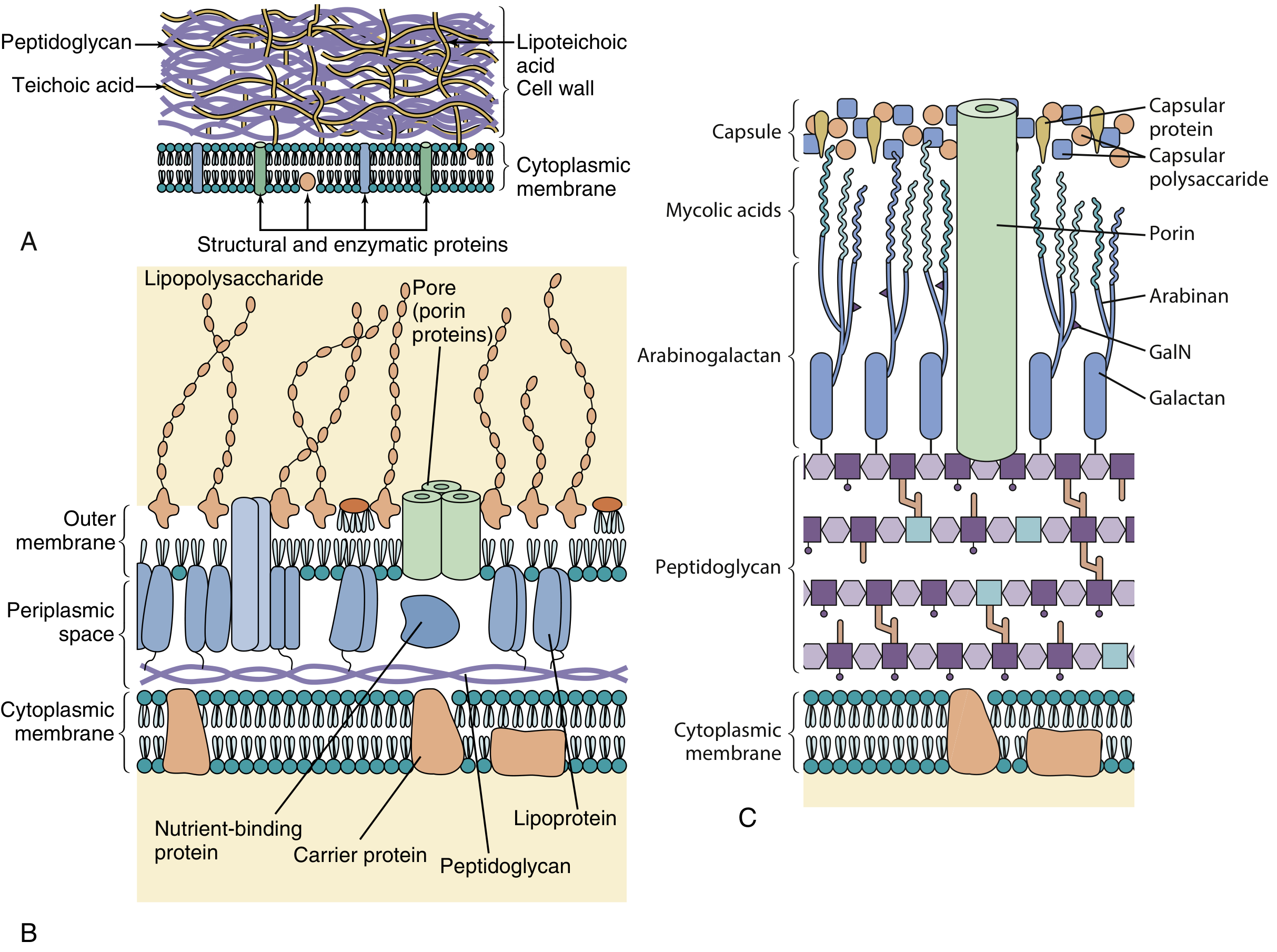
🟪 Side-by-side visual diagram
GRAM-POSITIVE BACTERIUM                  GRAM-NEGATIVE BACTERIUM
Outside                                  Outside
  │                                        │
  │   Teichoic acid                         │   O antigen
  │       ↑                                 │       ↑
  │  ╔═══════════════╗                      │  ╔═══════════════════╗
  │  ║ THICK         ║                      │  ║ OUTER MEMBRANE    ║
  │  ║ PEPTIDOGLYCAN ║                      │  ║ LPS + porins       ║
  │  ║ 20-80 nm      ║                      │  ╚═══════════════════╝
  │  ║               ║                      │       Periplasm
  │  ║               ║                      │  ─ Thin peptidoglycan
  │  ╚═══════════════╝                      │       Periplasm
  │  ═ Cytoplasmic membrane                 │  ═ Inner/cytoplasmic membrane
  │                                        │
Cytoplasm                                Cytoplasm
5. Gram-positive cell wall in detail
🟦 Structure
             OUTSIDE
                │
      ┌───────────────────────┐
      │  Thick peptidoglycan  │
      │  ███████████████████  │
      │  ███████████████████  │
      │  ███████████████████  │
      │  ███████████████████  │
      └───────────────────────┘
                │
      ═════════════════════════
        Cytoplasmic membrane
                │
             CYTOPLASM
✅ Teichoic acids
✅ Lipoteichoic acids
✅ One cytoplasmic membrane
❌ No outer membrane
❌ No LPS
❌ No lipid A endotoxin
Staphylococcus
Streptococcus
Enterococcus
Bacillus
Clostridium
Corynebacterium
Listeria
Actinomyces
Nocardia
Teichoic acids
A. Wall teichoic acid
Peptidoglycan
     │
     └──── Wall teichoic acid
B. Lipoteichoic acid
Cytoplasmic membrane
      │
      └──── Lipoteichoic acid
              extends outward through peptidoglycan
Functions of teichoic acids
| Function | Meaning |
|---|---|
| Negative surface charge | Helps determine surface chemistry |
| Cell-wall maintenance | Supports wall structure and growth |
| Adhesion | Can contribute to attachment to host tissues |
| Antigenicity | Can act as an antigenic marker |
| Inflammation | Can stimulate innate immune responses |
Teichoic acid is associated with gram-positive bacteria.
LPS is associated with gram-negative bacteria.
6. Why gram-positive organisms stain purple?
🟩 Gram stain procedure
1. Crystal violet       → all bacteria initially purple
2. Iodine               → makes a large crystal violet-iodine complex
3. Alcohol/acetone      → decolorizing step
4. Safranin             → pink counterstain
Gram-positive outcome
THICK peptidoglycan
        ↓
Alcohol dehydrates and tightens the thick wall
        ↓
Crystal violet-iodine complex cannot escape
        ↓
Cell remains PURPLE / BLUE
Gram-positive = Purple
Think: "Positive holds purple."
7. Gram-negative cell wall in detail
🟦 Structure
                  OUTSIDE
                     │
        ╔══════════════════════════╗
        ║ OUTER MEMBRANE           ║
        ║ LPS + phospholipid       ║
        ║ porins                   ║
        ╚══════════════════════════╝
                     │
             PERIPLASMIC SPACE
                     │
          ─ Thin peptidoglycan ─
                     │
             PERIPLASMIC SPACE
                     │
        ═══════════════════════════
          INNER / CYTOPLASMIC MEMBRANE
                     │
                  CYTOPLASM
✅ Cytoplasmic or inner membrane
✅ Outer membrane
✅ Periplasmic space
✅ LPS
✅ Porins
✅ Lipoprotein linkage to peptidoglycan
❌ Teichoic acids
Neisseria
E. coli
Klebsiella
Pseudomonas
Salmonella
Shigella
Vibrio
Haemophilus
Bacteroides
Legionella
The outer membrane: why it matters
GRAM-NEGATIVE ENVELOPE = "Double membrane"

Outer membrane
       ↓
Thin peptidoglycan
       ↓
Inner membrane
| Function | Clinical relevance |
|---|---|
| Acts as permeability barrier | Some antibiotics cannot enter easily |
| Contains LPS | Can cause endotoxin-mediated septic shock |
| Contains porins | Hydrophilic molecules and some antibiotics pass through these channels |
| Contains outer-membrane proteins | Important for nutrient transport, adhesion, and immune interactions |
| Adds structural protection | Greater resistance to detergents and some drugs |
Porins
Outside
   ↓
[ PORIN CHANNEL ]
   ↓
Periplasmic space
- nutrients,
- ions,
- and certain antibiotics.
Clinical correlation
Porin mutation or loss
        ↓
Antibiotic cannot enter effectively
        ↓
Antibiotic resistance
8. Periplasmic space: highly tested concept
Outer membrane
      │
      │  Periplasmic space
      │  ├─ thin peptidoglycan
      │  ├─ beta-lactamases
      │  ├─ transport proteins
      │  └─ degradative enzymes
      │
Inner membrane
Why is this clinically important?
Beta-lactam antibiotic enters through porin
                ↓
Periplasmic beta-lactamase breaks beta-lactam ring
                ↓
Drug cannot bind PBP effectively
                ↓
Cell wall synthesis continues
                ↓
Resistance
9. LPS: the endotoxin of gram-negative bacteria
🟩 Lipopolysaccharide anatomy
OUTSIDE

       O antigen
          │
     [ O-O-O-O-O ]  ← variable repeating polysaccharide
          │
       Core polysaccharide
          │
       Lipid A
          │
══════════════════════════
      Outer membrane

INSIDE
| LPS component | Function / clinical significance |
|---|---|
| Lipid A | Toxic component: endotoxin |
| Core polysaccharide | Structural connecting segment |
| O antigen | Variable antigen used in serotyping |
⭐ Lipid A = endotoxin
Gram-negative bacterium lyses or releases outer-membrane fragments
                         ↓
LPS enters blood
                         ↓
Lipid A binds immune recognition system
                         ↓
Macrophages release TNF-alpha, IL-1, IL-6
                         ↓
Fever + vasodilation + capillary leak
                         ↓
Hypotension + DIC + multiorgan failure
USMLE septic-shock sequence
Lipid A
   ↓
Macrophage activation
   ↓
TNF-alpha + IL-1
   ↓
Nitric oxide-mediated vasodilation
   ↓
Hypotension / shock

Also:
Complement + coagulation activation
   ↓
DIC
| Feature | Endotoxin | Exotoxin |
|---|---|---|
| Main source | Gram-negative outer membrane | Gram-positive or gram-negative bacteria |
| Chemical nature | LPS, specifically lipid A | Protein |
| Heat stability | Relatively heat stable | Usually heat labile |
| Secreted actively? | Usually released with membrane shedding/lysis | Often actively secreted |
| Toxoid vaccine possible? | No | Often yes |
| Examples | Gram-negative septic shock | Tetanus toxin, botulinum toxin, cholera toxin |
10. Why gram-negative bacteria stain pink?
Thin peptidoglycan layer
          +
Alcohol dissolves/disrupts outer membrane
          ↓
Crystal violet-iodine complex escapes
          ↓
Cell becomes colorless after decolorization
          ↓
Safranin counterstain enters
          ↓
Cell appears PINK / RED
Gram-negative = thin wall loses purple
                then takes pink counterstain
11. Gram-positive vs gram-negative: rapid comparison table
| Feature | 🟪 Gram-positive | 🟥 Gram-negative |
|---|---|---|
| Gram stain color | Purple / blue | Pink / red |
| Peptidoglycan | Thick | Thin |
| Outer membrane | No | Yes |
| Periplasm | Not prominent in the classic teaching model | Prominent |
| Teichoic acids | Yes | No |
| LPS | No | Yes |
| Endotoxin | No | Yes, lipid A |
| Porins | No | Yes, in outer membrane |
| Relative beta-lactam access | Often easier | Often limited by outer membrane |
| Lysozyme access | More accessible | Outer membrane partly protects |
| Examples | Staph, Strep, Bacillus, Clostridium | Neisseria, Enterobacterales, Pseudomonas |
12. Cell-wall synthesis: where antibiotics work
🟩 Build the wall in three broad locations
CYTOPLASM
  ↓ precursor formation
  ↓
CYTOPLASMIC MEMBRANE
  ↓ transport of building blocks
  ↓
OUTSIDE MEMBRANE
  ↓ glycan polymerization + peptide cross-linking
  ↓
MATURE PEPTIDOGLYCAN WALL
Antibiotic map
CYTOPLASM
│
├─ Fosfomycin
│  Blocks early NAM synthesis
│
├─ Cycloserine
│  Blocks D-alanine-related precursor formation
│
CYTOPLASMIC MEMBRANE
│
├─ Bacitracin
│  Blocks transport/recycling of bactoprenol carrier
│
OUTSIDE THE MEMBRANE
│
├─ Vancomycin
│  Binds D-Ala-D-Ala precursor
│  Blocks polymerization and cross-linking
│
└─ Beta-lactams
   Bind PBPs / transpeptidases
   Block cross-linking of peptidoglycan
13. Beta-lactams and PBPs: the most testable mechanism
Normal wall cross-linking
Peptidoglycan strand A          Peptidoglycan strand B

NAM-peptide                       NAM-peptide
    │                                  │
    └─────── PBP cross-link ───────────┘
Peptidoglycan strands
         +
PBP transpeptidation
         ↓
Strong rigid bacterial wall
What beta-lactam antibiotics do
- Penicillins
- Cephalosporins
- Carbapenems
- Monobactams
Beta-lactam resembles D-Ala-D-Ala
              ↓
Binds PBP / transpeptidase
              ↓
Transpeptidation blocked
              ↓
No peptide cross-linking
              ↓
Weak wall
              ↓
Autolysins continue breaking wall
              ↓
Bacterial lysis
Beta-lactams are most effective against actively dividing bacteria, because these bacteria are actively making new cell wall.
14. Vancomycin mechanism
Normal precursor:
NAM ─ peptide ─ D-Ala ─ D-Ala
Vancomycin binds D-Ala-D-Ala
              ↓
Prevents proper peptidoglycan assembly
              ↓
Cell-wall synthesis stops
Why vancomycin generally does not work against gram-negative bacteria
Vancomycin is large
      ↓
Cannot cross gram-negative outer membrane porins efficiently
      ↓
Cannot reach the peptidoglycan target
      ↓
Natural lack of activity against most gram-negative bacteria
15. Lysozyme: natural host defense against peptidoglycan
- tears,
- saliva,
- mucus,
- breast milk,
- neutrophil granules.
NAG ─ NAM ─ NAG ─ NAM
      ↑
Lysozyme breaks this glycosidic linkage
Peptidoglycan weakens
       ↓
Bacterial cell becomes vulnerable to osmotic lysis
16. Special bacterial envelopes you must know
A. Mycoplasma: no cell wall
Mycoplasma

Outside
   │
[ Sterol-containing cytoplasmic membrane ]
   │
Cytoplasm

NO PEPTIDOGLYCAN
NO GRAM STAIN APPEARANCE
NO BETA-LACTAM TARGET
Clinical implications
| Feature | Meaning |
|---|---|
| No peptidoglycan | Beta-lactams do not work |
| No rigid wall | Pleomorphic shape |
| Sterols in membrane | Provides membrane stability |
| No Gram staining | Cannot be reliably seen on Gram stain |
B. Mycobacteria: acid-fast, waxy wall
OUTSIDE
  │
  Waxy mycolic-acid-rich layer
  │
  Arabinogalactan
  │
  Peptidoglycan
  │
  Cytoplasmic membrane
  │
CYTOPLASM
Why it matters
Mycolic acid-rich envelope
          ↓
Resists decolorization by acid-alcohol
          ↓
Acid-fast staining positive
- slow nutrient entry,
- slow growth,
- relative resistance to many drugs and disinfectants,
- persistence in macrophages.
C. Chlamydia: unusual cell envelope
Chlamydia = obligate intracellular bacterium
           → cannot be cultured on standard artificial media
           → treat with doxycycline or azithromycin,
             not with beta-lactam monotherapy
17. One integrated visual memory story
GRAM-POSITIVE = ONE thick purple brick wall

Outside
  │
  │  Teichoic acids sticking outward
  │
█████████████████████████████
████ THICK PEPTIDOGLYCAN ████
█████████████████████████████
════════ CYTOPLASMIC MEMBRANE ════════
                Cytoplasm
GRAM-NEGATIVE = TWO membranes with a thin wall trapped between

Outside
  │
╔════ OUTER MEMBRANE: LPS + porins ════╗
       O antigen
       Core polysaccharide
       Lipid A = endotoxin
╚══════════════════════════════════════╝
       Periplasm
──────── Thin peptidoglycan ────────
       Periplasm with beta-lactamases
════════ INNER MEMBRANE ═══════════════
                Cytoplasm
🚨 USMLE traps and must-know associations
| If question says... | Think... |
|---|---|
| Lipid A | Gram-negative endotoxin |
| Teichoic acid | Gram-positive cell wall |
| D-Ala-D-Ala | Vancomycin target |
| PBP / transpeptidase | Beta-lactam target |
| No cell wall / sterol membrane | Mycoplasma |
| Acid-fast / mycolic acids | Mycobacterium |
| Porin loss | Gram-negative antibiotic resistance |
| Periplasmic beta-lactamase | Gram-negative beta-lactam resistance |
| Thick wall retains crystal violet | Gram-positive |
| Thin wall loses crystal violet and takes safranin | Gram-negative |
📌 Final exam-ready notes
BACTERIAL CELL WALL

• Main structural polymer = peptidoglycan (murein).
• Peptidoglycan consists of alternating NAG and NAM sugars.
• Short peptide side chains from NAM cross-link adjacent strands.
• PBPs perform transpeptidation, the final cross-linking step.
• Beta-lactams bind PBPs and block transpeptidation.
• Vancomycin binds D-Ala-D-Ala.

GRAM-POSITIVE
• Thick peptidoglycan.
• Teichoic acid and lipoteichoic acid.
• No outer membrane.
• No LPS/endotoxin.
• Retains crystal violet: purple.

GRAM-NEGATIVE
• Thin peptidoglycan.
• Inner membrane + outer membrane.
• Periplasm contains peptidoglycan and may contain beta-lactamases.
• Outer membrane has porins and LPS.
• LPS: lipid A = endotoxin; O antigen = serotype marker.
• Does not retain crystal violet; becomes pink with safranin.

SPECIAL
• Mycoplasma: no peptidoglycan, no beta-lactam target.
• Mycobacteria: peptidoglycan plus arabinogalactan and mycolic acids;
  acid-fast and relatively drug-resistant.