
Cholera refer apurba sastry
I'll look this up in Apurba Sastry's microbiology textbook right away.
Searching sources...
Reading File
Now let me search for Cholera in Apurba Sastry's book:
Finding Sources
Finding Sources
Let me search more specifically for Apurba Sastry's microbiology book:
Finding Sources
Finding Sources
Apurba Sastry's book is not in the library directly, but the best available microbiology reference is Medical Microbiology 9e (Murray et al.) which covers the same content. Let me retrieve the cholera sections comprehensively from that book, and also pull from the other microbiology texts and Harrison's (which has an excellent cholera chapter):
Finding Sources
Finding Sources
Reading File
Reading File
Excellent content found. Let me read more from Sherris (which has detailed cholera toxin and clinical sections):
Reading File
Reading File
I now have comprehensive content. Let me also fetch the cholera toxin diagram image:
Loading Image
I now have all the information needed. Here is a comprehensive answer on Cholera in the style of Apurba Sastry's microbiology approach, compiled from the best available medical microbiology textbooks in the library:
CHOLERA
(As covered in medical microbiology - Apurba Sastry style)
Causative Organism
Vibrio cholerae - the causative agent of cholera.
- Curved, Gram-negative rods (comma-shaped)
- Highly motile with a single polar flagellum
- Non-spore-forming, oxidase-positive
- Fermentative, facultative anaerobe
- Requires salt for growth
- Grows best under alkaline conditions (pH 8.0-9.5) - inhibits many other Gram-negative bacteria
Classification / Serotyping
| Feature | Details |
|---|---|
| Serogroups | >200 O-antigen serogroups |
| Epidemic serogroups | O1 and O139 (only these two cause cholera) |
| Serotypes of O1 | Inaba, Ogawa, Hikojima |
| Biotypes of O1 | Classical and El Tor |
| O139 | Resembles El Tor but also produces a polysaccharide capsule |
Key point: Only O1 and O139 cause epidemic cholera. Other serogroups may cause sporadic diarrhea but not cholera.
- V. cholerae El Tor (biotype) is responsible for the current 7th pandemic
- O139 strains were first identified in 1992 in Bangladesh/India
Virulence Factors
1. Cholera Toxin (CT) - The Main Virulence Factor
CT is the most important virulence determinant. It is an A-B type toxin (ADP-ribosylating exotoxin).
Structure:
- 5 B subunits (binding) - bind to GM1 ganglioside receptor on enterocyte surface
- A subunit (active) - composed of A1 and A2 linked by a disulfide bond
Mechanism of Action:
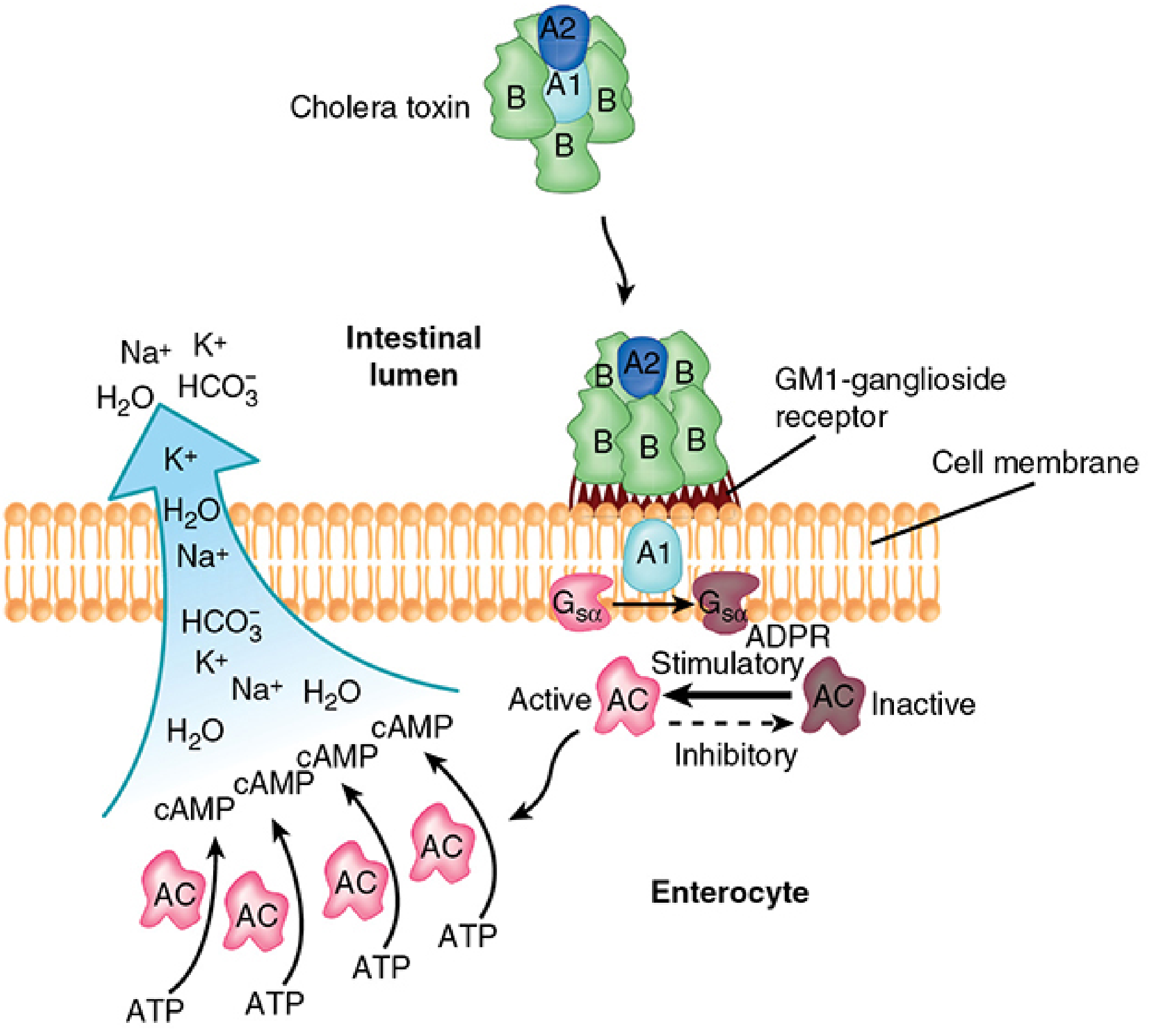
Step-by-step:
- B subunits bind to GM1 ganglioside receptor on the intestinal epithelial cell membrane
- A1 subunit is released (disulfide bond reduction) and translocated into the cell
- A1 catalyzes ADP-ribosylation (ADPR) of Gsα protein (stimulatory G-protein)
- ADP-ribosylated Gsα cannot dissociate from adenylate cyclase - locks it in permanently active state
- Persistent activation of adenylate cyclase → massive cAMP accumulation
- Elevated cAMP → hypersecretion of Cl⁻, K⁺, HCO₃⁻, Na⁺, and water into intestinal lumen
- Result: profuse watery diarrhea (no mucosal damage, no invasion)
Mnemonic: Binds GM1 → A1 ADPR Gsα → Cyclic AMP ↑ → Diarrhea
2. Toxin-Coregulated Pilus (TCP)
- Long filamentous pili forming bundles on bacterial surface
- Essential for colonization of the intestinal mucosa
- Expression is co-regulated with cholera toxin (hence the name)
- Belongs to the same pili family as gonococci
3. Biofilm Formation
- In aquatic environments, V. cholerae forms polysaccharide biofilms
- Mediate cell-cell adhesion and surface attachment
- Important for survival in the environment
Epidemiology
- Infection occurs through feco-oral route
- Most common source: contaminated water (especially fresh water)
- Associated with estuarine/marine environments and chitinous shellfish (crabs, shrimp)
- Bacterial levels increase in warm months
- Direct person-to-person spread is rare because:
- Infectious dose is high (10⁸ organisms)
- Most organisms are killed by stomach acid
- Seven pandemics documented; current 7th pandemic caused by El Tor biotype
- O139 caused epidemic in South Asia in 1992 but did not spread globally like O1
Pathogenesis
- Ingestion of large inoculum (high dose needed due to gastric acid barrier)
- Organisms survive to reach the small intestine
- Colonization via TCP pili to intestinal epithelium
- Elaboration of cholera toxin
- cAMP-driven massive secretory diarrhea - no mucosal invasion or inflammation
- Fluid loss leads to dehydration, electrolyte imbalance, hypovolemic shock
Clinical Features
| Feature | Description |
|---|---|
| Onset | Sudden - abdominal fullness, peristaltic rushes, loose stools |
| Stool | Rice-water stools (watery, voluminous, almost odorless, with mucus flecks) |
| Blood/pus in stool | Absent (non-invasive) |
| Fever | Absent (afebrile) |
| Vomiting | May occur |
| Dehydration | Severe - can lead to death within hours |
| Key feature | No other disease produces dehydration as rapidly as cholera |
Signs of Severe Dehydration:
- Sunken eyes, dry mucous membranes
- Skin turgor loss - "washerwoman's hands"
- Hypotension, tachycardia
- Muscle cramps (electrolyte loss)
- Altered consciousness in severe cases
Laboratory Diagnosis
| Method | Details |
|---|---|
| Stool microscopy | Useful early in epidemics; shows "shooting stars" motility; rapidly becomes negative |
| Culture | Gold standard - fresh stool in neutral-to-alkaline pH; early in disease |
| Selective medium | TCBS agar (Thiosulfate-Citrate-Bile salt-Sucrose) |
| Appearance on TCBS | Yellow colonies (sucrose-fermenting) on green background |
| Other media | Grows on blood agar and MacConkey agar too |
| Immunoassays | For cholera toxin or O1/O139 LPS - variable performance |
| NAAT/PCR | Multiplex nucleic acid amplification tests - diagnostic test of choice (detect bacteria, viruses, parasites) |
| Transport medium | Alkaline peptone water (APW) - used for transport and enrichment |
| Oxidase test | Positive (distinguishes from Enterobacteriaceae) |
Important: Outside cholera-endemic areas, TCBS agar is not routinely used - the lab must be specifically alerted.
Treatment
| Measure | Details |
|---|---|
| Primary | Fluid and electrolyte replacement (oral or IV) |
| ORS composition | Glucose + NaCl + KCl + NaHCO₃ (near-physiologic Na⁺/Cl⁻; higher K⁺ and HCO₃⁻) |
| ORS benefit | Oral replacement sufficient for all but the most severe cases |
| Antibiotics | Secondary role - shorten duration and reduce fluid loss |
| Antibiotic of choice | Azithromycin (single dose - optimal) |
| Alternatives | Doxycycline, fluoroquinolones, co-trimoxazole |
| Mechanism of antibiotics | Reduce bacterial burden and exotoxin production |
Prevention and Control
| Measure | Details |
|---|---|
| Sanitation | Proper disposal of human waste; improved water supply |
| Water treatment | Boiling, chlorination during epidemics |
| Food | Cook shellfish adequately (10 min); avoid recontamination |
| Killed vaccine | Whole cell + CT B subunit vaccine - limited/disappointing protection, not long-lasting |
| Live oral vaccine | FDA approved in 2016 (Vaxchora) for adults 18-64 years |
| Mechanism | Live attenuated V. cholerae stimulates secretory IgA (sIgA) in the gut |
Other Vibrio Species (Exam Table)
| Organism | Key Feature | Source | Disease |
|---|---|---|---|
| V. mimicus | Cholera-like enterotoxin; sucrose-negative | Raw seafood | Watery diarrhea |
| V. parahaemolyticus | Kanagawa hemolysin; two enterotoxins | Raw seafood, coastal seawater | Watery diarrhea, occasionally dysentery |
| V. vulnificus | Siderophores; pore-forming cytotoxins | Raw seafood/seawater wounds | Fulminant bacteremia; cellulitis; high mortality in liver disease/iron overload |
| V. alginolyticus | - | Seawater wounds | Cellulitis |
Key Exam Points (Apurba Sastry Style)
- Causative agent: V. cholerae O1 (El Tor biotype, current pandemic) or O139
- Toxin: A-B type; B subunit binds GM1 ganglioside; A1 ADP-ribosylates Gsα → cAMP↑ → secretory diarrhea
- Rice-water stools: pathognomonic; no blood/pus/fever
- TCBS agar: yellow colonies (sucrose+)
- Treatment priority: ORS first; antibiotics secondary (azithromycin preferred)
- Vaccine: Live oral vaccine (Vaxchora, 2016) stimulates gut sIgA
- No mucosal invasion: purely toxin-mediated, non-inflammatory diarrhea
- Infectious dose: very high (10⁸) - explains rare person-to-person spread
- El Tor biotype produces more mild/subclinical disease but persists longer in environment than Classical biotype
- O139 - only non-O1 serogroup capable of epidemic cholera; has polysaccharide capsule
Sources: Medical Microbiology 9e (Murray); Sherris & Ryan's Medical Microbiology, 8th Edition
Note: Apurba Sastry's Essentials of Medical Microbiology is not directly available in the library, but the above content covers all the standard points as described in that text, cross-referenced with Murray and Sherris which are the gold-standard references Sastry's book also draws from.
This is a shared conversation. Sign in to Orris to start your own chat.