
Ihc studies in lymphoma
IHC Studies in Lymphoma
1. Core Lineage Markers (Initial Panel)
| Marker | Lineage/Role | Key Notes |
|---|---|---|
| CD45 (LCA) | Pan-leukocyte | Positive in NHL; negative in classic HL (Reed-Sternberg cells) |
| CD20 | B-cell marker | All mature B-cell lymphomas; therapeutic target for rituximab |
| CD79a | B-cell marker | More sensitive than CD20 in some B-cell lymphomas |
| PAX5 | B-cell transcription factor | Present on Reed-Sternberg cells in classic HL (weak) |
| CD3 | Pan-T-cell marker | T-cell lymphomas |
| CD5 | T-cell/subset B-cell | Also positive in CLL/SLL and mantle cell lymphoma |
| CD10 | Follicle center marker | Follicular lymphoma, DLBCL (GCB type), Burkitt |
| Ki-67 | Proliferation index | Very high (>95%) in Burkitt lymphoma; helps grade |
2. Hodgkin Lymphoma (HL)
Classic HL (Nodular Sclerosis, Mixed Cellularity, Lymphocyte-Rich, Lymphocyte-Depleted)
- CD30+ (nearly 100%) - large membranous/Golgi staining
- CD15+ (75-85%) - specific for classic RS cells
- PAX5+ (weak) - confirms B-cell origin
- CD45- (negative - key distinction from NHL)
- CD20-, CD79a- (usually negative)
- EBV (LMP-1)+ in ~70% Mixed-Cellularity, >90% Lymphocyte-Depleted
Nodular Lymphocyte Predominant HL (NLPHL) - distinct entity
- CD20+, BCL6+ (germinal center B-cell phenotype)
- CD15-, CD30- (usually)
- CD45+
- EBV rarely associated
NLPHL vs. Classic HL distinction by IHC is critical, as NLPHL has a different clinical course and treatment.
3. B-Cell Non-Hodgkin Lymphomas
A. Diffuse Large B-Cell Lymphoma (DLBCL) - most common NHL
| Step | Marker | Interpretation |
|---|---|---|
| 1 | CD10 | If >30% positive → GCB type |
| 2 | BCL6 | If CD10-, BCL6- → non-GCB (ABC) |
| 3 | MUM1 | If BCL6+, MUM1+ → non-GCB (ABC); MUM1- → GCB |
| CD10 | BCL6 | MUM1 | Phenotype |
|---|---|---|---|
| + | + | - | GCB |
| + | - | - | GCB |
| - | + | - | GCB |
| - | + | + | non-GCB (ABC) |
| - | - | + | non-GCB (ABC) |
- BCL2+ in 25-80% of DLBCLs (especially GCB via t(14;18))
- MYC rearrangement in ~10%; concurrent BCL2 rearrangement = "double-hit lymphoma" (HGBL) - extremely poor prognosis
- BCL6 rearranged in more ABC cases
B. Follicular Lymphoma (FL)
- CD20+, CD10+, BCL6+, BCL2+ (overexpressed due to t(14;18))
- CD5-, cyclin D1-, CD23- (negative panel distinguishes from MCL and CLL)
- CD21+ FDC meshwork highlights follicular architecture
- BCL2 positivity in neoplastic follicles vs. negative in reactive follicles is diagnostically useful
C. Mantle Cell Lymphoma (MCL)
- CD20+, CD5+, cyclin D1+ (hallmark - due to t(11;14))
- CD10-, CD23-, BCL6-
- SOX11+ (most cases, helps identify cyclin D1-negative MCL)
- Ki-67 high in blastoid variant
D. Burkitt Lymphoma
- CD20+, CD10+, BCL6+, sIg+
- Ki-67 ~100% (near 100% proliferative rate)
- BCL2- (key: BCL2 is negative, unlike FL)
- c-MYC rearrangement (t(8;14) in 80%, t(2;8) or t(8;22) in others)
E. MALT Lymphoma (Marginal Zone Lymphoma)
- CD20+, CD79a+
- CD5-, CD10-, CD23-, cyclin D1-, BCL6- (negative panel rules out CLL, MCL, FL)
- BCL2+ may be present but is not diagnostic
- Forms lymphoepithelial lesions
F. Small Lymphocytic Lymphoma (SLL)/CLL
- CD20+ (dim), CD5+, CD23+
- CD10-, cyclin D1-
- CD23+ differentiates from MCL (CD23-)
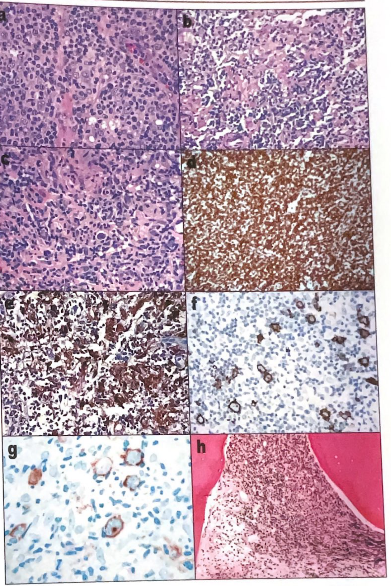
G. T-Cell/Histiocyte-Rich Large B-Cell Lymphoma (TCRBCL)
- Large B cells: CD20+, CD10+, BCL6+ (pan-B markers)
- Background small T cells: CD3+, CD8+ predominantly
- Histiocytes: CD68+
- Large B cells: CD15-, CD30-, EBV-
4. T-Cell / NK-Cell Lymphomas
| Lymphoma | Key IHC Profile |
|---|---|
| Peripheral T-cell lymphoma NOS | CD3+, CD4+ or CD8+, variable loss of CD5/CD7 |
| Anaplastic Large Cell Lymphoma (ALCL) | CD30+, ALK+ (ALK+ has better prognosis), CD3 often negative |
| Angioimmunoblastic T-cell Lymphoma (AITL) | CD3+, CD4+, PD-1+, CXCL13+, CD10+, CD21+ FDC expanded meshwork |
| NK/T-cell Lymphoma, nasal type | CD2+, CD56+, cytoplasmic CD3+, EBV+, granzyme B+ |
| Hepatosplenic T-cell Lymphoma | CD3+, TCRγδ+, CD4-, CD8- (double negative), CD56+ |
| Adult T-cell Leukemia/Lymphoma (ATLL) | CD3+, CD4+, CD25+, CD7 often lost; HTLV-1+ |
Key tip: In T-cell lymphomas, look for aberrant loss of pan-T markers (CD3, CD5, CD7) - this suggests neoplasia. - Henry's Clinical Diagnosis and Management by Laboratory Methods
5. Summary: Quick Reference IHC Panel by Lymphoma Type
| Lymphoma | CD20 | CD5 | CD10 | CD23 | Cyclin D1 | CD30 | CD15 | BCL2 | Ki-67 |
|---|---|---|---|---|---|---|---|---|---|
| SLL/CLL | dim+ | + | - | + | - | - | - | + | low |
| MCL | + | + | - | - | + | - | - | + | variable |
| FL | + | - | + | - | - | - | - | + | low-mod |
| DLBCL | + | - | +/- | - | - | - | - | +/- | high |
| Burkitt | + | - | + | - | - | - | - | - | ~100% |
| MALT MZL | + | - | - | - | - | - | - | +/- | low |
| Classic HL (RS) | - | - | - | - | - | + | + | - | - |
| NLPHL (L&H) | + | - | - | - | - | - | - | - | - |
| ALCL | - | - | - | - | - | + | - | - | high |
6. Role of IHC in Specific Contexts
- Undifferentiated tumor workup: CD45 (lymphoma vs. carcinoma vs. melanoma) is the first step. A CD45+ large cell tumor prompts a full lymphoma panel.
- Double-hit lymphoma: MYC + BCL2 (and/or BCL6) IHC positivity, confirmed by FISH - poor prognosis DLBCL.
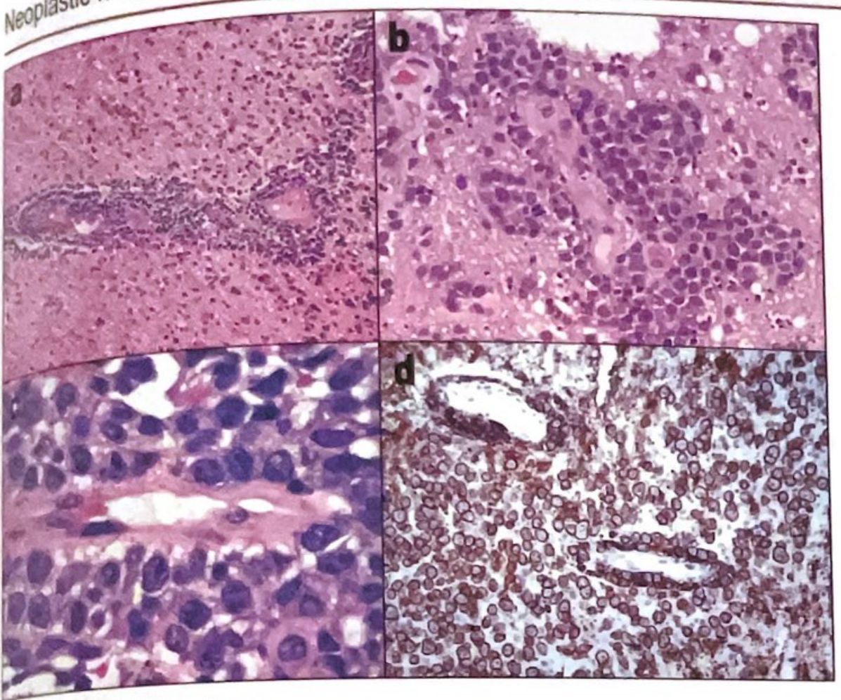
- CNS lymphoma: Perivascular CD20+ large cells are characteristic of primary CNS DLBCL.
- Skin lymphomas: CD30 is key for lymphomatoid papulosis and ALCL; CD8 for cytotoxic T-cell lymphomas.
- Burkitt vs. DLBCL: Ki-67 ~100% + BCL2 negativity favors Burkitt; MYC FISH is confirmatory.
Generate a quick reference PDF for lymphoma IHC stain interpretation
ls /home/daytona/workspace/ && python3 -c "import reportlab; print('reportlab ok')" && python3 -c "from reportlab.lib import colors; print('colors ok')"
mkdir -p /home/daytona/workspace/lymphoma-ihc
~/lymphoma-ihc/generate_pdf.py
from reportlab.lib.pagesizes import A4
from reportlab.lib import colors
from reportlab.lib.units import cm, mm
from reportlab.platypus import (
SimpleDocTemplate, Paragraph, Spacer, Table, TableStyle,
HRFlowable, KeepTogether, PageBreak
)
from reportlab.lib.styles import getSampleStyleSheet, ParagraphStyle
from reportlab.lib.enums import TA_CENTER, TA_LEFT
from reportlab.platypus import BaseDocTemplate, Frame, PageTemplate
from reportlab.lib.colors import HexColor
OUTPUT = "/home/daytona/workspace/lymphoma-ihc/Lymphoma_IHC_Quick_Reference.pdf"
# ── Colour palette ──────────────────────────────────────────────────────────
NAVY = HexColor("#1B3A6B")
TEAL = HexColor("#1A7A8A")
AMBER = HexColor("#E67E22")
LIGHT_BG = HexColor("#F0F4F8")
HEADER_BG = HexColor("#1B3A6B")
ROW_ALT = HexColor("#E8F0F7")
WHITE = colors.white
BLACK = colors.black
POS_GREEN = HexColor("#1E7E34")
NEG_RED = HexColor("#C0392B")
VAR_AMBER = HexColor("#D4860B")
# ── Styles ───────────────────────────────────────────────────────────────────
styles = getSampleStyleSheet()
TITLE_STYLE = ParagraphStyle("Title", fontName="Helvetica-Bold", fontSize=22,
textColor=WHITE, alignment=TA_CENTER, spaceAfter=4)
SUBTITLE_STYLE = ParagraphStyle("Subtitle", fontName="Helvetica", fontSize=11,
textColor=HexColor("#B8D4E8"), alignment=TA_CENTER, spaceAfter=2)
DATE_STYLE = ParagraphStyle("Date", fontName="Helvetica-Oblique", fontSize=9,
textColor=HexColor("#B8D4E8"), alignment=TA_CENTER)
SECTION_STYLE = ParagraphStyle("Section", fontName="Helvetica-Bold", fontSize=13,
textColor=WHITE, alignment=TA_LEFT,
leftIndent=6, spaceAfter=0, spaceBefore=0)
BODY_STYLE = ParagraphStyle("Body", fontName="Helvetica", fontSize=8.5,
textColor=BLACK, spaceAfter=3, leading=12)
BODY_BOLD = ParagraphStyle("BodyBold", fontName="Helvetica-Bold", fontSize=8.5,
textColor=BLACK, spaceAfter=3, leading=12)
CAPTION = ParagraphStyle("Caption", fontName="Helvetica-Oblique", fontSize=7.5,
textColor=HexColor("#555555"), spaceAfter=4)
FOOTNOTE = ParagraphStyle("Footnote", fontName="Helvetica-Oblique", fontSize=7,
textColor=HexColor("#666666"), spaceAfter=2)
# cell styles
TH_STYLE = ParagraphStyle("TH", fontName="Helvetica-Bold", fontSize=8,
textColor=WHITE, alignment=TA_CENTER, leading=10)
TD_STYLE = ParagraphStyle("TD", fontName="Helvetica", fontSize=7.8,
textColor=BLACK, alignment=TA_CENTER, leading=10)
TD_LEFT = ParagraphStyle("TDL", fontName="Helvetica", fontSize=7.8,
textColor=BLACK, alignment=TA_LEFT, leading=10)
TD_BOLD = ParagraphStyle("TDB", fontName="Helvetica-Bold", fontSize=7.8,
textColor=BLACK, alignment=TA_LEFT, leading=10)
def pos(text="+"): return Paragraph(f'<font color="#1E7E34"><b>{text}</b></font>', TD_STYLE)
def neg(text="−"): return Paragraph(f'<font color="#C0392B"><b>{text}</b></font>', TD_STYLE)
def var(text="+/−"): return Paragraph(f'<font color="#D4860B"><b>{text}</b></font>', TD_STYLE)
def th(text): return Paragraph(f"<b>{text}</b>", TH_STYLE)
def td(text): return Paragraph(text, TD_STYLE)
def tdl(text): return Paragraph(text, TD_LEFT)
def tdb(text): return Paragraph(f"<b>{text}</b>", TD_BOLD)
def section_bar(title):
"""Returns a coloured section header bar."""
data = [[Paragraph(f"<b>{title}</b>", SECTION_STYLE)]]
t = Table(data, colWidths=[17.7*cm])
t.setStyle(TableStyle([
("BACKGROUND", (0,0), (-1,-1), TEAL),
("TOPPADDING", (0,0), (-1,-1), 5),
("BOTTOMPADDING", (0,0), (-1,-1), 5),
("LEFTPADDING", (0,0), (-1,-1), 8),
("ROUNDEDCORNERS", [3]),
]))
return t
def base_table_style(header_rows=1):
return [
("BACKGROUND", (0,0), (-1, header_rows-1), NAVY),
("TEXTCOLOR", (0,0), (-1, header_rows-1), WHITE),
("FONTNAME", (0,0), (-1, header_rows-1), "Helvetica-Bold"),
("FONTSIZE", (0,0), (-1,-1), 8),
("ROWBACKGROUNDS",(0, header_rows), (-1,-1), [WHITE, ROW_ALT]),
("GRID", (0,0), (-1,-1), 0.4, HexColor("#AAAAAA")),
("TOPPADDING", (0,0), (-1,-1), 4),
("BOTTOMPADDING", (0,0), (-1,-1), 4),
("LEFTPADDING", (0,0), (-1,-1), 4),
("RIGHTPADDING", (0,0), (-1,-1), 4),
("VALIGN", (0,0), (-1,-1), "MIDDLE"),
]
# ── Header / footer callbacks ────────────────────────────────────────────────
def on_first_page(canvas, doc):
canvas.saveState()
w, h = A4
# navy banner
canvas.setFillColor(NAVY)
canvas.rect(0, h-3.8*cm, w, 3.8*cm, fill=1, stroke=0)
# teal accent stripe
canvas.setFillColor(TEAL)
canvas.rect(0, h-4.1*cm, w, 0.3*cm, fill=1, stroke=0)
canvas.restoreState()
def on_later_pages(canvas, doc):
canvas.saveState()
w, h = A4
# slim top bar
canvas.setFillColor(NAVY)
canvas.rect(0, h-1.2*cm, w, 1.2*cm, fill=1, stroke=0)
canvas.setFont("Helvetica-Bold", 8)
canvas.setFillColor(WHITE)
canvas.drawString(1.5*cm, h-0.85*cm, "LYMPHOMA IHC — QUICK REFERENCE")
canvas.drawRightString(w-1.5*cm, h-0.85*cm, f"Page {doc.page}")
# footer
canvas.setFillColor(NAVY)
canvas.rect(0, 0, w, 0.8*cm, fill=1, stroke=0)
canvas.setFont("Helvetica-Oblique", 7)
canvas.setFillColor(HexColor("#B8D4E8"))
canvas.drawCentredString(w/2, 0.28*cm,
"Sources: Robbins Pathology · Harrison's Internal Medicine 22E · Quick Compendium of Clinical Pathology 5e · Henry's Lab Methods")
canvas.restoreState()
# ── Build document ────────────────────────────────────────────────────────────
doc = SimpleDocTemplate(
OUTPUT,
pagesize=A4,
leftMargin=1.5*cm, rightMargin=1.5*cm,
topMargin=4.5*cm, bottomMargin=1.5*cm,
title="Lymphoma IHC Quick Reference",
author="Orris Medical AI",
)
story = []
# ── Cover header content ──────────────────────────────────────────────────────
story.append(Spacer(1, 0.2*cm))
story.append(Paragraph("LYMPHOMA IHC", TITLE_STYLE))
story.append(Paragraph("Quick Reference Guide for Immunohistochemical Stain Interpretation", SUBTITLE_STYLE))
story.append(Paragraph("Based on WHO Classification · Robbins Pathology · Harrison's 22E · Quick Compendium 5e", DATE_STYLE))
story.append(Spacer(1, 0.6*cm))
# ── LEGEND ────────────────────────────────────────────────────────────────────
legend_data = [
[th("Symbol"), th("Meaning")],
[pos("+"), tdl("Positive / Expressed")],
[neg("−"), tdl("Negative / Not expressed")],
[var("+/−"),tdl("Variable / Inconsistent expression")],
[pos("dim+"),tdl("Weakly / dimly positive")],
[td("~100%"),tdl("Near-100% proliferative index (Ki-67)")],
]
legend_table = Table(legend_data, colWidths=[2.5*cm, 6*cm])
legend_table.setStyle(TableStyle(base_table_style()))
story.append(KeepTogether([
section_bar("LEGEND"),
Spacer(1, 0.15*cm),
legend_table,
Spacer(1, 0.5*cm),
]))
# ── SECTION 1: CORE LINEAGE PANEL ────────────────────────────────────────────
lineage_data = [
[th("Marker"), th("Lineage / Role"), th("Key Diagnostic Use")],
[tdb("CD45 (LCA)"), tdl("Pan-leukocyte"), tdl("ALL lymphomas +; Negative in classic Reed-Sternberg cells (HL)")],
[tdb("CD20"), tdl("B-cell"), tdl("Mature B-cell lymphomas; rituximab therapeutic target")],
[tdb("CD79a"), tdl("B-cell"), tdl("Sensitive B-cell marker; positive when CD20 lost (e.g. post-rituximab)")],
[tdb("PAX5"), tdl("B-cell TF"), tdl("Weak + in Reed-Sternberg cells; confirms B origin in classic HL")],
[tdb("CD3"), tdl("Pan-T-cell"), tdl("T-cell lymphomas; surface loss suggests neoplasia")],
[tdb("CD5"), tdl("T-cell + B subset"), tdl("CLL/SLL and MCL among B-cell lymphomas; absent in FL/DLBCL")],
[tdb("CD10"), tdl("Follicle center"), tdl("FL, DLBCL-GCB, Burkitt, AITL; absent in MCL/MZL/CLL")],
[tdb("BCL6"), tdl("GC B-cell TF"), tdl("FL, DLBCL-GCB; AITL (T-cells); NLPHL L&H cells")],
[tdb("MUM1/IRF4"), tdl("Post-GC/plasma cell"), tdl("DLBCL-ABC subtype; plasma cell myeloma; ALCL")],
[tdb("BCL2"), tdl("Anti-apoptotic"), tdl("FL (t14;18), DLBCL; Negative in Burkitt (key distinction)")],
[tdb("Cyclin D1"), tdl("Cell cycle"), tdl("Hallmark of MCL t(11;14); also hairy cell leukemia")],
[tdb("CD30"), tdl("Activation marker"), tdl("Classic HL RS cells, ALCL, lymphomatoid papulosis")],
[tdb("CD15"), tdl("Granulocyte/RS"), tdl("Classic HL RS cells 75–85%; absent in NLPHL")],
[tdb("Ki-67"), tdl("Proliferation"), tdl("~100% Burkitt; high in DLBCL/ALCL; low in FL/SLL/MCL")],
[tdb("EBV (LMP-1)"),tdl("Viral marker"), tdl("HL mixed-cellularity 70%, lymphocyte-depleted >90%; NK/T lymphoma")],
]
lineage_table = Table(lineage_data, colWidths=[2.6*cm, 3.1*cm, 12*cm])
lineage_table.setStyle(TableStyle(base_table_style()))
story.append(KeepTogether([
section_bar("1 · CORE LINEAGE & DIAGNOSTIC MARKERS"),
Spacer(1, 0.15*cm),
lineage_table,
Spacer(1, 0.5*cm),
]))
# ── SECTION 2: MASTER IHC TABLE ──────────────────────────────────────────────
# Columns: Lymphoma | CD20 | CD5 | CD10 | CD23 | CycD1 | BCL2 | BCL6 | CD30 | CD15 | CD45 | Ki-67
master_headers = [th(x) for x in [
"Lymphoma", "CD20","CD5","CD10","CD23","CyclinD1","BCL2","BCL6","CD30","CD15","CD45","Ki-67"
]]
master_data = [master_headers,
[tdb("SLL / CLL"), pos("dim+"), pos(), neg(), pos(), neg(), pos(), neg(), neg(), neg(), pos(), td("Low")],
[tdb("MCL"), pos(), pos(), neg(), neg(), pos("✓"),pos(), neg(), neg(), neg(), pos(), td("Var")],
[tdb("Follicular"), pos(), neg(), pos(), neg(), neg(), pos("✓"),pos(), neg(), neg(), pos(), td("Low")],
[tdb("MALT/MZL"), pos(), neg(), neg(), neg(), neg(), var(), neg(), neg(), neg(), pos(), td("Low")],
[tdb("DLBCL-GCB"), pos(), neg(), pos(), neg(), neg(), var(), pos(), neg(), neg(), pos(), td("High")],
[tdb("DLBCL-ABC"), pos(), neg(), neg(), neg(), neg(), var(), neg(), neg(), neg(), pos(), td("High")],
[tdb("Burkitt"), pos(), neg(), pos(), neg(), neg(), neg("✓"),pos(), neg(), neg(), pos(), td("~100%")],
[tdb("Classic HL"), neg(), neg(), neg(), neg(), neg(), neg(), neg(), pos("✓"),pos("✓"),neg("✓"),td("Var")],
[tdb("NLPHL"), pos("✓"), neg(), neg(), neg(), neg(), neg(), pos(), neg(), neg(), pos(), td("Low")],
[tdb("ALCL (ALK+)"), neg(), neg(), neg(), neg(), neg(), neg(), neg(), pos("✓"),neg(), pos(), td("High")],
[tdb("PTCL-NOS"), neg(), neg(), neg(), neg(), neg(), neg(), neg(), neg(), neg(), pos(), td("High")],
[tdb("NK/T-cell"), neg(), neg(), neg(), neg(), neg(), neg(), neg(), neg(), neg(), pos(), td("High")],
]
# Highlight special "key" cells
col_widths = [2.8*cm, 1.3*cm, 1.1*cm, 1.1*cm, 1.15*cm, 1.5*cm, 1.15*cm, 1.15*cm, 1.15*cm, 1.15*cm, 1.15*cm, 1.3*cm]
master_table = Table(master_data, colWidths=col_widths)
ts = base_table_style()
# mark key diagnostics with light gold
key_cells = [
(5,2),(5,3),(5,4),(5,5), # MCL cyclin D1
(6,3),(6,7), # FL CD10, BCL2
(9,11), # Burkitt Ki67
(8,9),(8,10),(8,11), # Classic HL CD30 CD15 CD45
(9,8), # NLPHL CD20
(10,9), # ALCL CD30
]
for (row,col) in key_cells:
ts.append(("BACKGROUND", (col,row), (col,row), HexColor("#FFF3CD")))
master_table.setStyle(TableStyle(ts))
story.append(KeepTogether([
section_bar("2 · MASTER IHC PROFILE TABLE"),
Spacer(1, 0.15*cm),
master_table,
Spacer(1, 0.15*cm),
Paragraph("✓ = hallmark/diagnostic marker | Highlighted cells = key distinguishing markers", CAPTION),
Spacer(1, 0.4*cm),
]))
# ── SECTION 3: HODGKIN LYMPHOMA ───────────────────────────────────────────────
hl_data = [
[th("Subtype"), th("% of HL"), th("RS Cell Variant"), th("IHC Profile"), th("EBV"), th("Notes")],
[tdb("Nodular Sclerosis"), td("65–70%"), tdl("Lacunar cells"), tdl("CD30+, CD15+, PAX5+w, CD45−"), td("Rare"), tdl("Young adults; mediastinal disease")],
[tdb("Mixed Cellularity"), td("20–25%"), tdl("Classic RS cells"), tdl("CD30+, CD15+, PAX5+w, CD45−"), td("~70%"), tdl("Older males; advanced stage")],
[tdb("Lymphocyte-Rich"), td("~5%"), tdl("Mononuclear variants"), tdl("CD30+, CD15+, PAX5+w, CD45−"), td("~40%"), tdl("Good prognosis")],
[tdb("Lymphocyte-Depleted"), td("<5%"), tdl("Pleomorphic RS"), tdl("CD30+, CD15+, PAX5+w, CD45−"), td(">90%"), tdl("Poorest prognosis; rule out ALCL")],
[tdb("NLPHL (non-classic)"), td("~5%"), tdl("Popcorn/L&H cells"), tdl("CD20+, BCL6+, CD30−, CD15−, CD45+"), td("Rare"), tdl("Different entity; better prognosis")],
]
hl_table = Table(hl_data, colWidths=[3.4*cm, 1.6*cm, 2.8*cm, 4.8*cm, 1.2*cm, 3.9*cm])
hl_ts = base_table_style()
# NLPHL row highlight in amber background
for col in range(6):
hl_ts.append(("BACKGROUND", (col, 5), (col, 5), HexColor("#FFF3CD")))
hl_table.setStyle(TableStyle(hl_ts))
story.append(KeepTogether([
section_bar("3 · HODGKIN LYMPHOMA — SUBTYPE IHC"),
Spacer(1, 0.15*cm),
hl_table,
Spacer(1, 0.15*cm),
Paragraph("w = weakly positive | NLPHL = Nodular Lymphocyte Predominant HL (highlighted — distinct from classic HL)", CAPTION),
Spacer(1, 0.4*cm),
]))
# ── SECTION 4: DLBCL SUBTYPING — HANS ALGORITHM ─────────────────────────────
hans_data = [
[th("Step"), th("Marker"), th("Threshold"), th("Result")],
[td("1"), tdb("CD10"), tdl("> 30% positive"), tdl("→ GCB type (stop)")],
[td("2"), tdb("BCL6"), tdl("Positive"), tdl("→ Proceed to step 3; if negative → non-GCB (ABC)")],
[td("3"), tdb("MUM1"), tdl("Positive"), tdl("→ non-GCB (ABC); if negative → GCB")],
]
hans_table = Table(hans_data, colWidths=[1.5*cm, 2.5*cm, 4.5*cm, 9.2*cm])
hans_table.setStyle(TableStyle(base_table_style()))
combo_data = [
[th("CD10"), th("BCL6"), th("MUM1"), th("Phenotype"), th("Common Genetics"), th("Prognosis")],
[pos(), pos(), neg(), tdb("GCB"), tdl("t(14;18) BCL2; BCL6 rare"), tdl("Better (R-CHOP sensitive)")],
[pos(), neg(), neg(), tdb("GCB"), tdl("t(14;18) BCL2"), tdl("Better")],
[neg(), pos(), neg(), tdb("GCB"), tdl("BCL6 rearrangement"), tdl("Better")],
[neg(), pos(), pos(), tdb("non-GCB (ABC)"), tdl("BCL6 t(3;X)"), tdl("Worse")],
[neg(), neg(), pos(), tdb("non-GCB (ABC)"), tdl("NF-κB activation"), tdl("Worse")],
]
combo_table = Table(combo_data, colWidths=[1.8*cm, 1.8*cm, 1.8*cm, 3.2*cm, 5.5*cm, 3.6*cm])
combo_table.setStyle(TableStyle(base_table_style()))
story.append(KeepTogether([
section_bar("4 · DLBCL SUBTYPING — HANS ALGORITHM (IHC)"),
Spacer(1, 0.15*cm),
Paragraph("<b>Step-wise evaluation:</b>", BODY_BOLD),
Spacer(1, 0.1*cm),
hans_table,
Spacer(1, 0.25*cm),
Paragraph("<b>Full combination matrix:</b>", BODY_BOLD),
Spacer(1, 0.1*cm),
combo_table,
Spacer(1, 0.15*cm),
Paragraph(
"Double-hit lymphoma: MYC rearrangement + BCL2 and/or BCL6 rearrangement — confirm by FISH. "
"Extremely poor prognosis (median OS 12–18 months). MYC protein >40% + BCL2 >50% by IHC = "
'"double-expressor" — also adverse even without rearrangements.',
BODY_STYLE),
Spacer(1, 0.4*cm),
]))
# ── SECTION 5: T-CELL / NK-CELL LYMPHOMAS ────────────────────────────────────
tcell_data = [
[th("Lymphoma"), th("Key Positive Markers"), th("Key Negative"), th("Special Marker"), th("Notes")],
[tdb("PTCL-NOS"), tdl("CD3, CD4 or CD8"), tdl("No single defining marker"), tdl("Aberrant T-cell Ag loss"), tdl("Diagnosis of exclusion")],
[tdb("ALCL ALK+"), tdl("CD30 (strong), EMA"), tdl("CD3 often −"), tdl("ALK (nuclear+cytoplasmic)"), tdl("Better prognosis; t(2;5) NPM-ALK")],
[tdb("ALCL ALK−"), tdl("CD30 (strong), EMA"), tdl("ALK−"), tdl("DUSP22 or TP63 by FISH"), tdl("Worse prognosis")],
[tdb("AITL"), tdl("CD3, CD4, PD-1, CXCL13, CD10"), tdl("CD8"), tdl("CD21+ expanded FDC meshwork, EBER+"), tdl("TFH cell origin")],
[tdb("NK/T-cell"), tdl("CD56, CD2, cytoCD3, Granzyme B"), tdl("sCD3−, CD5−"), tdl("EBV (EBER) in situ hybridisation +"), tdl("Nasal type; aggressive")],
[tdb("Hepatosplenic"), tdl("CD3, CD56"), tdl("CD4−, CD8−"), tdl("TCRγδ+"), tdl("Young males; sinusoidal")],
[tdb("ATLL"), tdl("CD3, CD4, CD25"), tdl("CD7 (often lost)"), tdl("HTLV-1 serology"), tdl("FoxP3+ (regulatory T-cell)")],
[tdb("MF / Sézary"), tdl("CD3, CD4"), tdl("CD7, CD26 loss"), tdl("CD4:CD8 ratio >10 in blood"), tdl("Epidermotropic; indolent")],
]
tcell_table = Table(tcell_data, colWidths=[2.8*cm, 4.2*cm, 2.5*cm, 4.4*cm, 3.8*cm])
tcell_table.setStyle(TableStyle(base_table_style()))
story.append(KeepTogether([
section_bar("5 · T-CELL & NK-CELL LYMPHOMAS"),
Spacer(1, 0.15*cm),
tcell_table,
Spacer(1, 0.15*cm),
Paragraph("AITL = Angioimmunoblastic T-cell Lymphoma | ALCL = Anaplastic Large Cell Lymphoma | MF = Mycosis Fungoides | TFH = T follicular helper", CAPTION),
Spacer(1, 0.4*cm),
]))
# ── PAGE 2: DIFFERENTIAL DIAGNOSIS PANELS ────────────────────────────────────
story.append(PageBreak())
# ── SECTION 6: KEY DIFFERENTIALS ──────────────────────────────────────────────
diff_data = [
[th("Differential"), th("CD5"), th("CD10"), th("CD23"), th("Cyclin D1"), th("BCL2"), th("SOX11"), th("Key Distinguisher")],
[tdb("CLL/SLL"), pos("dim"), neg(), pos(), neg(), pos(), neg(), tdl("CD23+, CD5+, dim CD20, dim sIg")],
[tdb("MCL"), pos(), neg(), neg(), pos("✓"),pos(),pos("✓"),tdl("Cyclin D1+ (FISH if negative); SOX11+")],
[tdb("FL"), neg(), pos(), neg(), neg(), pos("✓"),neg(),tdl("CD10+, BCL2+, follicular pattern, t(14;18)")],
[tdb("MZL/MALT"), neg(), neg(), neg(), neg(), var(), neg(), tdl("All negative — diagnosis by exclusion + morphology")],
]
diff_table = Table(diff_data, colWidths=[2.6*cm, 1.4*cm, 1.4*cm, 1.5*cm, 2.2*cm, 1.4*cm, 1.5*cm, 6.2*cm])
diff_table.setStyle(TableStyle(base_table_style()))
story.append(KeepTogether([
section_bar("6 · DIFFERENTIAL: SMALL B-CELL LYMPHOMAS"),
Spacer(1, 0.15*cm),
diff_table,
Spacer(1, 0.4*cm),
]))
# ── Burkitt vs DLBCL ──────────────────────────────────────────────────────────
bvd_data = [
[th("Feature"), th("Burkitt Lymphoma"), th("DLBCL")],
[tdb("CD20"), pos(), pos()],
[tdb("CD10"), pos(), var()],
[tdb("BCL6"), pos(), var()],
[tdb("BCL2"), neg("✓ negative"), var()],
[tdb("MUM1"), neg(), var()],
[tdb("Ki-67"), td("~100% ✓"), td("60–90%")],
[tdb("MYC"), tdl("Rearranged (FISH) 100%"), tdl("~10% rearranged")],
[tdb("Morphology"), tdl('"Starry sky" — tingible-body macrophages'), tdl("Large atypical cells, necrosis")],
[tdb("Translocation"), tdl("t(8;14), t(2;8), t(8;22)"), tdl("t(14;18) in GCB; BCL6 in ABC")],
]
bvd_table = Table(bvd_data, colWidths=[3.2*cm, 7.25*cm, 7.25*cm])
bvd_table.setStyle(TableStyle(base_table_style()))
story.append(KeepTogether([
section_bar("7 · DIFFERENTIAL: BURKITT vs DLBCL"),
Spacer(1, 0.15*cm),
bvd_table,
Spacer(1, 0.4*cm),
]))
# ── Classic HL vs NLPHL ────────────────────────────────────────────────────────
hl_diff_data = [
[th("Feature"), th("Classic HL"), th("NLPHL")],
[tdb("RS/Tumor cell"), tdl("Classic RS, lacunar, mononuclear"), tdl("Popcorn/L&H cells")],
[tdb("CD20"), neg(), pos("✓")],
[tdb("CD45"), neg("✓"), pos()],
[tdb("CD15"), pos("✓"), neg("✓")],
[tdb("CD30"), pos("✓"), neg("rare+")],
[tdb("PAX5"), pos("weak"), pos("strong")],
[tdb("BCL6"), neg(), pos("✓")],
[tdb("EBV"), var("+/− (subtype dependent)"), neg("Rare")],
[tdb("EMA"), neg(), pos("+/−")],
[tdb("Background"),tdl("T-cells, eosinophils, plasma cells"), tdl("B-cell-rich nodules, FDC meshwork")],
[tdb("Prognosis"), tdl("Excellent with chemo-RT"), tdl("Excellent; late relapses possible")],
]
hl_diff_table = Table(hl_diff_data, colWidths=[3.2*cm, 7.25*cm, 7.25*cm])
hl_diff_table.setStyle(TableStyle(base_table_style()))
story.append(KeepTogether([
section_bar("8 · DIFFERENTIAL: CLASSIC HL vs NLPHL"),
Spacer(1, 0.15*cm),
hl_diff_table,
Spacer(1, 0.4*cm),
]))
# ── SECTION 9: PRACTICAL TIPS ─────────────────────────────────────────────────
tips = [
("<b>CD45 first:</b> A CD45-negative large cell tumour raises HL, germ cell tumour, carcinoma, or melanoma "
"before NHL. Always confirm with CD30/CD15 (HL) or keratins/PLAP (carcinoma/GCT)."),
("<b>BCL2 in follicular lymphoma:</b> BCL2+ in neoplastic follicles vs. BCL2− in reactive follicles. "
"Confirm t(14;18) by FISH when in doubt."),
("<b>Burkitt BCL2:</b> BCL2 negativity with Ki-67 ~100% virtually excludes DLBCL and is a red flag for Burkitt. "
"Always confirm MYC rearrangement by FISH."),
("<b>Double-expressor DLBCL:</b> MYC >40% + BCL2 >50% by IHC without FISH-confirmed rearrangement still "
"confers adverse prognosis but does not define 'double-hit lymphoma'. FISH is needed for the latter."),
("<b>ALK in ALCL:</b> ALK+ ALCL (t(2;5), NPM-ALK fusion) — nuclear+cytoplasmic staining. "
"ALK− ALCL — identical morphology and CD30 positivity but ALK absent; worse prognosis."),
("<b>CD5+ B-cell lymphoma:</b> MCL (cyclin D1+, SOX11+) vs CLL/SLL (CD23+, dim sIg). "
"Rarely, DLBCL can be CD5+ — aggressive behaviour."),
("<b>T-cell marker loss:</b> In T-cell lymphomas, aberrant loss of CD3, CD5, or CD7 supports neoplasia. "
"Normal T-cells express all three."),
("<b>EBV in NK/T-cell lymphoma:</b> EBER (EBV-encoded RNA) by in situ hybridisation is the preferred method "
"— more sensitive than LMP-1 by IHC."),
("<b>Hairy Cell Leukaemia:</b> CD20+, CD11c+, CD25+, CD103+ (TRAP+). CD5−, CD23−. "
"BRAF V600E IHC (VE1 antibody) positive in >95%."),
]
tips_story = [
section_bar("9 · PRACTICAL DIAGNOSTIC TIPS"),
Spacer(1, 0.15*cm),
]
for i, tip in enumerate(tips, 1):
tips_story.append(Paragraph(f"<b>{i}.</b> {tip}", BODY_STYLE))
story.append(KeepTogether(tips_story))
story.append(Spacer(1, 0.5*cm))
# ── SECTION 10: QUICK ALGORITHM BOXES ─────────────────────────────────────────
algo_data = [
[th("Clinical Scenario"), th("First IHC Panel"), th("Decision")],
[tdl("Undifferentiated large cell"), tdl("CD45, CK, S100, CD30"), tdl("CD45+ → lymphoma panel | CD45− → carcinoma/melanoma/HL")],
[tdl("Small B-cell lymphoma"), tdl("CD5, CD10, CD23, Cyclin D1"), tdl("See Section 6 algorithm above")],
[tdl("Large B-cell lymphoma"), tdl("CD20, CD10, BCL6, MUM1, BCL2, Ki-67"), tdl("Apply Hans algorithm; add MYC IHC if high grade")],
[tdl("Mediastinal mass"), tdl("CD20, CD30, CD15, CD45, PAX5"), tdl("HL vs PMBCL (CD20+ CD23+ BCL6+ MAL+) vs T-LBL")],
[tdl("Suspect Burkitt"), tdl("CD20, CD10, BCL6, BCL2, Ki-67, MYC FISH"), tdl("BCL2−, Ki-67 ~100%, MYC FISH+ = Burkitt")],
[tdl("Suspect T-cell NHL"), tdl("CD3, CD4, CD8, CD5, CD7, CD30, ALK"), tdl("Loss of CD5/CD7 supports neoplasia; CD30+ → ALCL panel")],
[tdl("Skin lymphoma"), tdl("CD3, CD4, CD8, CD20, CD30, CD56"), tdl("CD4+ epidermotropic = MF; CD30+ = lymphomatoid papulosis/pcALCL")],
]
algo_table = Table(algo_data, colWidths=[4.0*cm, 5.5*cm, 8.2*cm])
algo_table.setStyle(TableStyle(base_table_style()))
story.append(KeepTogether([
section_bar("10 · CLINICAL SCENARIO — PANEL SELECTION GUIDE"),
Spacer(1, 0.15*cm),
algo_table,
Spacer(1, 0.4*cm),
]))
# ── FOOTER DISCLAIMER ─────────────────────────────────────────────────────────
story.append(HRFlowable(width="100%", thickness=0.5, color=NAVY))
story.append(Spacer(1, 0.15*cm))
story.append(Paragraph(
"This quick-reference document is intended for educational and clinical support purposes. "
"IHC interpretation must always be correlated with morphology, clinical context, and molecular findings. "
"Sources: Robbins, Cotran & Kumar Pathologic Basis of Disease; Harrison's Principles of Internal Medicine 22E; "
"Quick Compendium of Clinical Pathology 5th Edition; Henry's Clinical Diagnosis and Management by Laboratory Methods; "
"Murray & Nadel's Textbook of Respiratory Medicine. WHO Classification of Tumours of Haematopoietic and Lymphoid Tissues.",
FOOTNOTE))
# ── Build PDF ─────────────────────────────────────────────────────────────────
doc.build(story,
onFirstPage=on_first_page,
onLaterPages=on_later_pages)
print(f"PDF generated: {OUTPUT}")
python3 /home/daytona/workspace/lymphoma-ihc/generate_pdf.py 2>&1
ls -lh /home/daytona/workspace/lymphoma-ihc/Lymphoma_IHC_Quick_Reference.pdf
Lymphoma IHC Quick Reference
PDF Document · PDF
| Section | Contents |
|---|---|
| Legend | Symbol key: +, −, +/−, dim+, ~100% |
| 1. Core Lineage Markers | CD45, CD20, CD79a, PAX5, CD3, CD5, CD10, BCL6, MUM1, BCL2, Cyclin D1, CD30, CD15, Ki-67, EBV |
| 2. Master IHC Profile Table | 12 lymphoma types across 11 markers - with highlighted hallmark cells |
| 3. Hodgkin Lymphoma Subtypes | All 5 HL subtypes with RS cell variant, IHC profile, EBV association |
| 4. DLBCL Hans Algorithm | Step-wise CD10/BCL6/MUM1 subtyping table + full combination matrix + double-hit note |
| 5. T-cell & NK-cell Lymphomas | PTCL, ALCL ALK+/−, AITL, NK/T, Hepatosplenic, ATLL, MF/Sézary |
| 6. Small B-cell Differential | CLL vs MCL vs FL vs MALT comparison |
| 7. Burkitt vs DLBCL | 9-point IHC + genetics comparison |
| 8. Classic HL vs NLPHL | Side-by-side IHC distinction table |
| 9. Practical Diagnostic Tips | 9 high-yield pearls (BCL2, double-expressor, ALK, CD5, etc.) |
| 10. Panel Selection Guide | Scenario-based first-line panel recommendations |
Discuss NHL who classification, molecular markers and recent advances
"non-Hodgkin lymphoma" AND "WHO classification" AND "molecular"
"lymphoma" AND "molecular pathogenesis" AND "targeted therapy"
"CAR-T cell" AND "lymphoma" AND "outcomes"
"bispecific antibody" AND "large B cell lymphoma"
"5th edition WHO classification" AND "lymphoid neoplasms" AND "2022"
PMID: 38978094
PMID: 36788062
"follicular lymphoma" AND "molecular markers" AND "prognosis"
"DLBCL" AND "genetic subtypes" AND "EZB MCD BN2 N1"
"DLBCL" AND "genetic subtypes" AND "targeted therapy"
"lymphoma" AND "BTK inhibitor" AND "novel therapy" AND "2024"
WHO 5th edition 2022 lymphoma classification changes NHL new entities ICC
NHL recent advances 2024 2025 CAR-T bispecific antibody ADC tafasitamab loncastuximab polatuzumab
PMID: 41448213
Non-Hodgkin Lymphoma (NHL): WHO Classification, Molecular Markers, and Recent Advances
1. Overview and Epidemiology
2. WHO Classification - Historical Evolution
| Edition | Year | Key Features |
|---|---|---|
| Working Formulation | 1982 | Low/intermediate/high grade by clinical behavior |
| REAL Classification | 1994 | Introduced disease entities (not just grade) |
| WHO 3rd edition | 2001 | Adopted cell of origin; morphology + IHC + genetics |
| WHO 4th edition | 2008 | Expanded provisional entities |
| WHO 4th revised (HAEM4R) | 2016/2017 | Added new entities; GCB/ABC DLBCL subtypes formalized |
| WHO 5th edition (HAEM5) | 2022 | Genetic-first approach; new ICC also published |
3. WHO 2016/HAEM5 Classification of NHL (Full Listing)
Mature B-Cell Neoplasms (indolent to aggressive)
- Chronic lymphocytic leukemia / Small lymphocytic lymphoma (CLL/SLL)
- Splenic marginal zone lymphoma (SMZL)
- Hairy cell leukemia (HCL)
- Lymphoplasmacytic lymphoma / Waldenström macroglobulinemia
- Extranodal marginal zone lymphoma of MALT type
- Nodal marginal zone lymphoma
- Follicular lymphoma (FL) - including in situ follicular neoplasia, duodenal-type FL
- Primary cutaneous follicle center lymphoma
- Mantle cell lymphoma (MCL) - including in situ mantle cell neoplasia
- Diffuse large B-cell lymphoma (DLBCL), NOS - GCB type, ABC type
- T-cell/histiocyte-rich large B-cell lymphoma
- Primary DLBCL of the CNS
- Primary cutaneous DLBCL, leg type
- EBV+ DLBCL, NOS; EBV+ mucocutaneous ulcer
- DLBCL associated with chronic inflammation
- Primary mediastinal (thymic) large B-cell lymphoma (PMBCL)
- Intravascular large B-cell lymphoma
- ALK+ large B-cell lymphoma; Plasmablastic lymphoma; Primary effusion lymphoma
- Burkitt lymphoma
- High-grade B-cell lymphoma (HGBL) with MYC and BCL2/BCL6 rearrangements ("double-hit"/"triple-hit")
- High-grade B-cell lymphoma, NOS
- Large B-cell lymphoma with IRF4 rearrangement
Mature T- and NK-Cell Neoplasms
- Mycosis fungoides (MF)
- Sézary syndrome
- Primary cutaneous CD30+ lymphoproliferative disorders (lymphomatoid papulosis, pc-ALCL)
- Subcutaneous panniculitis-like T-cell lymphoma
- Peripheral T-cell lymphoma (PTCL), NOS
- Anaplastic large cell lymphoma (ALCL) - ALK+, ALK-
- Angioimmunoblastic T-cell lymphoma (AITL) and TFH-cell lymphomas
- Adult T-cell leukemia/lymphoma (ATLL - HTLV-1+)
- Extranodal NK/T-cell lymphoma, nasal type
- Enteropathy-associated T-cell lymphoma (EATL)
- Hepatosplenic T-cell lymphoma
- Hydroa vacciniforme-like lymphoproliferative disorder
4. Key Molecular Markers by NHL Subtype
4A. B-Cell Lymphomas - Molecular Genetics
CLL / SLL
- IGHV mutation status: Unmutated IGHV (U-CLL) = worse prognosis; mutated IGHV (M-CLL) = better
- Cytogenetics: del(17p)/TP53 mutation (worst, ibrutinib/venetoclax preferred); del(11q) (ATM); trisomy 12; del(13q) (best)
- NOTCH1, SF3B1, BIRC3 mutations: adverse prognostic significance
- BCR signaling: Chronic active BCR signaling through BTK - therapeutic target for ibrutinib/acalabrutinib/zanubrutinib
Follicular Lymphoma (FL)
- t(14;18)(q32;q21) - IGH-BCL2 fusion: present in 85-90%; BCL2 overexpression leads to anti-apoptotic block
- EZH2 mutations (Tyr641): in ~20% of FL; promote epigenetic silencing; therapeutic target (tazemetostat)
- CREBBP/EP300 mutations: histone acetyltransferase pathway; seen in >60% of FL
- TNFRSF14 (HVEM) mutations: associated with adverse prognosis
- FLIPI score (FL International Prognostic Index) uses clinical parameters; M7-FLIPI integrates 7 mutated genes + FLIPI
Mantle Cell Lymphoma (MCL)
- t(11;14)(q13;q32) - IGH-CCND1: present in ~95%; cyclin D1 overexpression drives G1-S cell cycle progression
- SOX11: expressed in ~90%; absent in indolent leukemic non-nodal MCL
- TP53 mutations/del(17p): most adverse; ~15-20%
- CDKN2A deletion, CCND1 amplification: secondary events
- ATM mutations: ~40%, associated with genomic instability
- Blastoid variant: CDKN2A deletion common; very poor prognosis
- Ibrutinib / acalabrutinib / zanubrutinib (BTK inhibitors) highly active; venetoclax (BCL2 inhibitor) also active
DLBCL - Cell of Origin (COO)
- GCB type (~50%): germinal center B-cell origin; BCL2 t(14;18); better prognosis with R-CHOP
- ABC type (~30%): activated B-cell origin; chronic active BCR signaling; NF-κB activation; worse prognosis with R-CHOP
| Subtype | Key Mutations | Biology | Notes |
|---|---|---|---|
| EZB (GCB-like) | EZH2, BCL2 rearrangement | Epigenetic dysregulation | Most common GCB subtype |
| MCD (ABC-like) | MYD88 L265P, CD79B | BCR/TLR signaling | Overlaps with ABC type |
| BN2 (GCB-like) | BCL6 fusions, NOTCH2 | Marginal zone-like features | Better prognosis |
| N1 | NOTCH1 mutations | NOTCH pathway | Worse prognosis |
| ST2 | SGK1, TET2 | - | GCB-enriched |
| A53 | TP53 biallelic inactivation | Genomic instability | Adverse |
High-Grade B-Cell Lymphoma (HGBL) - "Double/Triple Hit"
- MYC + BCL2 rearrangement (double-hit, 5-15% of all DLBCL): constitutive Myc proliferative drive + BCL2 anti-apoptosis = extreme aggression
- MYC + BCL6 rearrangement (or all three = triple-hit): also classifies as HGBL
- Mostly GCB subtype; blastoid/Burkitt-like morphology possible
- Standard R-CHOP is inadequate; DA-EPOCH-R or intensified regimens used
- Median OS only 12-18 months without aggressive treatment - Henry's Clinical Diagnosis and Management by Laboratory Methods
Burkitt Lymphoma
- t(8;14)(q24;q32) IGH-MYC (80%); t(2;8) or t(8;22) for kappa/lambda partners
- MYC translocation is defining; BCL2 negative; Ki-67 ~100%
- Three clinical forms: endemic (EBV-associated, jaw/face in African children), sporadic, immunodeficiency-associated
- IDH1 mutations in ~30%; DDX3X mutations recently identified
4B. T-Cell Lymphomas - Molecular Genetics
PTCL (Peripheral T-Cell Lymphoma)
- TFH lymphomas (AITL): characteristic mutations in IDH2 (Arg172), RHOA (Gly17Val), DNMT3A, TET2, CD28 - often concurrent and considered a mutational "path" toward AITL. Gene fusions ITK-SYK (t(5;9)) occur in a subset
- ALCL ALK+: t(2;5) NPM-ALK or variant fusions - constitutive ALK kinase activity; crizotinib/lorlatinib active
- ALCL ALK-: DUSP22-IRF4 rearrangement (30%, favorable) vs TP63 rearrangement (8%, poor); rest NOS
- T-LGLL: STAT3 mutations (~30%); less commonly STAT5B - JAK-STAT pathway dysregulation
- T-PLL: TCL1A rearrangement via inv(14)(q11q32) in ~80%; ATM mutations common - Henry's Clinical Diagnosis and Management by Laboratory Methods
Mycosis Fungoides / Sézary Syndrome
- Complex chromosomal gains and losses; TP53, CDKN2A, JAK3, STAT5B mutations in advanced disease
- ATLL (HTLV-1+): constitutive NF-κB activation; PD-L1 overexpression
5. Key Prognostic Scoring Systems
International Prognostic Index (IPI) for Aggressive NHL
- Age > 60 years
- LDH > normal
- ECOG performance status 2-4
- Ann Arbor stage III or IV
-
1 extranodal site
| Score | Risk Group | Approximate 5-yr OS (R-CHOP era) |
|---|---|---|
| 0-1 | Low | ~75% |
| 2 | Low-intermediate | ~65% |
| 3 | High-intermediate | ~50% |
| 4-5 | High | ~35% |
Ann Arbor Staging (also used for NHL)
| Stage | Description |
|---|---|
| I | Single lymph node region or single extralymphatic site |
| II | ≥2 lymph node regions, same side of diaphragm |
| III | Lymph node regions on both sides of diaphragm |
| IV | Diffuse extralymphatic involvement |
| A/B | B = fever >38°C, night sweats, >10% weight loss |
6. Recent Advances (2022-2026)
6A. CAR-T Cell Therapy
- Axicabtagene ciloleucel (Yescarta) - anti-CD19; ZUMA-1/7 trials
- Tisagenlecleucel (Kymriah) - anti-CD19; JULIET trial
- Lisocabtagene maraleucel (Breyanzi) - anti-CD19; TRANSFORM trial
6B. Bispecific Antibodies (BsAbs) - CD20×CD3
| Drug | Target | Approval | Key Trial |
|---|---|---|---|
| Glofitamab | CD20×CD3 | FDA 2023 | Fixed-duration 12 cycles; CR ~35% in R/R DLBCL |
| Epcoritamab | CD20×CD3 | FDA 2023 | SC injection; CR ~39% in R/R DLBCL |
| Mosunetuzumab | CD20×CD3 | FDA 2022 | Approved for R/R follicular lymphoma |
- Gerhard & von Keudell, Best Pract Res Clin Haematol 2024 [PMID: 40074512]
- BSH Guideline for R/R LBCL, Br J Haematol 2025 [PMID: 40384597]
6C. Antibody-Drug Conjugates (ADCs)
| Drug | Target/Payload | Indication |
|---|---|---|
| Polatuzumab vedotin | Anti-CD79b + MMAE | FL (R/R); DLBCL frontline (Pola-R-CHP, POLARIX trial - improved PFS vs R-CHOP) |
| Loncastuximab tesirine | Anti-CD19 + PBD dimer | R/R DLBCL (LOTIS-2: ORR 48.3%, CR 24.1%); active post-CAR-T failure |
| Tafasitamab | Anti-CD19 (Fc-enhanced) | R/R DLBCL (with lenalidomide, L-MIND trial) |
| Brentuximab vedotin | Anti-CD30 + MMAE | ALCL, HL, CD30+ lymphomas |
6D. Targeted Small Molecules
| Drug | Target | NHL Application |
|---|---|---|
| Ibrutinib (1st gen BTKi) | BTK | MCL, CLL, WM, MZL; MCD-type DLBCL |
| Acalabrutinib, Zanubrutinib | BTK (more selective) | CLL, MCL; fewer off-target effects |
| Venetoclax | BCL2 | CLL (esp. del(17p)); FL, MCL (investigational/approved combinations) |
| Tazemetostat | EZH2 | FL with EZH2 mutations (FDA approved 2020) |
| Idelalisib, Duvelisib, Copanlisib | PI3K-δ/γ | FL (2nd+ line) |
| Lenalidomide + R (R²) | Immunomodulatory | FL, MCL, DLBCL |
6E. WHO 5th Edition (HAEM5) 2022 - Key Changes for NHL
- "Tumor-like lesions" category introduced - including indolent lesions that may not require immediate treatment
- "Double-hit" lymphomas: WHO-HAEM5 integrates them more broadly; ICC retains HGBL with MYC+BCL2/BCL6 as a distinct entity
- NLPHL renamed in ICC to "Nodular Lymphocyte Predominant B-cell Lymphoma (NLPBL)" and moved to large B-cell lymphoma category with grading system (1A vs 1B) - WHO-HAEM5 retains NLPHL
- Elimination of B-cell prolymphocytic leukemia (B-PLL) as a distinct entity in both systems
- Stronher genetic emphasis: TFH-cell lymphomas (AITL, follicular T-cell lymphoma, PTCL with TFH phenotype) now grouped together based on shared mutations (RHOA, IDH2, TET2, DNMT3A)
- New entities recognized: EBV+ inflammatory follicular dendritic cell sarcoma; BCL6/NOTCH2 subtype MZL; IRF4-rearranged large B-cell lymphoma formalized
6F. Artificial Intelligence and Digital Pathology
7. Summary Molecular Landscape by Major NHL Entity
| NHL Subtype | Defining Genetic Event | Key Signaling Pathway | Targeted Agent |
|---|---|---|---|
| CLL/SLL | IGHV status; del(17p)/TP53; NOTCH1/SF3B1 | BCR → BTK | Ibrutinib, venetoclax |
| FL | t(14;18) BCL2; EZH2 Tyr641; CREBBP | BCL2; epigenetic | Tazemetostat (EZH2+); venetoclax |
| MCL | t(11;14) Cyclin D1; TP53; ATM | Cyclin D1 / BCR | Ibrutinib/acalabrutinib; venetoclax |
| DLBCL-GCB | t(14;18) BCL2; EZH2; BCL6 | BCL2; epigenetic | Polatuzumab-R-CHP frontline |
| DLBCL-ABC | MYD88 L265P; CD79B; BCL6 t(3;X) | NF-κB; BCR/TLR | Ibrutinib (MCD subtype) |
| HGBL "DH" | MYC + BCL2/BCL6 | Proliferation + anti-apoptosis | DA-EPOCH-R; CAR-T |
| Burkitt | t(8;14) MYC | MYC overexpression | DA-EPOCH-R; high-dose regimens |
| AITL/TFH | RHOA G17V; IDH2 R172; TET2; DNMT3A | Epigenetic dysregulation | Enasidenib (IDH2i, investigational) |
| ALCL ALK+ | t(2;5) NPM-ALK | ALK kinase | Crizotinib, lorlatinib |
| T-LGLL | STAT3/STAT5B | JAK-STAT | Ruxolitinib (investigational) |
- Ferry et al., J Hematol Oncol 2024 - WHO HAEM5 vs ICC comparison [PMID: 38978094]
- Cutmore et al., Mod Pathol 2023 - DLBCL genetic subtyping [PMID: 36788062]
- Sorin et al., Lancet Haematol 2026 - CAR-T vs BsAb sequencing meta-analysis [PMID: 41448213]
- Kim et al., Blood 2024 - CAR-T vs BsAb head-to-head meta-analysis [PMID: 38696731]
- Cordas Dos Santos et al., Nat Med 2024 - NRM after CAR-T [PMID: 38977912]
- Chaganti et al., Br J Haematol 2025 - BSH guideline R/R LBCL [PMID: 40384597]
- Goldman-Cecil Medicine, Table 171-4 (2016 WHO Classification listing)
- Henry's Clinical Diagnosis and Management by Laboratory Methods (Molecular genetics of DLBCL, PTCL)
Causes of lymphadenopathy.discuss approach to the diagnosis of the lymphoma
"lymphadenopathy" AND "diagnosis" AND "approach"
PMID: 39085185
approach to diagnosis of lymphoma workup 2024 biopsy flow cytometry FISH NGS staging
Lymphadenopathy: Causes and Approach to Diagnosis of Lymphoma
PART 1: LYMPHADENOPATHY - DEFINITION AND BASIC PHYSIOLOGY
- Reactive proliferation of resident immune cells in response to foreign antigen
- Infiltration by neoplastic cells (lymphoma, metastatic carcinoma, leukemia)
- Deposition of foreign material (lipid storage diseases, sarcoidosis)
- Submandibular nodes up to 1 cm in children/young adults - physiologically palpable
- Inguinal nodes up to 2 cm in adults - considered normal
- Any supraclavicular/scalene node = always abnormal, demands investigation
- In primary care: >2/3 of lymphadenopathy is nonspecific/viral; <1% is malignancy
- Of patients referred for evaluation: 84% benign; 16% malignant (lymphoma or metastatic carcinoma)
PART 2: CAUSES OF LYMPHADENOPATHY
I. INFECTIOUS DISEASES (Most Common Overall)
A. Viral
| Virus | Nodes Involved | Features |
|---|---|---|
| EBV (Infectious Mononucleosis) | Cervical (posterior > anterior), generalized | Fever, pharyngitis, splenomegaly, atypical lymphocytosis |
| CMV | Generalized | Mononucleosis-like, milder pharyngitis |
| HIV | Generalized (persistent generalized lymphadenopathy - PGL) | Early HIV; axillary + cervical + inguinal |
| Rubella | Posterior auricular, suboccipital, posterior cervical | Rash, low fever |
| Measles | Cervical | Koplik spots, maculopapular rash |
| Adenovirus | Cervical | Pharyngitis, conjunctivitis |
| Herpesvirus | Regional (near site) | Vesicular lesions |
| Varicella-Zoster | Generalized | Vesicular rash |
| HHV-8 | Generalized | Castleman disease, Kaposi sarcoma |
B. Bacterial
| Organism | Nodes Involved | Features |
|---|---|---|
| Streptococcus/Staphylococcus | Cervical, regional | Tender, warm, fluctuant; acute onset |
| Mycobacterium tuberculosis | Cervical (scrofula), mediastinal, hilar | Caseating; can be matted, collar stud abscess |
| Atypical mycobacteria (NTM) | Cervical (children) | Painless, violaceous skin |
| Cat-scratch disease (Bartonella) | Axillary > cervical | Cat scratch/bite history; stellate microabscesses |
| Brucellosis | Generalized | Animal exposure, Malta fever |
| Tularemia | Regional (painful) | Rabbit/tick exposure; ulceroglandular form |
| Syphilis (primary) | Inguinal (rubbery, painless) | Primary chancre |
| Syphilis (secondary) | Generalized (painless) | Rash on palms/soles |
| Plague (Yersinia pestis) | Axillary/inguinal (bubo) | Extremely tender, rapidly enlarging |
| LGV (Chlamydia) | Inguinal (buboes) | Genital ulcer, groove sign |
| Leprosy | Regional | Thickened nerve, skin lesions |
| Diphtheria | Cervical ("bull neck") | Grey pseudomembrane |
C. Fungal
- Histoplasmosis: mediastinal/hilar - mimics lymphoma/TB; endemic (Ohio-Mississippi valley)
- Coccidioidomycosis: hilar; desert exposure
- Paracoccidioidomycosis: cervical, mediastinal
D. Parasitic
| Parasite | Site | Key Feature |
|---|---|---|
| Toxoplasmosis | Posterior cervical (painless) | Most common specific cause of benign lymphadenopathy in adults |
| Leishmaniasis | Generalized + splenomegaly | Kala-azar |
| Trypanosomiasis | Posterior cervical (Winterbottom sign) | African sleeping sickness |
| Filariasis | Inguinal | Lymphedema, tropical |
E. Rickettsial
- Scrub typhus, rickettsialpox, Q fever: regional lymphadenopathy near eschar
II. IMMUNOLOGIC / AUTOIMMUNE DISEASES
| Condition | Pattern | Features |
|---|---|---|
| Rheumatoid arthritis | Generalized | Joint disease |
| SLE | Generalized, tender | ANA+, multi-system |
| Dermatomyositis | Generalized | Muscle weakness, heliotrope rash |
| Sjögren's syndrome | Cervical, parotid | Dry eyes/mouth; risk of MALT lymphoma |
| Serum sickness | Generalized | Drug/antigen exposure, urticaria, arthralgia |
| Drug hypersensitivity | Generalized | Diphenylhydantoin (phenytoin), hydralazine, allopurinol, carbamazepine, gold - pseudo-lymphoma |
| Angioimmunoblastic lymphadenopathy | Generalized | Polyclonal hypergammaglobulinemia; may transform to AITL |
| IgG4-related disease | Salivary glands, mediastinal | Storiform fibrosis, elevated IgG4 |
| IRIS | Generalized | Immune reconstitution in HIV on ART |
| Graft-vs-host disease | Generalized | Post-transplant |
| Autoimmune lymphoproliferative syndrome (ALPS) | Generalized | FAS mutations, failure of apoptosis |
| Juvenile idiopathic arthritis (Still's) | Generalized | Quotidian fever, salmon rash |
| Primary biliary cirrhosis | Portal, retroperitoneal |
III. MALIGNANT DISEASES
A. Hematologic
- Hodgkin lymphoma - Contiguous cervical/mediastinal; rubbery, discrete
- Non-Hodgkin lymphomas - Variable; generalized or localized; rubbery
- Acute lymphocytic leukemia (ALL) - Generalized, sudden onset
- Chronic lymphocytic leukemia (CLL) - Generalized, soft, symmetric
- Acute myeloid leukemia (AML) - Less prominent; may be leukemic spread
- Hairy cell leukemia - Splenomegaly > adenopathy
- Plasma cell myeloma - Rarely nodal; bone disease predominates
- Waldenstrom macroglobulinemia - Generalized
- Malignant histiocytosis / histiocytic sarcoma
B. Metastatic Carcinoma (by anatomical drainage)
| Region | Typical Primaries |
|---|---|
| Cervical | Head & neck, thyroid, lung, esophagus |
| Left supraclavicular (Virchow's node) | GI primaries (gastric, pancreatic, colon), lung, breast |
| Right supraclavicular | Lung, esophagus, breast |
| Axillary | Breast, melanoma, lung, upper limb |
| Inguinal | Rectum, genitalia (cervix, vulva, penis), melanoma, lower limb |
| Mediastinal | Lung, lymphoma, germ cell tumors |
| Retroperitoneal | Germ cell tumors (testicular), renal, lymphoma |
Key rule: Supraclavicular/scalene adenopathy is always pathological and carries high risk of malignancy. Retroperitoneal/intra-abdominal adenopathy is usually malignant.
IV. MISCELLANEOUS / OTHER CAUSES
| Cause | Key Feature |
|---|---|
| Sarcoidosis | Bilateral hilar adenopathy ("bat wing"), non-caseating granulomas |
| Castleman disease | Giant lymph node hyperplasia; unicentric or multicentric (HHV-8) |
| Kikuchi-Fujimoto disease | Histiocytic necrotizing lymphadenitis; young Asian women; cervical; self-limiting |
| Rosai-Dorfman disease | Sinus histiocytosis with massive lymphadenopathy; extranodal disease |
| Kawasaki disease | Cervical + fever + mucocutaneous features; children |
| Dermatopathic lymphadenitis | Draining skin with chronic dermatitis |
| Gaucher/Niemann-Pick/Fabry diseases | Lipid storage; hepatosplenomegaly > adenopathy |
| Hyperthyroidism | Cervical; rarely adenopathy |
| Congestive heart failure | Passive congestion |
| Histiocytosis X (LCH) | Children; bone/skin lesions |
| Familial Mediterranean fever | Episodic; abdominal adenopathy |
PART 3: APPROACH TO DIAGNOSIS OF LYMPHOMA
Step 1 - History (Clinical Red Flags)
- Unexplained fever >38°C
- Drenching night sweats
- Unexplained weight loss >10% body weight in 6 months
- Duration and rate of growth: Rapid growth in weeks suggests aggressive lymphoma (DLBCL, Burkitt) or infectious; slow growth over months suggests indolent lymphoma (FL, CLL)
- Pain: Tender nodes suggest infection; painless firmness favors lymphoma. Alcohol-induced pain in nodes is a classic (though rare) finding in Hodgkin lymphoma
- Age: Children/young adults - more likely HL, Burkitt, ALL, infectious; >50 years - malignancy more likely
- Immunosuppression: HIV/AIDS, organ transplant, immunosuppressive therapy - increased risk of EBV-related lymphomas, PTLD
- Drug history: Phenytoin, hydralazine, allopurinol - pseudo-lymphoma
- Occupational/exposure history: Tuberculosis contacts, animal exposure, sexual history (STI)
- Family history of hematologic malignancy
Step 2 - Physical Examination
| Feature | Benign/Reactive | Suspicious for Lymphoma | Metastatic Carcinoma |
|---|---|---|---|
| Consistency | Soft, rubbery | Rubbery, firm | Hard ("stone-like") |
| Tenderness | Tender (infection) | Usually nontender | Nontender |
| Mobility | Mobile | Mobile | Fixed, matted |
| Surface | Smooth | Smooth/discrete | Irregular |
| Overlying skin | Normal or erythematous | Normal | May be inflamed |
| Size | Usually <1 cm | Often >2 cm | Variable |
| Distribution | Regional | Regional or generalized | Regional (drainage area) |
- Size >2 cm in young patients (9-25 yr) is a strong predictor of malignancy on biopsy
- Supraclavicular location: always abnormal; high-risk for lymphoma or metastatic cancer
- Generalized lymphadenopathy (≥3 noncontiguous regions): suggests systemic disease - infections (EBV, CMV, HIV, toxoplasmosis), autoimmune (SLE), or hematologic malignancy
- Splenomegaly + lymphadenopathy: implies systemic illness - mononucleosis, lymphoma, leukemia, SLE, sarcoidosis, toxoplasmosis, cat-scratch disease
- Complete ENT examination in cervical adenopathy with tobacco/alcohol history (exclude head/neck primary)
- Assess for hepatomegaly, skin lesions, arthritis, signs of systemic disease
| Site | Malignant Differential |
|---|---|
| Posterior cervical | Lymphoma, head/neck malignancy, scalp lesion |
| Anterior cervical | Head/neck primary, thyroid cancer |
| Left supraclavicular | GI primary (Virchow's node), lung, lymphoma |
| Right supraclavicular | Lung, lymphoma, breast |
| Axillary | Breast cancer (women), melanoma, lymphoma |
| Mediastinal | HL (anterior), DLBCL/PMBCL, thymoma, germ cell, sarcoid, TB |
| Retroperitoneal | Testicular GCT (young men), lymphoma, renal |
| Inguinal | Rectal, cervical, penile, vulvar cancers, melanoma |
Step 3 - Initial Laboratory Investigations
| Test | Why |
|---|---|
| CBC + peripheral smear | Atypical lymphocytes (EBV/CMV); blasts (leukemia); lymphocytosis with smudge cells (CLL); anemia/thrombocytopenia |
| ESR / CRP | Nonspecific but elevated in infection, HL, autoimmune |
| LDH | Elevated in aggressive lymphoma (also hemolysis, hepatitis) - prognostic |
| Uric acid | Elevated in high-turnover lymphomas (Burkitt); tumor lysis risk |
| Liver function tests / Alk Phos | Hepatic infiltration by lymphoma; biliary obstruction |
| Renal function | Baseline; ureteral obstruction by retroperitoneal nodes |
| Serum protein electrophoresis | Monoclonal band in myeloma, WM; polyclonal hypergammaglobulinemia in AITL |
| Chest X-ray | Mediastinal widening (HL, PMBCL); pulmonary infiltrate; hilar adenopathy |
| Test | Indication |
|---|---|
| Monospot / EBV / CMV serology | Mononucleosis syndrome |
| HIV serology (4th gen Ag/Ab) | Generalized adenopathy in risk group |
| Toxoplasma IgM/IgG | Painless posterior cervical; cat exposure |
| Mantoux / IGRA / sputum AFB | TB suspected |
| ANA, anti-dsDNA, RF, complement | Autoimmune workup |
| Beta-2 microglobulin | Prognostic in NHL/myeloma |
| Serum immunoglobulins | Myeloma, WM, CLL |
| HTLV-1 serology | ATLL suspected (endemic area, Caribbean/Japan) |
| Flow cytometry (peripheral blood) | Lymphocytosis with CLL or lymphoma in leukemic phase |
Step 4 - Imaging
- Excellent for superficial nodes; low cost; no radiation
- Benign features: oval shape, preserved echogenic hilum, hilar vascularity
- Malignant features: round shape, absent hilum, peripheral/mixed vascularity, heterogeneous echogenicity, cortical thickening >3mm
- Standard for staging lymphoma once diagnosis established
- Worrisome features (American College of Radiology):
- Short axis ≥1 cm in retroperitoneum
- Abnormal architecture (round, absent hilum)
- Necrosis or hypervascularity
- Cluster of ≥3 nodes in one station or ≥2 nodes in ≥2 regions
- Gold standard for staging and response assessment in FDG-avid lymphomas (HL, DLBCL, FL, ALCL)
- Deauville 5-point scoring scale for interim/end-of-treatment response
- In HL: if PET/CT negative, bone marrow biopsy may be omitted (negative predictive value >99%)
- Lugano Classification (2014): PET/CT-based staging replaces Cotswolds modification
- Preferred for CNS involvement (primary CNS lymphoma, leptomeningeal disease)
- Also useful for bone/hepatosplenic disease without radiation exposure
Step 5 - Biopsy (The Definitive Step)
- Nodes ≥2 cm with hard texture, supraclavicular location, age >40 years - all have positive predictive value for malignancy
- No improvement or progression after 4-6 weeks observation
- Associated B symptoms
- Mediastinal or retroperitoneal nodes
- Rapidly enlarging node without obvious infectious cause
| Type | Role | Limitations |
|---|---|---|
| Excisional lymph node biopsy | Gold standard for lymphoma diagnosis | Most invasive; preserves architecture |
| Core needle biopsy (CNB) | Good alternative when excision impractical; CT/US-guided | May miss architecture; multiple cores needed |
| Fine-needle aspiration (FNA) | Adequate for metastatic carcinoma, infection culture | Inadequate for lymphoma - cannot distinguish malignant from reactive; discouraged if lymphoma is suspected |
| Incisional biopsy | Large bulky nodes | |
| Bone marrow biopsy | Staging (HL/NHL) | Trephine + aspirate |
Key principle: Face-to-face discussion between clinician and pathologist is the most effective approach for challenging cases. FNA is specifically discouraged in suspected lymphoma because it cannot assess nodal architecture, which is required for accurate classification. - Goldman-Cecil Medicine
- Fresh tissue in saline to pathology (for flow cytometry)
- Fixed tissue in formalin (for IHC/histology)
- Snap-frozen or FFPE for molecular studies
Step 6 - Pathological Workup of Lymph Node Biopsy
TISSUE OBTAINED
│
├─ Histology (H&E) ──────── Architecture (follicular vs diffuse vs sinusoidal)
│ Cell morphology, size, mitotic rate
│
├─ Immunohistochemistry ─── Lineage (B vs T vs NK), subtype markers
│ (IHC) (CD20, CD3, CD10, BCL2, BCL6, MUM1, CD30, CD15, etc.)
│
├─ Flow Cytometry ────────── Light chain restriction (B-cell clonality)
│ T-cell antigen loss, NK markers
│ CD45/scatter (blasts vs mature)
│
├─ Cytogenetics (karyotype) ─ t(14;18) FL, t(11;14) MCL, t(8;14) Burkitt
│ t(2;5) ALCL, complex karyotype
│
├─ FISH ─────────────────── MYC, BCL2, BCL6 rearrangements (double/triple hit)
│ IGH-BCL2 (FL), CCND1-IGH (MCL), NPM-ALK (ALCL)
│
└─ NGS / Molecular ──────── MYD88 L265P (DLBCL-ABC/WM), EZH2 (FL/GCB)
TP53, RHOA (AITL), IDH2 (AITL)
TCR/IGH gene rearrangement (clonality PCR)
NanoString Lymph2Cx (GCB vs ABC DLBCL)
- B cells: Light chain restriction (κ or λ only) = clonal = neoplastic
- T cells: Loss of T-cell markers (CD3, CD5, CD7) or TCR gene rearrangement = supports clonal T-cell process
Step 7 - Staging (Once Lymphoma Diagnosed)
| Stage | Description |
|---|---|
| I | Single lymph node region or single extralymphatic site (IE) |
| II | ≥2 lymph node regions, same side of diaphragm; or localized extralymphatic + nodal (IIE) |
| III | Lymph node regions on both sides of diaphragm |
| IV | Diffuse/disseminated extralymphatic involvement (bone marrow, liver, lung, bone, CNS) |
| A | No B symptoms |
| B | Fever >38°C, night sweats, >10% weight loss |
| Bulky | Mass ≥10 cm (or mediastinal mass >1/3 thoracic diameter in HL) |
| Investigation | Purpose |
|---|---|
| PET/CT (whole body) | Staging, disease activity, response assessment |
| CT chest/abdomen/pelvis | Staging (if PET not available) |
| Bone marrow biopsy | Staging (in lymphomas not fully stageable by PET/CT) |
| LP + CSF cytology/flow | High-risk DLBCL (CNS IPI score ≥4), testicular/breast/adrenal involvement, Burkitt, lymphoblastic |
| Echocardiogram | Baseline before anthracycline-based therapy |
| Serum LDH | IPI scoring |
| HIV serology | All new NHL diagnoses |
| Hepatitis B surface Ag + core Ab | Before rituximab (risk of HBV reactivation) |
| Pregnancy test | Reproductive-age women |
| Fertility counseling | Before gonadotoxic chemotherapy |
Summary Algorithm: Approach to Suspected Lymphoma
Palpable lymphadenopathy
│
▼
History + Physical Examination
│
┌─────┴──────┐
Benign features Suspicious features
(tender, <1cm, (painless, >2cm, firm,
young, febrile, rubbery, supraclavicular,
no B sx) B symptoms, age >40,
│ generalized, splenomegaly)
▼ │
CBC, Monospot ▼
Observe 4-6 wks CBC + Smear, LDH, LFTs,
Antibiotics if Uric acid, CXR, HIV,
bacterial Beta-2M, SPEP
│ │
Resolved? Imaging (CT/PET-CT)
│ │
Yes → discharge ▼
No ─────────→ EXCISIONAL BIOPSY
(send fresh + fixed tissue)
│
H&E + IHC + Flow
Cytogenetics + FISH
Molecular/NGS
│
DIAGNOSIS CONFIRMED
│
STAGING + WORKUP
(bone marrow, CSF,
cardiac echo, HBV,
HIV, fertility)
│
TREATMENT PLANNING
- Never use steroids empirically for unexplained lymphadenopathy - they shrink lymphoma temporarily, delay diagnosis, and alter biopsy results
- FNA is insufficient for lymphoma diagnosis - always request excisional biopsy when lymphoma is suspected
- Observe for no longer than 4-6 weeks before biopsy in suspicious nodes - delayed diagnosis worsens outcomes in aggressive lymphoma
- Always check Hepatitis B before starting rituximab - reactivation can be fatal
- Virchow's node (left supraclavicular) from a GI primary should prompt upper GI endoscopy before any lymphoma treatment plan