
Cannabis use a brain atrophy
cannabis marijuana brain atrophy gray matter volume
cannabis brain atrophy neuroimaging evidence 2024 2025
cannabis brain structure neuroimaging
https://www.frontiersin.org/journals/psychiatry/articles/10.…
cannabis marijuana brain MRI gray matter atrophy
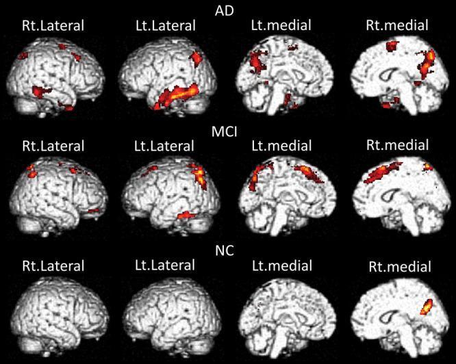
This diagnostic comparison chart utilizes Voxel-Based Morphometry (VBM) on brain MRI renderings to illustrate the correlation between BF-227 PET uptake (amyloid-beta deposition) and gray matter atrophy across three clinical groups: Alzheimer's Disease (AD), Mild Cognitive Impairment (MCI), and Normal Controls (NC). The visualization presents four views per group: Right Lateral, Left Lateral, Left Medial, and Right Medial. Significant areas of gray matter volume decrease are highlighted with a heat map (red to yellow). In the AD group, extensive atrophy is visible globally, particularly in the lateral temporal gyrus, precuneus, and cingulate cortex. The MCI group shows a similar but less extensive distribution of atrophy, primarily localized to the medial temporal and superior medial surfaces. In the NC group, atrophy is minimal and highly localized, primarily restricted to the posterior medial regions. This progression illustrates the neuroanatomical evolution of Alzheimer’s disease, highlighting the relationship between amyloid deposition and structural brain volume loss in key regions like the precuneus and temporal lobes.
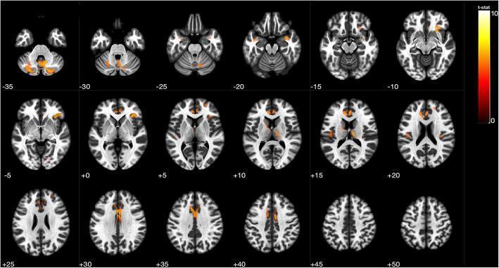
This diagnostic image is a series of 18 axial MRI brain slices displaying Voxel-Based Morphometry (VBM) results. The slices are arranged sequentially from inferior to superior, with z-axis coordinates labeled from -35 to +50. The background provides anatomical detail in grayscale, while a heat-map overlay (red-to-yellow) highlights areas of statistically significant gray matter atrophy in patients with Cerebral Small Vessel Disease (CSVD) compared to healthy controls. A color-coded t-statistic scale (ranging from 0 to 10) indicates the degree of volume loss, with yellow representing the highest significance (p < 0.0001). Key anatomical regions exhibiting atrophy include the bilateral cerebellum (visible at z = -35 to -25), the right thalamus and inferior frontal gyrus (z = +5 to +15), and the anterior and medial cingulate cortices (z = +25 to +40). This visual serves as a neuroimaging demonstration of structural brain changes associated with CSVD and potential cognitive or autonomic dysfunction.
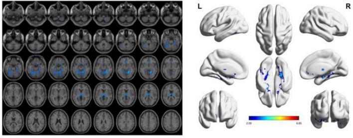
This diagnostic image displays Voxel-Based Morphometry (VBM) results derived from functional and structural MRI, illustrating gray matter atrophy in patients with Vascular Dementia (VD) compared to those with Vascular Cognitive Impairment No Dementia (VCIND). The left panel consists of a montage of axial brain slices displaying statistically significant areas of atrophy (p < 0.05) highlighted in blue. These blue-shaded regions are primarily concentrated in subcortical structures, including the thalamus and basal ganglia, as well as portions of the cerebellum. The right panel shows three-dimensional cortical surface renderings from lateral, medial, dorsal, and ventral perspectives. These renderings further localize atrophy to the bilateral fusiform gyri, lingual gyri, and inferior temporal regions, with a color scale bar indicating statistical intensity ranging from 2.09 to 6.85. The visualization demonstrates the pattern of progressive neurodegeneration characteristic of vascular cognitive decline, highlighting both deep gray matter and cortical involvement.
Cannabis Use and Brain Atrophy
Overview
The Endocannabinoid System: Why the Brain is Vulnerable
- Prefrontal cortex - executive function, decision-making
- Hippocampus - memory formation and consolidation
- Amygdala - emotional processing and stress regulation
- Basal ganglia / nucleus accumbens - reward and motivation
- Cerebellum - motor coordination
Structural Brain Changes: What the Evidence Shows
1. Cortical Thinning
- 80% (8/10) reported decreased cortical thickness in cannabis users vs. non-using controls
- Thinning was most consistently found in:
- Frontal lobes (most replicated finding)
- Hippocampal cortex
- Cingulate cortex
2. Gray Matter Volume Reduction
- Hippocampus - one study found diminished hippocampal gray matter in cannabis users, with possible protective effects from CBD
- Prefrontal and orbitofrontal cortex
- Parahippocampal and fusiform gyri
- Amygdala (findings less consistent)
3. White Matter Integrity
4. Functional Connectivity
- 63% of heavy lifetime cannabis users showed reduced brain activity during working memory tasks
- 68% of recent users showed a similar impairment
The Critical Causal Question
- Lifetime cannabis use was associated with structural brain differences and neurocognitive changes
- However, Mendelian randomization analysis (genetic data) found NO significant causal associations between genetically predicted cannabis use/dependence and neurocognitive decline or brain structure changes
- Pre-existing brain differences predispose individuals to cannabis use (reverse causation)
- Confounding factors (socioeconomic status, comorbid substance use, psychiatric conditions) drive the observed differences
- The brain changes observed may reflect a complex interaction rather than direct toxicity
Adolescent Brain: The Most Vulnerable Window
"Children and adolescents are also more vulnerable to the harmful effects of cannabis... Brain imaging studies have revealed that use of cannabis at this stage is associated with structural and functional brain changes... often in the form of reduced brain connectivity and cortical thickness."
- ECS signaling is critical for neuronal migration, synaptogenesis, and connectivity during development
- Perturbation during fetal or adolescent development can produce lasting structural changes
- ABCD study data suggest pre-adolescent cannabis exposure correlates with lower episodic, verbal, inhibitory, and working memory task performance
Prenatal Exposure
- Increased intracranial volumes with blunted visuospatial processing development
- White matter integrity changes in the fornix
- Cognitive and memory impairments in offspring
- The American College of Obstetricians and Gynecologists recommends against cannabis use in pregnancy or when planning pregnancy
Cognitive Consequences of Structural Changes
- Working memory (prefrontal-hippocampal circuit)
- Verbal memory and learning (hippocampus)
- Executive function (prefrontal cortex)
- Episodic memory (hippocampal-fornix circuit)
- Processing speed (white matter tracts)
Summary Table
| Brain Region | Structural Change | Functional Impact |
|---|---|---|
| Prefrontal cortex | Cortical thinning (dose-dependent) | Executive function, impulse control |
| Hippocampus | Volume reduction, cortical thinning | Memory formation and retrieval |
| Cingulate cortex | Cortical thinning | Attention, conflict monitoring |
| White matter (fornix) | Reduced integrity | Memory, emotional processing |
| Amygdala | Inconsistent volume changes | Emotional regulation |
Bottom Line
- Early-onset users (adolescents), where dose-dependent prefrontal thinning is well-documented
- Heavy/frequent users, with cumulative effects on hippocampal and cortical volumes
- Prenatal exposure, with lasting effects on offspring brain development