I am making content for you to post on Instagram so everybody will know what you can do Now I need the best diagrams you can prepare which are easier to understand One of them has to be of brachial plexus
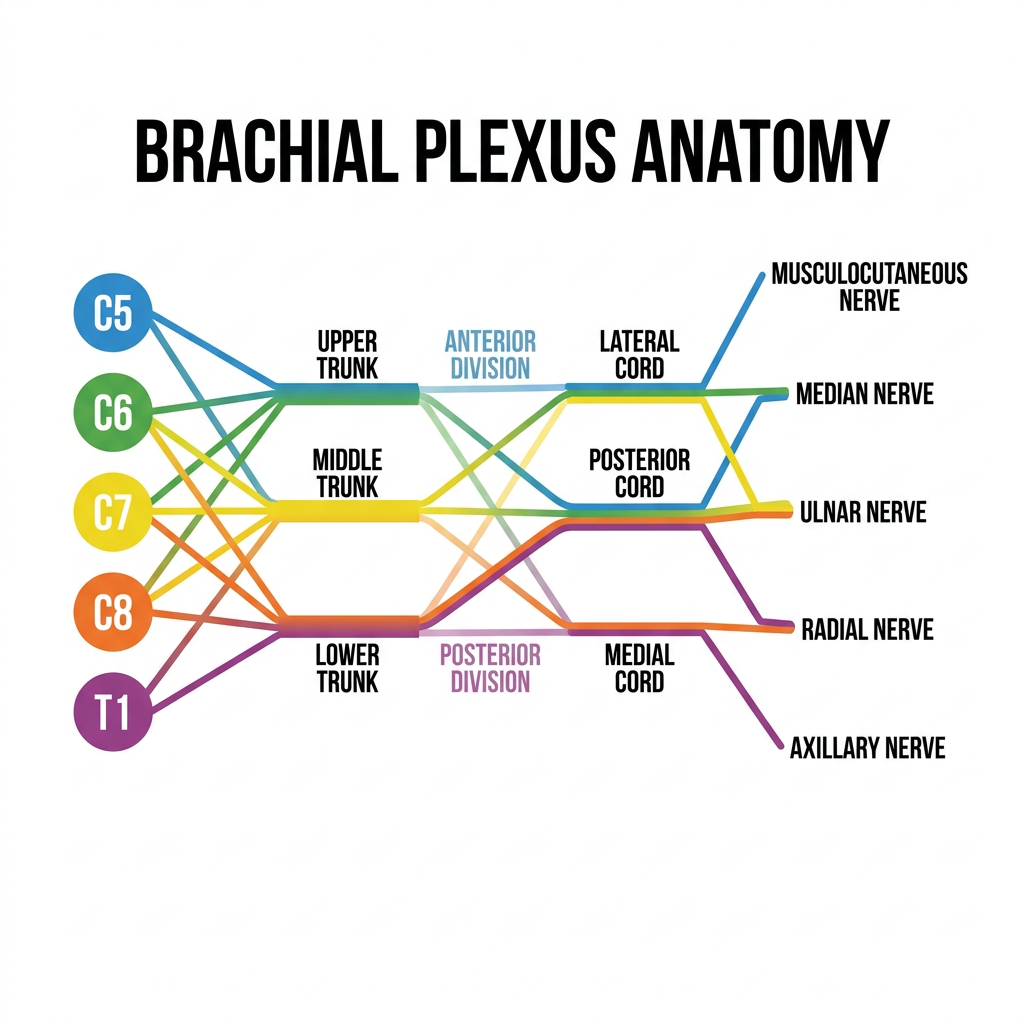
Brachial plexus anatomy diagram for Instagram, clean minimal style, white background. Shows roots C5-T1 emerging from spine, clearly labeled trunks (superior, middle, inferior), divisions (anterior/posterior), cords (lateral, medial, posterior), and terminal branches (musculocutaneous, median, ulnar, radial, axillary nerves). Color-coded by level: roots in blue, trunks in green, divisions in orange, cords in purple, branches in red. Bold clean labels, medical education infographic style, easy to read, square format 1:1 ratio.
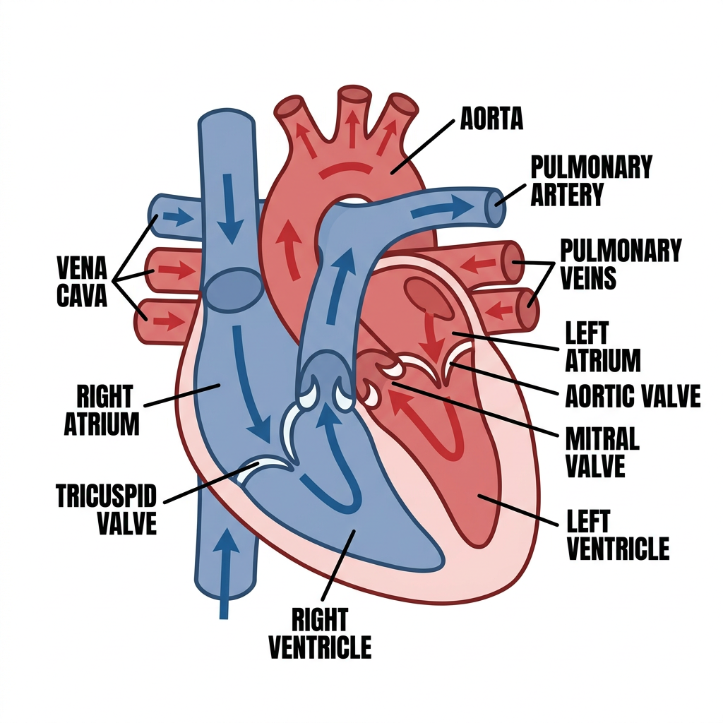
Heart anatomy diagram for Instagram, clean minimal style, white background. Cross-section of the heart showing all 4 chambers (right atrium, left atrium, right ventricle, left ventricle), valves (tricuspid, mitral, aortic, pulmonary), major vessels (aorta, pulmonary artery, pulmonary veins, vena cava), and blood flow direction arrows (blue for deoxygenated, red for oxygenated). Bold clean labels, color-coded chambers, medical education infographic style, square format 1:1 ratio.
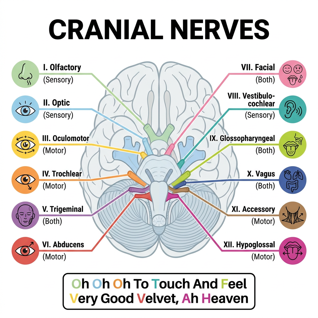
Cranial nerves diagram for Instagram, clean minimal style, white background. Shows the base of the brain with all 12 cranial nerves labeled by number (I-XII) and name (Olfactory, Optic, Oculomotor, Trochlear, Trigeminal, Abducens, Facial, Vestibulocochlear, Glossopharyngeal, Vagus, Accessory, Hypoglossal), each color-coded and with a small icon showing its function (sensory, motor, or both). Memory mnemonic included at bottom. Bold clean labels, medical education infographic, square 1:1 ratio.
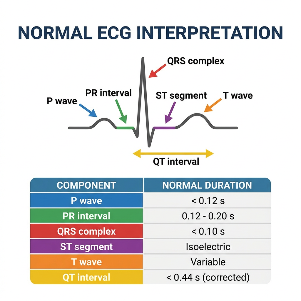
ECG interpretation diagram for Instagram, clean minimal style, white background. Shows a single clear normal ECG complex with every wave labeled: P wave, PR interval, QRS complex, ST segment, T wave, QT interval. Below the wave, a reference table shows normal values for each component. Color-coded annotations, easy-to-read font, arrows pointing to each part, medical education infographic style, square 1:1 ratio.
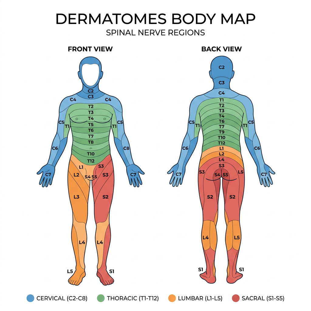
Dermatomes body map diagram for Instagram, clean minimal style, white background. Front and back view of human body outline showing all spinal nerve dermatome regions color-coded by spinal level: cervical (C2-C8) in blue shades, thoracic (T1-T12) in green shades, lumbar (L1-L5) in orange shades, sacral (S1-S5) in red shades. Each region clearly labeled with spinal level. Clean bold labels, medical education infographic, square 1:1 ratio.
Brachial plexus anatomy educational diagram, clean white background, minimal style. Shows nerve roots C5 C6 C7 C8 T1 on the left side, connecting to three trunks (upper middle lower), then anterior and posterior divisions, then three cords (lateral posterior medial), then five terminal nerve branches (musculocutaneous nerve, median nerve, ulnar nerve, radial nerve, axillary nerve) on the right. Each level color coded differently. All parts clearly labeled with bold text. Medical school teaching diagram, infographic style, square format.
Make more diagrams , and then combine all of them in one pdf
Respiratory system anatomy diagram, clean minimal style, white background. Shows full respiratory tract from nose/mouth down to alveoli. Labels: nasal cavity, pharynx, larynx, trachea, bronchi, bronchioles, alveoli. Inset close-up of alveolar gas exchange showing O2 entering blood and CO2 leaving. Color coded: airways in blue, lung tissue in pink, blood vessels in red. Bold clean labels, medical education infographic, square 1:1 format.
Cardiac cycle diagram, clean minimal style, white background. Shows phases: systole and diastole with pressure-volume loop. Includes a timeline bar showing: atrial systole, isovolumetric contraction, ventricular ejection, isovolumetric relaxation, ventricular filling. Pressure curves for aorta, left ventricle, left atrium overlaid. Valve opening and closing events marked. Bold labels, color coded curves (blue for atrium, red for ventricle, green for aorta), medical education infographic, square 1:1 format.
Drug mechanism of action overview diagram, clean minimal style, white background. Shows a cell membrane with 4 types of drug targets illustrated side by side: 1) Ion channel (ligand-gated), 2) G-protein coupled receptor with cAMP pathway, 3) Enzyme inhibition, 4) Nuclear receptor with gene transcription. Each type labeled and color coded. Examples of drug classes listed under each. Bold clean labels, medical pharmacology infographic, educational style, square 1:1 format.
Coagulation cascade diagram, clean minimal style, white background. Shows intrinsic pathway (factors XII, XI, IX, VIII) on left, extrinsic pathway (factor VII, tissue factor) on right, converging at factor X to common pathway leading to fibrin clot. Each factor labeled with Roman numerals. Arrows show activation sequence. Color coded: intrinsic in blue, extrinsic in orange, common pathway in red. Warfarin and heparin inhibition sites labeled. Bold clean labels, medical education infographic, square 1:1 format.
Nephron anatomy and function diagram, clean minimal style, white background. Shows a single nephron with all segments labeled: glomerulus, Bowman capsule, proximal convoluted tubule, loop of Henle (descending and ascending), distal convoluted tubule, collecting duct. Arrows show filtration, reabsorption, secretion at each segment. Key substances (Na, K, H2O, glucose, HCO3) shown at relevant segments. Color coded by function. Bold labels, medical education infographic style, square 1:1 format.
429 Too Many Requests. {'message': '{\n "error": {\n "code": 429,\n "message": "Resource exhausted. Please try again later. Please refer to https://cloud.google.com/vertex-ai/generative-ai/docs/error-code-429 for more details.",\n "status": "RESOURCE_EXHAUSTED"\n }\n}\n', 'status': 'Too Many Requests'}
We encountered an error while processing your request. Please try again or reach out to us for help.
Give more images
An error occurred (ValidationException) when calling the ConverseStream operation: Expected toolResult blocks at messages.12.content for the following Ids: tooluse_O6wQUJ9k9ZBjagwLGTB7lg, tooluse_FtOsUyTv0Z8Zqqj4FyPLUV, tooluse_smDJtv4dftpvi6C3gsi5uD
We encountered an error while processing your request. Please try again or reach out to us for help.