Here is your complete, well-organized topic note on Monitoring of the Critically Ill Patient - written simply with a built-in dictionary for hard medical words.
🏥 Monitoring of the Critically Ill Patient
Complete Topic Note with Hard Words Dictionary
What is "Monitoring" in the ICU?
Monitoring means watching the patient's body functions closely and continuously to detect problems early and guide treatment. A critically ill patient cannot maintain their own body stability, so every major organ system must be tracked around the clock.
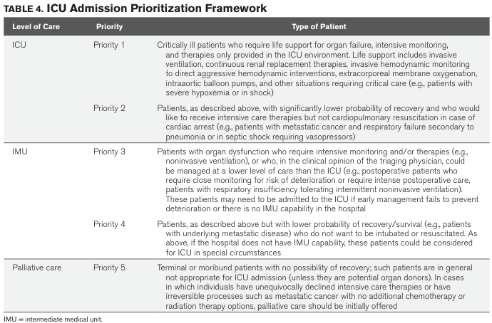
ICU Admission Priority Table
| Priority | Type of Patient |
|---|
| Priority 1 | Needs life support - mechanical ventilation, hemodynamic monitoring, vasopressors |
| Priority 2 | Same as above but may not want CPR |
| Priority 3 | Organ dysfunction needing close monitoring (e.g. noninvasive ventilation) |
| Priority 4 | Same but lower survival chance, declined intubation |
| Priority 5 | Terminal/palliative - not appropriate for ICU |
1. RESPIRATORY MONITORING
A. Physical Examination (Looking with Eyes)
- Respiratory rate: Normal = 12-20 breaths/minute in adults
- Tachypnea (>35 breaths/min) = warning sign of respiratory failure → may need ventilator
- Bradypnea (slow breathing) = too many narcotics or depressed brain drive
- Accessory muscle use (scalene, trapezius, sternocleidomastoid) = patient is struggling to breathe
- Tripod sign: Patient sits leaning forward using neck muscles - means diaphragm is not working well
- Hoover sign: Lower ribs pull inward during breathing - seen in emphysema
- Cyanosis: Blue color of lips/nails = low oxygen. Appears when 5 g/dL of hemoglobin is desaturated
📖 Hard Words Dictionary - Section 1A:
| Word | Simple Meaning |
|---|
| Tachypnea | Fast breathing (>20/min) |
| Bradypnea | Slow breathing |
| Scalene muscles | Neck muscles that help breathe when you're struggling |
| Sternocleidomastoid | Big neck muscle used as backup when diaphragm is weak |
| Diaphragm | Main breathing muscle below the lungs |
| Tripod sign | Leaning forward on arms to breathe easier |
| Hoover sign | Lower ribcage sucks inward instead of expanding |
| Cyanosis | Blue color due to low oxygen |
| Desaturated hemoglobin | Hemoglobin not carrying oxygen |
| Polycythemia | Too many red blood cells |
B. Pulse Oximetry (SpO₂)
- A probe on the finger shines light through the skin and measures oxygen saturation
- Normal SpO₂ = 95-100%
- Non-invasive (no needle needed)
- Limitation: Does NOT measure CO₂ (carbon dioxide)
- A normal SpO₂ is not reassuring if the patient is breathing very fast - they may be working hard just to keep it normal
C. Arterial Blood Gas (ABG)
- Blood taken from an artery (usually radial artery at wrist)
- Measures:
- PaO₂ = oxygen pressure in blood (normal ~80-100 mmHg)
- PaCO₂ = carbon dioxide in blood = reflects ventilation (normal 35-45 mmHg)
- pH = acid-base balance (normal 7.35-7.45)
- Alveolar-arterial (A-a) gradient: difference between expected oxygen in lungs and what is actually in blood. A high A-a gradient means lungs are not transferring oxygen properly.
Key formula:
PaCO₂ = (K × CO₂ production) / Alveolar ventilation
If CO₂ rises → ventilation is falling (patient not breathing enough)
D. Capnometry (ETCO₂ Monitoring)
- Measures CO₂ in exhaled breath using infrared light
- End-tidal CO₂ (ETCO₂) = CO₂ at end of each breath
- Always slightly lower than PaCO₂ (because of dead space - some air doesn't reach gas exchange areas)
- Uses:
- Detect esophageal intubation (tube in wrong place → no CO₂ waveform)
- Detect ventilator disconnection
- Monitor CPR effectiveness (ETCO₂ rises when compressions are effective)
- Detect pulmonary embolism (rising dead space)
📖 Hard Words Dictionary - Section 1D:
| Word | Simple Meaning |
|---|
| Capnometry | Measuring CO₂ in exhaled breath |
| ETCO₂ | CO₂ at the very end of breathing out |
| Dead space | Parts of lung that are ventilated but have no blood flow - wasted ventilation |
| Capnogram | Graph showing CO₂ vs time during breathing |
| Esophageal intubation | Tube accidentally placed in food pipe, not windpipe |
| Pulmonary embolism | Blood clot in lung blood vessels |
| Alveolar plateau | Flat part of capnogram showing pure lung gas |
2. HEMODYNAMIC MONITORING
Hemodynamic = related to blood flow and pressure
A. Blood Pressure (BP) Monitoring
Non-Invasive:
- Sphygmomanometry (regular BP cuff) - intermittent readings
- Oscillometry - electronic cuff
- Photoplethysmography - continuous beat-to-beat BP using infrared light on a finger; less accurate in hypotension or cold patients
Invasive (Intra-Arterial Catheter):
- A small tube placed inside an artery (usually radial at wrist, or femoral artery)
- Connected to an external strain-gauge transducer → gives continuous waveform
- Shows systolic, diastolic, and mean arterial pressure (MAP) at all times
- MAP is the most reliable number - accurate even in underdamped or overdamped systems
Key targets:
| Situation | MAP Target |
|---|
| Septic shock | ≥65 mmHg |
| Chronic hypertension with sepsis | 75-85 mmHg (to protect kidneys) |
| Hemorrhagic shock (trauma) | Systolic 80-90 mmHg (permissive hypotension) |
| Traumatic brain injury | Systolic >110 mmHg |
| TBI + hemorrhagic shock | MAP >80 mmHg |
Complications of arterial line:
- Thrombosis (clot) - minimize with 20-gauge or smaller catheter
- Distal ischemia (tissue death beyond clot)
- Retrograde air embolism to brain - never flush with air in line
📖 Hard Words Dictionary - Section 2A:
| Word | Simple Meaning |
|---|
| Hemodynamic | Related to blood flow and pressure |
| Sphygmomanometry | BP measurement with a cuff and stethoscope |
| Oscillometry | Electronic method of measuring BP |
| Photoplethysmography | Uses infrared light to measure blood volume and pressure continuously |
| Intra-arterial catheter | Small tube placed inside an artery for continuous BP |
| Strain-gauge transducer | Device that converts pressure into an electrical signal |
| MAP (Mean Arterial Pressure) | Average pressure in arteries; normal ~70-100 mmHg |
| Underdamped system | Recording system with too much bounce → overestimates systolic BP |
| Overdamped system | Recording system that is too sluggish → underestimates systolic BP |
| Seldinger technique | Inserting a catheter over a guidewire |
| Permissive hypotension | Allowing a lower BP in trauma to reduce bleeding and complications |
| Retrograde embolism | Air/clot traveling backwards into brain vessels |
| Ischemia | Reduced blood supply causing tissue damage |
| Thrombosis | Clot formation inside a blood vessel or catheter |
B. Central Venous Pressure (CVP) Monitoring
- CVP = back pressure in the large veins entering the heart (right side)
- Measured via central venous catheter (CVC) placed in jugular, subclavian, or femoral vein
- Normal CVP = 2-8 mmHg
- Low CVP = patient is dehydrated (hypovolemia) or too much vasodilation
- High CVP = fluid overload, right heart failure, cardiac tamponade
Contributors to CVP:
- Central venous blood volume
- Venous tone
- Right ventricular compliance
- Tricuspid valve disease
- Intrathoracic pressure (rises with mechanical ventilation)
- Dysrhythmias (atrial fibrillation, junctional rhythm)
Limitation: A single CVP number has limited value - use trends and clinical context together.
📖 Hard Words Dictionary - Section 2B:
| Word | Simple Meaning |
|---|
| CVP | Pressure in the large veins near the heart |
| Central venous catheter | Tube placed in a major vein |
| Hypovolemia | Too little blood/fluid in the body |
| Cardiac tamponade | Fluid around the heart compressing it |
| Venous compliance | How easily veins expand |
| Dysrhythmia | Abnormal heart rhythm |
| Atrial fibrillation | Irregular fast beating of the heart's upper chambers |
C. Cardiac Output Monitoring
Cardiac Output (CO) = how much blood the heart pumps per minute (Normal: ~5 L/min)
Methods:
| Method | How it works | Invasive? |
|---|
| Pulmonary Artery Catheter (PAC) - Swan-Ganz | Thermodilution: cold saline injected, temperature drop measured | Yes (highly) |
| Transpulmonary thermodilution (TPTD) | Cold saline via central line, measured at femoral artery | Moderately |
| Pulse contour analysis | Analyses the shape of arterial waveform to estimate CO | Minimally |
| Transthoracic echocardiography (TTE/TEE) | Ultrasound of heart to estimate flow | Non/minimally invasive |
Mixed Venous Oxygen Saturation (SvO₂):
- Measured from pulmonary artery blood
- Normal SvO₂ = 65-75%
- Low SvO₂ means tissues are extracting more oxygen (cardiac output is falling, or oxygen delivery is failing)
- Formula:
SvO₂ = SaO₂ - VO₂ / (Cardiac Output × Hemoglobin × 1.36)
Low SvO₂ causes: low cardiac output, low hemoglobin (anemia), low SaO₂ (lung failure), high metabolic demand (fever, seizures)
Central Venous O₂ Saturation (ScvO₂):
- Easier to measure (from central line, not pulmonary artery)
- Approximates SvO₂
- Target in sepsis resuscitation: >70%
📖 Hard Words Dictionary - Section 2C:
| Word | Simple Meaning |
|---|
| Cardiac output (CO) | Amount of blood heart pumps per minute |
| Thermodilution | Cold fluid injected → computer calculates blood flow from temperature change |
| Swan-Ganz / PAC | Catheter floated into the lung artery to measure pressures and CO |
| Pulse contour analysis | Using the shape of the BP wave to estimate CO |
| Transthoracic echocardiography (TTE) | Heart ultrasound from outside the chest |
| Transesophageal echocardiography (TEE) | Heart ultrasound probe swallowed into food pipe (closer to heart) |
| SvO₂ | Oxygen saturation in mixed venous (pulmonary artery) blood |
| ScvO₂ | Oxygen saturation in central venous blood (easier to get) |
| Fick equation | Formula: CO = VO₂ / (CaO₂ - CvO₂) |
| VO₂ | Oxygen consumed by the body per minute |
3. OXYGENATION & OXYGEN DELIVERY MONITORING
Arterial Oxygen Content (CaO₂):
CaO₂ = (1.36 × Hemoglobin × SaO₂/100) + (0.0031 × PaO₂)
- Most of the oxygen in blood is carried by hemoglobin, not dissolved
Systemic Oxygen Delivery (DO₂):
DO₂ = CO × CaO₂ × 10
- If delivery falls → tissues become hypoxic → lactic acidosis develops
Lactate Monitoring:
- Blood lactate = byproduct of anaerobic (without oxygen) metabolism
- Normal < 2 mmol/L
- Lactate > 4 mmol/L = severe shock, high mortality
- Trending lactate clearance (lactate going down) = sign of improvement
📖 Hard Words Dictionary - Section 3:
| Word | Simple Meaning |
|---|
| CaO₂ | Oxygen content in arterial blood |
| DO₂ | Total oxygen delivered to tissues per minute |
| Hemoglobin | Red blood cell protein that carries oxygen |
| Anaerobic metabolism | Energy production without oxygen - produces lactic acid |
| Lactic acidosis | Blood becomes acidic from lactic acid buildup |
| Lactate clearance | How fast the body removes lactic acid (sign of recovery) |
4. NEUROLOGICAL MONITORING
| Parameter | Method | Normal |
|---|
| GCS (Glasgow Coma Scale) | Eyes + Verbal + Motor response | 15 = fully awake |
| Pupils | Size, symmetry, light reflex | Equal, reactive |
| ICP (Intracranial Pressure) | Intraventricular catheter or bolt | < 20 mmHg |
| Cerebral Perfusion Pressure (CPP) | MAP - ICP | 60-70 mmHg |
| EEG | Electrical activity of brain | Continuous in ICU for seizures |
EVD (External Ventricular Drain): Tube placed into brain ventricles to drain CSF and monitor ICP - placed when intracranial pressure is raised (e.g. after subarachnoid hemorrhage, head injury)
📖 Hard Words Dictionary - Section 4:
| Word | Simple Meaning |
|---|
| GCS | Score (3-15) measuring consciousness |
| ICP | Pressure inside the skull |
| CPP | Pressure actually driving blood into the brain |
| EVD | Tube into brain to drain fluid and measure ICP |
| CSF | Clear fluid surrounding the brain and spinal cord |
| EEG | Machine recording brain electrical activity |
| Subarachnoid hemorrhage | Bleeding in the space around the brain |
5. RENAL MONITORING
| Parameter | How | Target |
|---|
| Urine output | Urinary catheter (Foley) | >0.5 mL/kg/hour |
| Serum creatinine | Blood test | Rising = kidney injury |
| BUN (Blood Urea Nitrogen) | Blood test | Elevated in poor kidney function |
| Fluid balance | Inputs minus outputs | Avoid fluid overload |
Acute Kidney Injury (AKI) in the ICU is very common and worsens survival.
6. TEMPERATURE MONITORING
- Core temperature monitored (rectal, esophageal, bladder probe)
- Hypothermia (<36°C) in ICU = worsens coagulopathy, cardiac irritability
- Hyperthermia/Fever = sign of infection OR post-brain injury
- Target core temperature after ICU admission (e.g. open abdomen): 37°C within 4 hours
7. LABORATORY & METABOLIC MONITORING (Daily/Frequent)
| Test | What it tells you |
|---|
| ABG | Oxygenation, ventilation, acid-base |
| CBC | Hemoglobin, infection (WBC), platelets |
| Electrolytes (Na, K, Cl, Mg) | Salt balance, arrhythmia risk |
| Coagulation (PT, INR, APTT, TEG) | Bleeding/clotting tendency |
| Lactate | Tissue oxygenation/shock marker |
| Blood glucose | Hyperglycemia worsens ICU outcomes |
| Procalcitonin / CRP | Infection/sepsis markers |
| Liver enzymes | Liver failure |
8. QUICK SUMMARY TABLE
| System | Key Monitor | Critical Value / Action |
|---|
| Respiratory | SpO₂, ABG, ETCO₂ | SpO₂ <90%, PaCO₂ rising → ventilator support |
| Cardiovascular | MAP, CVP, CO, lactate | MAP <65 → fluids/vasopressors |
| Renal | Urine output, creatinine | UO <0.5 mL/kg/hr → fluid challenge or dialysis |
| Neurological | GCS, ICP, pupils | ICP >20 → osmotherapy, EVD |
| Temperature | Core temp | <36°C → warming; fever → antibiotics/cooling |
| Metabolic | Glucose, electrolytes | Glucose target 140-180 mg/dL in ICU |
MASTER HARD WORDS DICTIONARY (All Terms)
| Word | Simple Meaning |
|---|
| Critically ill | Patient whose life is at immediate risk |
| ICU | Intensive Care Unit - highest level of hospital monitoring |
| Hemodynamic | Relating to blood flow and pressure |
| MAP | Mean Arterial Pressure - average pressure in arteries |
| SpO₂ | Pulse oximeter oxygen saturation reading |
| SaO₂ | Actual arterial oxygen saturation from ABG |
| PaO₂ | Partial pressure of oxygen dissolved in arterial blood |
| PaCO₂ | Partial pressure of CO₂ in arterial blood = breathing efficiency |
| ETCO₂ | CO₂ at end of each exhaled breath |
| ABG | Arterial Blood Gas - full blood chemistry from artery |
| CVP | Central Venous Pressure - pressure in veins entering heart |
| CO | Cardiac Output - blood pumped per minute |
| SvO₂ | Mixed venous oxygen saturation |
| ScvO₂ | Central venous oxygen saturation (easier to obtain) |
| ICP | Intracranial Pressure - pressure inside skull |
| CPP | Cerebral Perfusion Pressure = MAP - ICP |
| GCS | Glasgow Coma Scale (3-15) - measures consciousness |
| EVD | External Ventricular Drain - brain pressure monitoring tube |
| Tachypnea | Breathing too fast |
| Bradypnea | Breathing too slow |
| Cyanosis | Bluish skin from low oxygen |
| Pulse oximetry | Fingertip device measuring blood oxygen |
| Capnometry | Measuring CO₂ in exhaled air |
| Dead space | Lung area ventilated but not perfused |
| Thermodilution | Method to measure cardiac output using cold fluid |
| Lactate | Acid produced when tissues run out of oxygen |
| Anaerobic | Without oxygen |
| Vasopressors | Drugs that raise blood pressure (e.g. norepinephrine) |
| Transducer | Device converting pressure into electrical signal |
| Thrombosis | Blood clot inside catheter or vessel |
| Ischemia | Reduced blood flow causing tissue damage |
| Permissive hypotension | Deliberately allowing lower BP in trauma to reduce bleeding |
| Seldinger technique | Placing a catheter using a guidewire |
| Echocardiography (TTE/TEE) | Ultrasound imaging of the heart |
| Coagulopathy | Failure of blood to clot properly |
| TEG (Thromboelastography) | Test measuring the whole clotting process |
| Procalcitonin | Blood protein that rises in bacterial infection |
| Diuretics | Drugs that make the kidneys produce more urine |
| Colloid | IV fluids with large molecules (e.g. albumin) |
| Crystalloid | Simple IV saline or saline-based fluids |
| Septic shock | Life-threatening response to infection causing low BP |
| ARDS | Acute Respiratory Distress Syndrome - severe lung failure |
| Pulmonary embolism | Blood clot in lung blood vessels |
| Subarachnoid hemorrhage | Bleeding around the brain |
| CPR | Cardiopulmonary Resuscitation - chest compressions + breathing |
Sources: Goldman-Cecil Medicine; Schwartz's Principles of Surgery; Tintinalli's Emergency Medicine; Fishman's Pulmonary Diseases; Sabiston Textbook of Surgery