
What is urachus
urachus anatomy diagram median umbilical ligament bladder
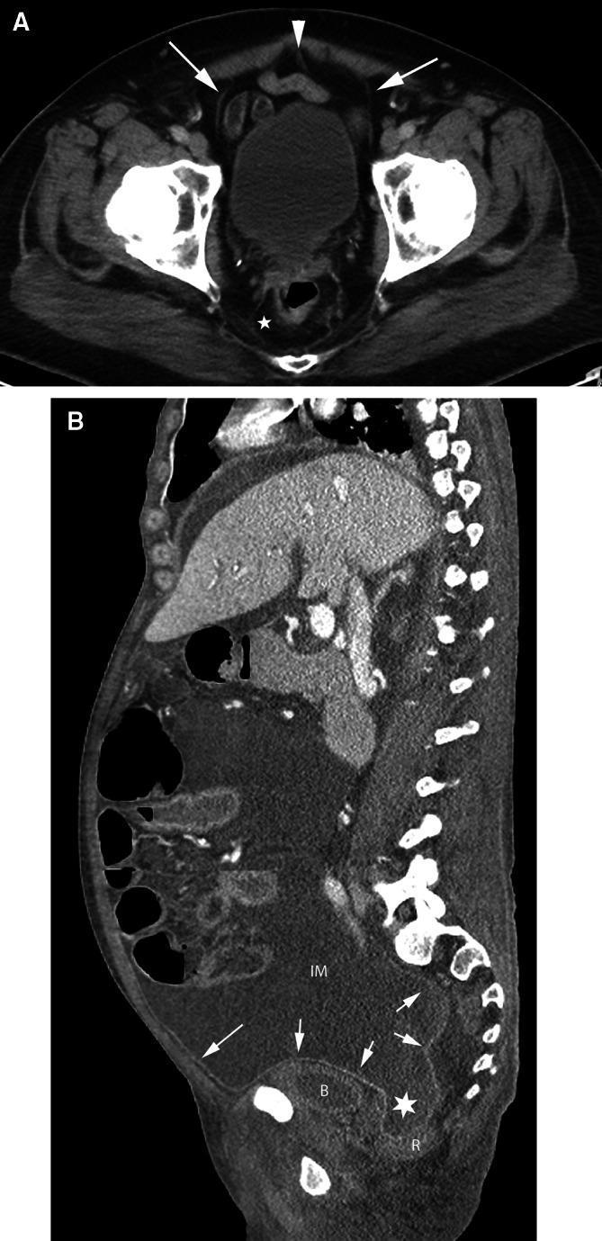
This educational clinical imaging figure consists of two CT scans (axial and sagittal) demonstrating the anatomy of pelvic spaces and peritoneal reflections. Image A is an axial CT scan of the pelvis at the level of the urinary bladder. An arrowhead identifies the urachus (median umbilical ligament), and arrows point to the obliterated umbilical arteries (medial umbilical folds). These structures are encased within the umbilicovesical fascia, defining the perivesical space. The prevesical space is visible anterior and lateral to the bladder, while a star indicates the perirectal space. Image B is a sagittal CT scan showing the course of the peritoneum (arrows) along the anterior abdominal wall as it reflects over the bladder (B) and rectum (R). The image illustrates a large volume of peritoneal fluid in the inframesocolic compartment (IM) communicating with the rectovesical recess (star). The bladder is seen displaced inferiorly by the fluid. This material is designed to teach the distinction between intraperitoneal and subperitoneal spaces, crucial for understanding pathways of disease spread in the pelvis.
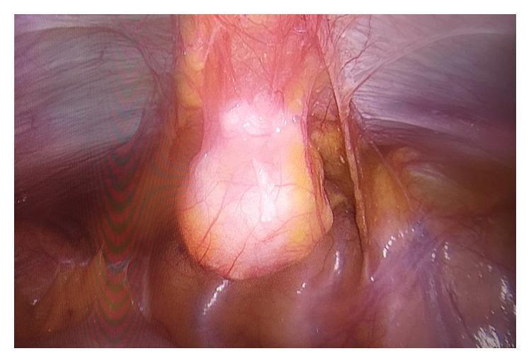
This clinical photograph is an intraoperative laparoscopic view of the pelvic cavity, specifically demonstrating a urachal cyst. Centrally, a cystic mass is visible within the median umbilical ligament. The mass is ovoid, translucent pink to yellow in color, and exhibits a smooth, glistening surface with prominent branching superficial vasculature. Anatomically, the lesion is situated superior to the dome of the bladder, approximately 2 cm from the bladder wall. Surrounding peritoneal folds and retroperitoneal fatty tissue are visible, providing orientation within the preperitoneal space. This image serves as an educational example of urachal pathology, illustrating the typical appearance and anatomical relationship of a mucinous cystic tumor of the urachus during surgical excision. The visual features—specifically the translucent quality and location along the midline embryonic remnant—are characteristic of urachal cysts or neoplasms of low malignant potential.
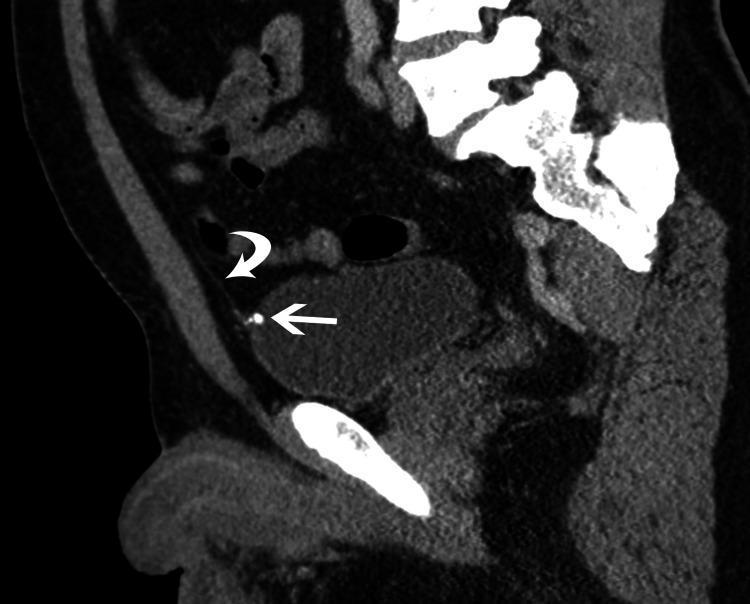
This diagnostic image is a sagittal reformatted CT scan of the lower abdomen and pelvis. It highlights a midline cord-like structure (curved arrow) identified as the median umbilical ligament, which represents a urachal remnant. This ligament is seen extending superiorly from the apex of the urinary bladder toward the umbilicus. At the point of insertion onto the anterior inner wall of the urinary bladder, a distinct hyperdense, punctate focus (straight arrow) is visible, measuring approximately 7mm, which represents a calcification of the urachal remnant. The anterior bladder wall is smooth and demonstrates no evidence of focal outpouching or diverticulum formation at the site of ligamentous attachment. This image is clinically significant for diagnosing incidental urachal calcifications and serves to differentiate the condition from urachal diverticula or other umbilical abnormalities. The content is suitable for medical students and radiology residents studying embryological remnants and pelvic anatomy.
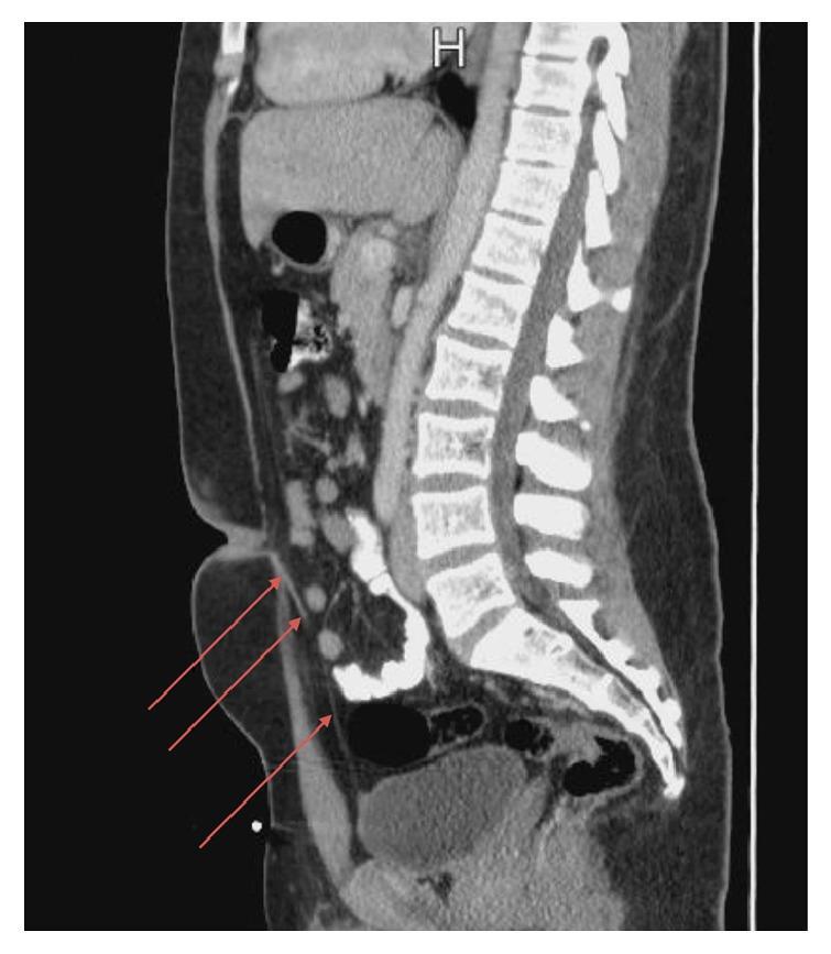
A sagittal view computed tomography (CT) scan of the abdomen and pelvis illustrating the normal anatomy and a specific embryological remnant. The posterior aspect displays the segmented lumbar and sacral vertebral column. Anteriorly, the abdominal cavity contains loops of bowel and the bladder in the pelvic region. Red arrows highlight a thin, linear fibrous band extending from the superior-anterior margin of the urinary bladder toward the umbilicus. This structure is identified as a urachal remnant, representing an obliterated urachus (median umbilical ligament). The surrounding subcutaneous fat and anterior abdominal wall muscles appear normal, with no evidence of associated fluid collections, inflammation, or cystic changes. This diagnostic image is used to teach medical students and radiologists how to identify urachal abnormalities and differentiate them from other abdominal pathologies.
The Urachus
Embryology and Development
- Smith and Tanagho's General Urology, 19th Edition - "By the 20th week, the bladder is well separated from the umbilicus, and the stretched urachus becomes the middle umbilical ligament."
- The Developing Human: Clinically Oriented Embryology - "The apex of the bladder in adults is continuous with the median umbilical ligament, which extends posteriorly along the posterior surface of the anterior abdominal wall."
Anatomy
- Runs in the space of Retzius (retropubic / prevesical space), between the peritoneum and the anterior abdominal wall
- Lies in the midline, flanked on either side by the obliterated umbilical arteries (medial umbilical folds)
- These three structures are enclosed within the umbilicovesical fascia, defining the perivesical space
Urachal Anomalies (Congenital)
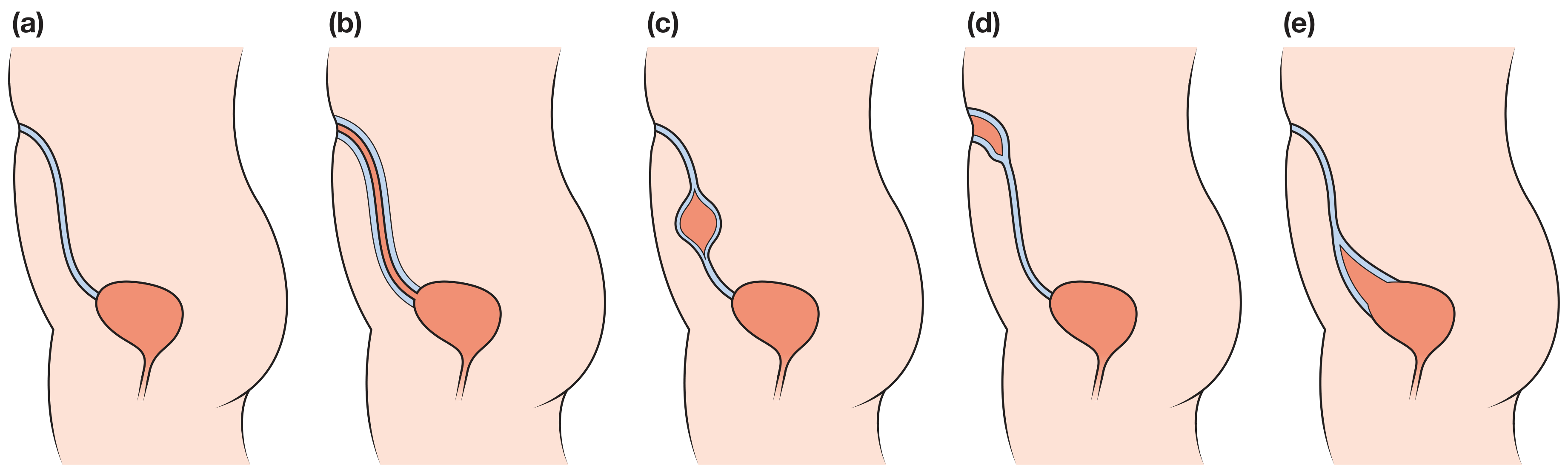
| Anomaly | Description |
|---|---|
| Patent urachus | Entire urachus remains open - direct communication between bladder and umbilicus; urine leaks from the umbilicus |
| Urachal cyst | Middle portion fails to obliterate but both ends close - no communication with bladder or umbilicus; can become infected |
| Urachal sinus | Fails to obliterate near the umbilicus - blind-ending tract opening at the umbilicus |
| Urachal diverticulum | Fails to obliterate near the bladder - blind-ending outpouching from the bladder dome |
- Bailey and Love's Short Practice of Surgery 28th Edition - covers all four types with clinical features.
- The Developing Human - In approximately 50% of cases, a remnant lumen is continuous with the bladder cavity.
Investigation
- Children: Ultrasound or micturating cystourethrogram (MCUG)
- Adults/adolescents: MRI (best delineates the tract and any associated mass)
- If UTI or haematuria: renal tract imaging + cystoscopy
Clinical Significance
- Urachal carcinoma: Urachal remnants carry a small but real risk of malignant transformation to adenocarcinoma (arising at the bladder dome/anterior wall), with high mortality. This is why complete surgical excision is recommended for symptomatic or incidentally discovered urachal anomalies.
- Infected urachal cyst: Can present as a midline infraumbilical tender mass, mimicking an abscess.
- Laparoscopic excision offers good visualization of the urachus-to-bladder dome junction and is the preferred surgical approach in many centres.