
96 hour chick embryo transverse section histological diagram
96 hour chick embryo transverse section histological diagram labels neural tube notochord somites
chick embryo development
96 hour chick embryo transverse section diagram histology
https://embryology.med.unsw.edu.au/embryology/images/thumb/3…
An accurate clean educational histological diagram of a 96-hour chick embryo in transverse section through the trunk, dorsal at top and ventral at bottom. Clearly label neural tube with central canal, neural crest, dorsal root ganglion, notochord, paired somites divided into dermatome, myotome and sclerotome, dorsal aorta, cardinal veins, coelom, gut tube, endoderm, lateral plate mesoderm with somatic and splanchnic layers, ectoderm, amnion, chorion, and extraembryonic blood vessels. Include a title and leader-line labels. Use textbook embryology illustration style.
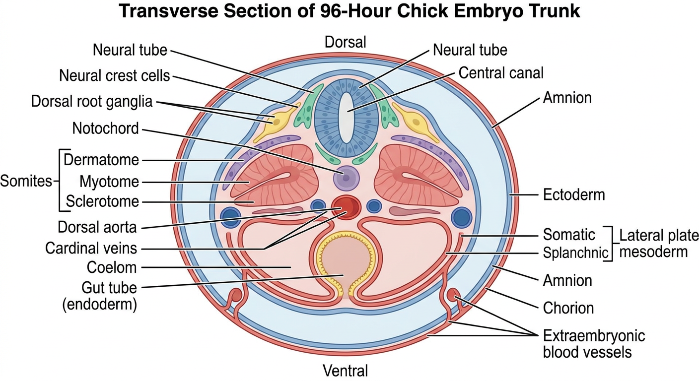
- Dorsal side: neural tube, neural crest, and paired dorsal root ganglia
- Midline: notochord immediately ventral to the neural tube
- Paraxial mesoderm: paired somites, differentiating into dermatome, myotome, and sclerotome
- Vessels: paired dorsal aortae and cardinal veins
- Ventral region: endoderm-lined gut tube and intraembryonic coelom
- Lateral plate mesoderm: splits into somatic and splanchnic layers
- Outer coverings: ectoderm, amnion, and chorion
Provide a clean, fully labelled exam diagram
96 hour chick embryo transverse section labels neural tube somite gut diagram
chick embryo development
Produce a clean, accurate, fully labelled exam diagram titled “T.S. of 96-hour Chick Embryo (Trunk Region)”. It must be a simple black-and-white pen-and-ink embryology line drawing, not a photomicrograph, on a plain white background, optimized for reproduction in an anatomy practical exam. Cross section is bilateral and symmetrical, dorsal at top, ventral at bottom. Draw the embryo and surrounding extraembryonic membranes. Use crisp straight leader lines to clear labels arranged outside the drawing, without overlaps. Include exactly these labels, with legible spelling: Amnion, Chorion, Neural tube, Central canal, Neural crest, Dorsal root ganglion, Notochord, Somite, Dermatome, Myotome, Sclerotome, Dorsal aorta, Posterior cardinal vein, Intraembryonic coelom, Somatic mesoderm, Splanchnic mesoderm, Gut tube, Endoderm, Ectoderm, Extraembryonic coelom, Vitelline blood vessel. Ensure the developing spinal cord is central dorsally, notochord immediately beneath it, paired somites lateral to it, paired dorsal aortae beneath the notochord, coelomic cavities laterally, and gut tube in the ventral midline. Use only exam-standard structures, no decorative text.
T.S. of 96-hour Chick Embryo (Trunk Region)
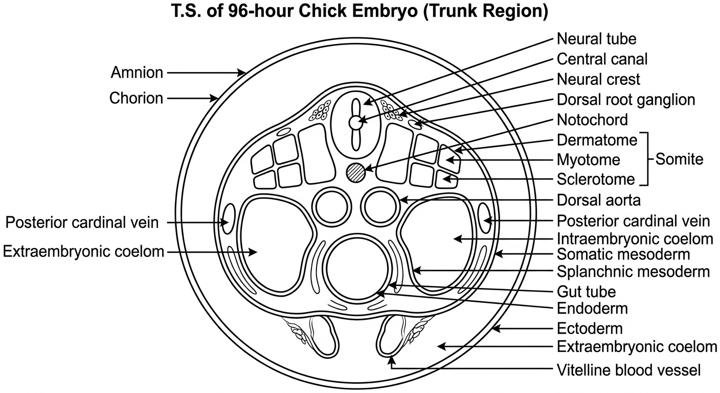

Diagram of this
Create a clean, fully labelled black-and-white exam line diagram based on a transverse histological section of the spinal cord. It should closely match the supplied micrograph: an oval transverse cord with a narrow elongated central canal running vertically in the center, surrounded by a butterfly-like central grey matter region and a broad peripheral white matter region. The ventral side is at the bottom, indicated by a deep anterior median fissure; dorsal side at the top by a shallow posterior median sulcus. Draw the grey commissure around the canal and distinct anterior horns, posterior horns, lateral funiculi, anterior funiculi, posterior funiculi. Use neat leader lines outside the drawing with clearly legible labels: Posterior median sulcus, Posterior funiculus (white matter), Posterior horn of grey matter, Central canal, Grey commissure, Lateral funiculus (white matter), Anterior horn of grey matter, Anterior white commissure, Anterior funiculus (white matter), Anterior median fissure. Title: “T.S. of Spinal Cord”. Plain white background, professional anatomy practical exam diagram, no colour, no shading, no extra structures or decorative elements.
Create a clean fully labelled black-and-white anatomy practical exam diagram of a transverse histological section through the embryonic neural tube, based on a low-power micrograph. Show an oval neural tube with a tall narrow central neural canal. Dorsal side is at the top and ventral side at the bottom. Show the dorsal roof plate, ventral floor plate, a thick mantle layer around the canal, a thin peripheral marginal layer, and the outer limiting membrane. Show the neural canal centrally, lined by the ependymal (ventricular) layer. Use a simple cross-section line drawing with neat external leader-line labels and no colour. Include title exactly: “T.S. of Developing Neural Tube”. Labels exactly: Roof plate, Neural canal, Ependymal layer, Mantle layer, Marginal layer, Floor plate, Outer limiting membrane. Add orientation labels Dorsal and Ventral. Plain white background, clean legible handwritten-exam style, no adult spinal-cord horns and no decorative elements.
T.S. of Developing Neural Tube
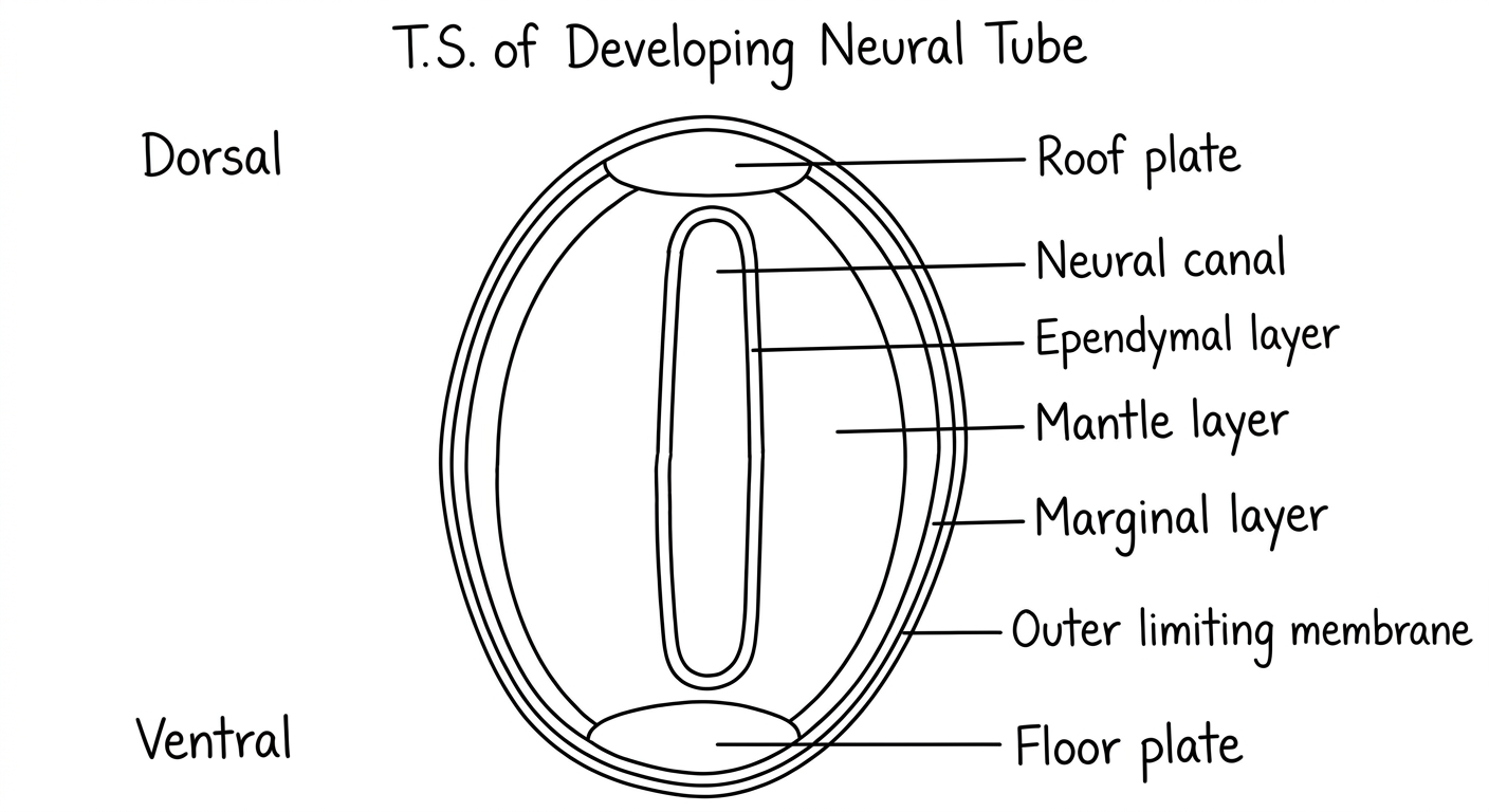
- Neural canal (neurocoel)
- Roof plate
- Mantle layer
- Marginal layer
Create a cleaner labelled version matching the uploaded image
neural tube development
developing neural tube transverse section roof plate mantle marginal layer diagram
Create a clean, high-resolution, fully labelled educational histology diagram that MATCHES a transverse section of a developing neural tube seen at low power. Portrait square composition. The neural tube must have the same overall anatomy as the reference: a rounded-oval tube, shallow dorsal midline indentation at the top, a long narrow vertical neural canal running almost the full height centrally, broad purple-pink cellular mantle layer surrounding the canal, a thin paler outer marginal layer, and a clearly thicker ventral basal region at the bottom. Dorsal at top, ventral at bottom. Use soft textbook histology colors inspired by H&E: purple-pink neural tissue, pale yellow canal and surrounding mesenchyme, cream background. No unrelated spinal cord features. Place clean black leader lines and labels outside the tissue without overlaps. Title exactly: “T.S. of Developing Neural Tube”. Labels exactly: Roof plate; Neural canal (neurocoel); Ependymal layer; Mantle layer; Marginal layer; Floor plate; Dorsal; Ventral. Include small numbered callouts inside the tissue matching the original image: 1 in the neural canal, 2 at roof plate, 3 in mantle layer, 4 in marginal layer. Professional practical-exam diagram, highly legible.
Clean labelled version matching the uploaded section
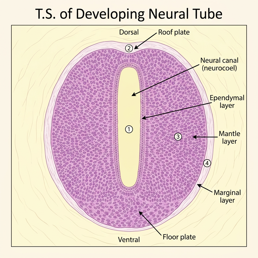
- Neural canal (neurocoel)
- Roof plate
- Mantle layer
- Marginal layer