
Papilary edema
papilledema optic disc swelling fundoscopy
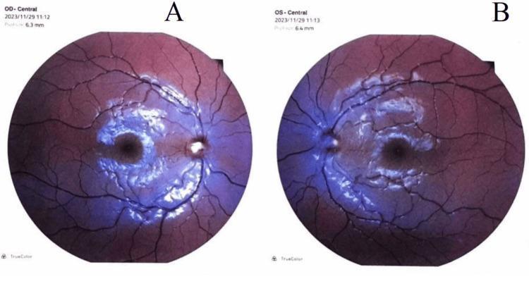
This diagnostic image displays bilateral fundus photographs of the right eye (OD, labeled A) and left eye (OS, labeled B). The images demonstrate severe bilateral papilledema, characterized by significant optic disc swelling with blurred, indistinct disc margins and a peripapillary halo. In both eyes, the optic nerves appear elevated and hyperemic, with a loss of the physiological cup. The retinal vasculature exhibits marked tortuosity and engorgement, with several vessels becoming obscured as they cross the disc margin due to the overlying edema. The surrounding retinal tissue shows evidence of compromised perfusion, with a general hyperemic hue and a lack of clarity in the macula and peripapillary region. These visual findings are consistent with Stage 4 papilledema, often associated with increased intracranial pressure. The modality is fundoscopy (retinal imaging), serving as a critical educational tool for identifying neuro-ophthalmological signs of intracranial hypertension.
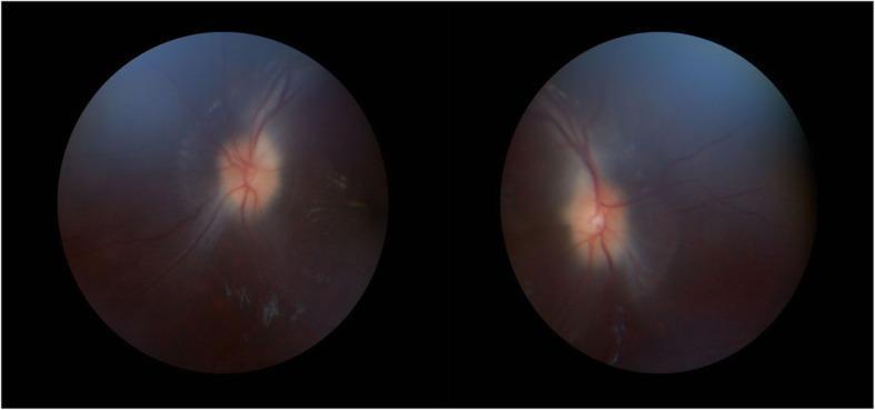
This diagnostic image consists of two fundoscopy circular frames showing the bilateral retinal fundi. The primary clinical finding is bilateral optic disc edema, consistent with grade 2 papilledema. The optic discs demonstrate significant blurring of the margins and visible elevation. Vascular changes include moderate engorgement and increased tortuosity of the retinal veins as they emerge from the disc. The physiological cups appear obscured due to the swelling. The surrounding retinal background maintains a normal red-orange hue, although there is a subtle presence of whitish spots suggestive of exudates in the peripheral field of the left eye. No obvious retinal hemorrhages are visualized. These findings are characteristic of increased intracranial pressure, as seen in conditions like idiopathic intracranial hypertension or space-occupying lesions. This content is intended for medical education in ophthalmology and neurology to demonstrate clinical signs of optic nerve head swelling.
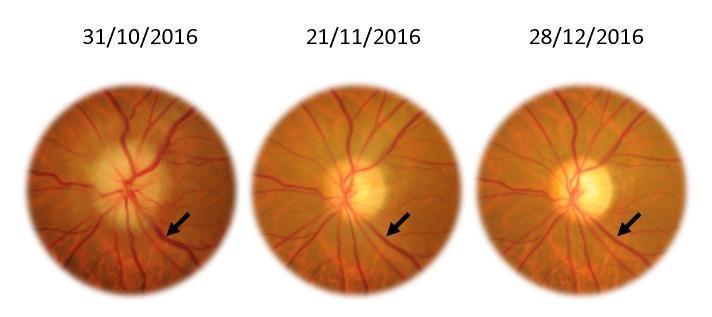
This Comparison Chart consists of three sequential color fundus photographs of the left eye, documenting the progression of ophthalmic findings over a two-month period (31/10/2016 to 28/12/2016). The images demonstrate the resolution of papilledema (optic disc edema) and venous congestion. In the initial photograph, the optic disc exhibits blurred margins, elevation, and obscuration of peripapillary vessels, characteristic of acute swelling. The retinal veins, highlighted by black arrows, appear significantly engorged and tortuous (venous turgescence). Subsequent images show a chronological improvement: the optic disc margins become progressively more distinct and sharp, indicating a reduction in edema. Simultaneously, the caliber of the retinal veins decreases, showing a marked reduction in turgidity and fullness. This timeline serves as a clinical illustration of treatment response or the natural history of intracranial pressure resolution. Key concepts include optic neuropathy, fundoscopy, venous caliber changes, and the morphological stages of papillary edema remission.
papilledema intracranial pressure diagnosis
Papilledema (Papillary Edema)
Definition
Pathophysiology
Stages
| Stage | Features |
|---|---|
| Early | Edema most prominent at superior and inferior poles (where nerve fiber layer is thickest); mild disc hyperemia; slight venous distension |
| Fully developed (acute) | Uniform disc elevation; blurred margins; flame hemorrhages; cotton-wool spots; absent spontaneous venous pulsations (SVPs); peripapillary/radial retinal folds |
| Chronic (weeks-months) | Gliotic, pale disc; fewer hemorrhages; "champagne cork" appearance with pseudodrusen (extruded axoplasm); collateral vessels |
| Atrophic | Optic nerve atrophy with disc pallor but reduced swelling; severe visual field constriction; ominous sign (dying nerve) |
Clinical Features
- Visual acuity is typically preserved in early/acute papilledema - the hallmark mismatch is disc edema with relatively intact visual function
- Transient visual obscurations (TVOs): Classic symptom - lasting seconds, monocular or binocular, triggered by posture changes or spontaneous; prolonged/spontaneous obscurations indicate more threatening papilledema
- Visual field defects: Enlarged blind spot (earliest), arcuate defects (inferonasal), concentric constriction; macula involvement causes metamorphopsia and acuity loss
- Headache: Common but not invariable
- Papilledema is typically bilateral but may be asymmetric due to anatomical variation in optic nerve subarachnoid septations
Fundoscopic Findings
- Arterial phase: absent/delayed fluorescence due to disc swelling
- Arteriovenous phase: dilated capillaries and microaneurysms
- Venous phase: fluorescein leakage from dilated capillaries
Causes
- Mass lesions - brain tumor, abscess, large infarction, intracranial hemorrhage; posterior fossa lesions especially likely to cause papilledema
- Venous sinus thrombosis - especially in pregnancy and hypercoagulable states
- Hydrocephalus
- Meningitis/encephalitis - Cryptococcal meningitis is the infectious cause most commonly associated with significant papilledema
- Idiopathic intracranial hypertension (IIH / pseudotumor cerebri) - most common in obese women; also with tetracycline antibiotics, vitamin A derivatives
- Malignant hypertension - produces bilateral disc edema indistinguishable from papilledema; accompanied by peripapillary cotton-wool spots and retinal hemorrhages
- Subarachnoid hemorrhage
- Cerebral edema (stroke, trauma)
Evaluation
- Neuroimaging first (MRI/CT) - exclude an intracranial mass before lumbar puncture; MR venography useful to exclude dural venous sinus thrombosis
- Lumbar puncture (if neuroimaging negative) - measure opening pressure in lateral decubitus position; elevated pressure with normal CSF = pseudotumor cerebri (IIH)
- Visual field testing - track progression
- OCT (optical coherence tomography) - quantifies retinal nerve fiber layer (RNFL) thickness; useful to monitor response to treatment
Management
| Approach | Detail |
|---|---|
| Weight loss | Mandatory; even 5-15% weight reduction helps; GLP-1 receptor agonists recommended when diet alone fails |
| Acetazolamide | Carbonic anhydrase inhibitor; reduces CSF production; improves symptoms and disc swelling in mild visual loss |
| Shunting (LP or VP) | Preferred for severe/progressive visual loss; lumboperitoneal shunt preferred |
| Transverse sinus stenting | Endovascular placement at transverse-sigmoid junction (where stenosis is typically found); emerging option |
| Optic nerve sheath fenestration | Less effective; does not address headache; reserved for ocular protection when shunting not possible |
| Emergency surgery | Fulminant papilledema causing rapid blindness requires urgent shunt placement |
Pseudopapilledema
Pt has vomiting while having headache
Headache with Vomiting - Clinical Approach
Why Vomiting Accompanies Headache
- Raised intracranial pressure (ICP) - stimulates the vomiting center in the medulla directly; characteristically effortless vomiting without preceding nausea or GI complaints - this is the key distinguishing feature of raised ICP vomiting
- Autonomic activation - nausea and vomiting from migraine occur via trigeminovascular activation and release of vasoactive peptides; these are typically accompanied by nausea and GI symptoms
Key clinical pearl: "Headache with effortless vomiting but no GI complaints is characteristic of elevated intracranial pressure." - Tintinalli's Emergency Medicine
Differential Diagnosis
1. Migraine (Most Common Benign Cause)
- Unilateral, throbbing/pulsatile, moderate-to-severe
- Nausea and vomiting common (part of diagnostic criteria)
- Also has photophobia, phonophobia
- Worsened by routine physical activity
- Relief in dark, quiet room
- Duration 4-72 hours (adults); 1-48 hours (children)
2. Raised Intracranial Pressure (Serious)
- Morning headache that is worse on waking, on straining, Valsalva, or recumbency
- Effortless, projectile vomiting - without nausea, no GI prodrome
- Progressive worsening over days to weeks
- May have papilledema on fundoscopy
- Causes: brain tumor, hydrocephalus, cerebral abscess, venous sinus thrombosis, idiopathic intracranial hypertension
3. Subarachnoid Hemorrhage (SAH) - Life-threatening
- Thunderclap headache - explosive, instantaneous onset ("worst headache of my life"), like a "clap of thunder" or "being hit on the head"
- Rapidly generalizes; accompanied by neck and back pain
- Vomiting accompanies the headache and may aggravate pain
- Loss of consciousness may occur
- Photophobia and phonophobia (mimics migraine - dangerous trap)
- Movement aggravates pain
- Confirm with unenhanced CT scan first; LP only if CT negative
4. Meningitis / Encephalitis
- Headache + fever + neck stiffness (meningism)
- Nausea and vomiting common
- Photophobia, altered consciousness
- Kernig's and Brudzinski's signs
- Viral or bacterial; HSV encephalitis if headache + altered mental status + seizures
5. Cerebral Abscess
- Pyrexia, persistent headache, raised ICP features (vomiting, papilledema)
- Focal neurological signs; seizures
- Bradycardia as ICP rises (Cushing's response)
6. Cluster Headache
- Severe periorbital/retro-orbital unilateral pain
- Autonomic features (lacrimation, rhinorrhea, ptosis, conjunctival injection)
- Restlessness/agitation (unlike migraine where patient prefers stillness)
- Nausea/vomiting less prominent than migraine
7. Hypertensive Emergency (Malignant Hypertension)
- Severe headache + nausea/vomiting + markedly elevated BP
- May have papilledema, encephalopathy, visual disturbance
- Bilateral disc edema, cotton-wool spots, flame hemorrhages on fundoscopy
Red Flags - Must Not Miss
| Red Flag | Likely Cause |
|---|---|
| Sudden onset / thunderclap headache | Subarachnoid hemorrhage (SAH) |
| Morning headache + early morning vomiting (effortless) | Raised ICP - brain tumor, hydrocephalus |
| Fever + neck stiffness + headache | Meningitis |
| Headache worsened by Valsalva / recumbency | Raised ICP |
| Progressive frequency and severity over weeks | Space-occupying lesion |
| Headache waking patient from sleep | Raised ICP, posterior fossa tumor |
| First/worst headache ever | SAH until proved otherwise |
| Headache + focal neurology / seizures | Tumor, AVM, abscess, hemorrhage |
| Headache + altered mental status | Encephalitis, SAH, herniation |
| Headache + papilledema | Raised ICP (any cause) |
Approach to Evaluation
History
- Onset: sudden (thunderclap = SAH) vs. gradual (tumor, IIH)
- Time of day: morning vomiting + headache = raised ICP
- Character of vomiting: effortless/projectile (raised ICP) vs. nausea-preceded (migraine, meningitis)
- Associated symptoms: fever, neck stiffness, visual changes, focal deficits, photophobia
- Pattern: progressive worsening? Episodic? New vs. long-standing?
- Aggravating factors: straining, coughing, bending forward, lying down
Examination
- Vitals: BP (hypertensive emergency), temperature (meningitis/abscess)
- Fundoscopy: papilledema (raised ICP), flame hemorrhages (hypertension)
- Meningism: neck stiffness, Kernig's, Brudzinski's
- Neurological exam: focal deficits, cranial nerve palsies (especially VI nerve - false localizing sign of raised ICP)
- Level of consciousness
Investigations
| Priority | Test | Indication |
|---|---|---|
| Urgent CT head (non-contrast) | First line | SAH, hemorrhage, mass lesion, hydrocephalus |
| Lumbar puncture | After negative CT | SAH (xanthochromia), meningitis (CSF cells, culture) |
| MRI ± MRV/MRA | If CT negative but high suspicion | Venous sinus thrombosis, early SAH, posterior fossa lesions |
| Blood cultures + CBC + CRP | Suspected meningitis | Before antibiotics if no LP delay |
| BP measurement | All patients | Hypertensive emergency |
Neuroimaging should always precede lumbar puncture when there is headache with red flags, early morning headaches + vomiting, progressive headaches, or focal neurological findings. - Rosen's Emergency Medicine
Management Summary by Cause
| Cause | Immediate Action |
|---|---|
| SAH | CT head urgent; neurosurgery; nimodipine; no LP if CT positive |
| Meningitis | Blood cultures then IV antibiotics + dexamethasone; LP when safe |
| Brain tumor / Raised ICP | Dexamethasone (if vasogenic edema); neurosurgery referral |
| Migraine | NSAIDs, triptans, antiemetics (metoclopramide/prochlorperazine); dark quiet room |
| IIH | Acetazolamide; weight loss; shunting if severe |
| Hypertensive emergency | Controlled IV BP reduction (labetalol, nitroprusside) |
Key points to note for papilary edema
Papilledema - Key Points
Definition (Get This Right)
- Papilledema = optic disc edema ONLY due to raised ICP
- Any other cause of disc swelling = called "optic disc edema" (not papilledema)
- This distinction is clinically and terminologically important
Pathophysiology (One Line)
Raised ICP → transmitted via optic nerve sheath (communicates with subarachnoid space) → blocks axoplasmic flow → axoplasm accumulates → disc swells
Classic Features (High Yield)
| Feature | Detail |
|---|---|
| Bilaterality | Almost always bilateral (asymmetry can occur due to anatomical variation) |
| Visual acuity | Preserved in early/acute phase - hallmark mismatch |
| Transient visual obscurations (TVOs) | Seconds-long visual blackouts; triggered by posture change or spontaneous |
| Blind spot | Enlarged - earliest visual field defect |
| SVPs | Spontaneous venous pulsations are absent with raised ICP |
| Headache | Common but NOT invariable |
4 Stages (Must Know)
| Stage | Key Finding |
|---|---|
| 1. Early | Superior + inferior pole swelling (thickest nerve fiber layer here) |
| 2. Acute/Fully developed | Uniform disc elevation; hemorrhages; cotton-wool spots; absent SVPs |
| 3. Chronic (weeks-months) | "Champagne cork" appearance; pseudodrusen; less hemorrhage; gliotic disc |
| 4. Atrophic | Disc pallor without swelling; severe field loss; ominous sign |
In atrophic stage - reduction in disc swelling = bad sign (dying nerve), NOT improvement
Fundoscopic Findings (Acute)
- Blurred disc margins (especially superior and inferior)
- Disc hyperemia + elevation
- Engorged, tortuous retinal veins
- Flame-shaped hemorrhages (peripapillary)
- Cotton-wool spots
- Peripapillary retinal folds (Paton's lines)
- Absent spontaneous venous pulsations
Key Causes (Raised ICP)
- Brain tumor / abscess / hemorrhage (especially posterior fossa - most likely to cause papilledema)
- Venous sinus thrombosis (especially pregnancy, hypercoagulable states)
- Cryptococcal meningitis - most common infectious cause of significant papilledema
- IIH (pseudotumor cerebri) - obese women; tetracyclines; vitamin A
- Hydrocephalus
- Malignant hypertension (produces identical disc edema)
IIH - Specific Points
- Obese women most commonly
- Risk factors: tetracyclines, vitamin A derivatives, modest weight gain (even 5-15%)
- Treatment ladder: weight loss → acetazolamide → shunt (LP preferred) → transverse sinus stenting → optic nerve sheath fenestration
- GLP-1 agonists (e.g., semaglutide) recommended if diet alone fails
- Fenestration = least effective; does not help headache
Investigation Sequence
- MRI/CT brain first - exclude mass lesion before LP
- MR venography - if sinus thrombosis suspected
- LP (lateral decubitus) - measure opening pressure; normal CSF + raised pressure = IIH
- Fluorescein angiography - leakage in venous phase confirms true papilledema (vs. pseudopapilledema)
- OCT - quantifies RNFL thickness; monitors treatment response
Pseudopapilledema vs. True Papilledema
| Feature | Pseudopapilledema (Drusen) | True Papilledema |
|---|---|---|
| SVPs | Present | Absent |
| Fluorescein leakage | Absent | Present |
| Hemorrhages | Absent | Present |
| Detection | B-ultrasound (hyperechoic), CT, OCT | Clinical + imaging |
One-Liners to Remember
- "Worst visual field defect is in atrophic stage" - when disc swelling resolves
- "Posterior fossa tumors" - most likely to cause papilledema (CSF pathway obstruction)
- "Cryptococcal meningitis" - the infection most associated with papilledema
- "TVOs" - classic symptom; prolonged/spontaneous TVOs = more dangerous papilledema
- "Never LP before CT" - risk of herniation if mass present
- "Champagne cork + pseudodrusen" = chronic papilledema
Does the patient should be dilated??
should you dilate pupil papilledema raised intracranial pressure fundus examination safety
Should the Patient with Papilledema Be Dilated?
Short Answer: YES - Dilation is required for proper examination, BUT with critical caveats
Why You SHOULD Dilate
| Benefit of Dilation | Detail |
|---|---|
| Better disc visualisation | Dilated fundus exam allows full characterisation of optic nerve head - margins, elevation, hemorrhages, Paton's lines |
| See SVPs clearly | Spontaneous venous pulsations are best viewed over the disc - requires good view through dilated pupil |
| Grade the papilledema | Frisén scale grading (0-5) requires detailed disc view only possible with dilation |
| Monitor progression | OCT + dilated fundus photography is the gold standard for follow-up |
| Detect pseudodrusen | Buried drusen causing pseudopapilledema may only be clearly visible on dilated exam |
"The optic disc is optimally viewed through a dilated pupil via a direct ophthalmoscope." - Papilledema and IIH, PMC Review
"A dilated fundus examination allows characterisation of the optic nerve head appearance, and the presence and degree of optic nerve edema are essential for guiding diagnosis, follow-up, and therapy." - Frontiers in Pediatrics, 2026
When You MUST NOT Dilate (Contraindications)
1. Active Neurological Monitoring (Most Important Contraindication)
- In any patient with suspected or confirmed raised ICP who is being monitored neurologically, the pupillary light reflex is the primary clinical indicator of brainstem herniation
- A fixed, dilated pupil = CN III compression = uncal herniation - this is an emergency sign
- Pharmacologically dilating the pupils abolishes this sign and can mask the most critical early warning of herniation
- Do NOT dilate if the patient has: altered consciousness, suspected herniation, acute head injury, rapidly deteriorating neurology, or is being observed in ICU/emergency
2. Narrow-Angle Glaucoma / Angle-Closure Risk
- Mydriatics can precipitate acute angle-closure glaucoma
- Screen with penlight oblique illumination or gonioscopy before dilating
3. Allergy to mydriatic agents
Practical Decision Framework
Patient with suspected papilledema
              ↓
Is the patient neurologically stable?
(Alert, no herniation signs, not being actively monitored)
              ↓
        YES                         NO
         ↓                           ↓
   DILATE for full            DO NOT DILATE
   fundus exam              (pupil monitoring is
   (tropicamide              critical for safety)
   ± phenylephrine)         Use indirect method /
                            undilated if needed,
                            or defer to stable phase
Agents Used for Dilation (when safe)
| Agent | Type | Onset | Duration |
|---|---|---|---|
| Tropicamide 0.5-1% | Antimuscarinic | 20-30 min | 4-6 hours |
| Phenylephrine 2.5% | Sympathomimetic | 15-20 min | 3-5 hours |
| Combined (tropicamide + phenylephrine) | Both | Faster, better dilation | Standard practice |
- Tropicamide alone is sufficient in most cases
- Avoid phenylephrine 10% (risk of systemic hypertension)
- Reverse with pilocarpine 1% if needed urgently after exam
Key One-Liners
- "Dilate to diagnose, don't dilate if monitoring"
- Undilated direct ophthalmoscopy misses a large amount - the disc looks through a "keyhole"
- Pharmacologic dilation lasts only hours and does not cause actual ICP rise
- The concern is NOT that dilation raises ICP - it does not
- The concern is that dilation masks pupillary signs of herniation in the acute neurological patient
- Once the patient is stable and imaging done, dilated fundus exam is part of standard workup for papilledema
Power n+10
What context is 'Power n+10' referring to?