
Rmv
- Remove something - from a file, list, or document?
- RMV - as in the Registry of Motor Vehicles?
- Something else entirely?
Respiratory minute volume
Respiratory Minute Volume (RMV)
Definition and Formula
RMV = Tidal Volume (TV) × Respiratory Rate (RR)
- Guyton and Hall Textbook of Medical Physiology, p. 1654
- Ganong's Review of Medical Physiology, p. 4135
Normal Values at Rest
| Parameter | Normal Value |
|---|---|
| Tidal Volume (TV) | ~500 mL/breath |
| Respiratory Rate (RR) | ~12 breaths/min |
| RMV | ~6 L/min |
Range and Extremes
- Minimum survivable: RMV as low as 1.5 L/min (RR of 2-4 breaths/min) is compatible with life for a short period.
- Maximum possible: RR can rise to 40-50 breaths/min, and tidal volume can approach vital capacity (~4,600 mL in a young adult male), giving an RMV exceeding 200 L/min - more than 30 times normal. However, this cannot be sustained beyond ~1 minute.
- Guyton and Hall, p. 1656
RMV vs. Alveolar Ventilation
Alveolar Ventilation = (TV - Dead Space) × RR = (500 - 150) × 12 = ~4,200 mL/min (4.2 L/min)
RMV vs. Maximal Voluntary Ventilation (MVV)
| Measurement | Normal Value | What it assesses |
|---|---|---|
| RMV | ~6 L/min | Resting ventilation |
| MVV | 140-180 L/min (men) | Maximum voluntary breathing capacity |
Regulation - What Changes RMV?
- Arterial PCO2 is normally maintained at 40 mmHg
- Rising PCO2 stimulates ventilation; RMV increases linearly with rising alveolar PCO2
- This response is due to increases in both depth and rate of respiration
- Above 7% inspired CO2: alveolar PCO2 rises abruptly causing CO2 narcosis (headache, confusion, coma)
- Ganong's, p. 834
- Decreased O2 stimulates RMV, but the effect is minimal above PO2 of 60 mmHg
- Marked stimulation only occurs at lower PO2 values
- Mediated by carotid and aortic chemoreceptors
- Hypothermia initially stimulates respiration, then progressively decreases RMV
- CO2 production decreases ~50% with an 8°C drop in body temperature
- Rosen's Emergency Medicine, p. 1753
Clinical Relevance
| Situation | Effect on RMV |
|---|---|
| Exercise | Markedly increased |
| Mechanical ventilation | Adjusted by changing RR and/or TV to target PaCO2 and pH |
| Salicylate poisoning | Elevated RMV maintained to compensate for metabolic acidosis |
| Hypothermia | Progressive decrease |
| Lung disease | Reduced MVV; dyspnea when RMV approaches half the maximal breathing capacity |
Minute volume = RR × TV (in liters) Adjust minute volume to modify PaCO2 and pH. - Pfenninger and Fowler's Procedures for Primary Care, p. 6174
Dyspnoeic index
dyspnoeic index RMV MVV respiratory physiology formula
dyspnoeic index = RMV/MVV x 100 normal value clinical significance
Dyspnoeic Index (DI)
Definition and Formula
Dyspnoeic Index = (RMV / MVV) × 100
- RMV = Respiratory Minute Volume (actual ventilation, ~6 L/min at rest)
- MVV = Maximal Voluntary Ventilation (maximum breathing capacity, ~140-180 L/min in healthy adult men)
Normal Value
- RMV ≈ 6 L/min
- MVV ≈ 140-180 L/min
- DI ≈ 3-5%
Critical Threshold for Dyspnoea
When actual ventilation reaches 30-40% of MVV, dyspnoea is inevitable.
- Fishman's Pulmonary Diseases and Disorders, p. 3578
The Breathing Reserve (related concept)
Breathing Reserve = MVV - RMV
- Normal: ~140-180 L/min (i.e., vast reserve)
- A low breathing reserve correlates with breathlessness in lung disease
- However, Fishman's notes this determination is "now primarily of historic interest" as a standalone measure - Fishman's Pulmonary Diseases, p. 694
Why MVV Matters: Disease Relationships
| Condition | Effect | Consequence |
|---|---|---|
| COPD / Obstructive disease | MVV markedly reduced | DI rises steeply; dyspnea at low activity |
| Restrictive lung disease | MVV reduced | Less breathing reserve |
| Healthy exercise | RMV increases | DI rises proportionally |
| Left ventricular failure | MVV near normal but mild RMV rise | Dyspnea out of proportion to DI - other mechanisms dominate |
Two Distinct "Dyspnea Indices" in Clinical Use
| Type | What it is |
|---|---|
| Classic physiological DI (RMV/MVV × 100) | Objective spirometric ratio used in pulmonary function assessment |
| Mahler Baseline Dyspnea Index (BDI) | Rater-administered clinical scale rating magnitude of task, magnitude of effort, and functional impairment - used in COPD outcome measurement |
| Dyspnea Index questionnaire (Gartner-Schmidt 2014) | A 10-item patient-reported scale for upper airway-related dyspnea (ENT context) |
Summary Table
| Parameter | Normal (rest) |
|---|---|
| RMV | ~6 L/min |
| MVV | ~140-180 L/min |
| Dyspnoeic Index | ~3-5% |
| Threshold for dyspnoea | >30-40% of MVV |
| Breathing Reserve | MVV - RMV (~135-175 L/min) |
Draw respiratory membrane
Detailed anatomical diagram of the respiratory membrane showing all layers through which gas exchange occurs between alveolar air and pulmonary capillary blood. Label each layer clearly: 1) Alveolar fluid layer with surfactant, 2) Alveolar epithelium (Type I pneumocyte), 3) Epithelial basement membrane, 4) Thin interstitial space, 5) Capillary basement membrane, 6) Capillary endothelium. Show an alveolus on the left side and a pulmonary capillary on the right side with red blood cells inside. Include arrows showing O2 diffusing from alveolus into blood and CO2 diffusing from blood into alveolus. Also label the total thickness as approximately 0.2-0.6 micrometers. Clean medical illustration style with a white background, clear labels and leader lines.
404 Not Found. {'message': '{\n "error": {\n "code": 404,\n "message": "Publisher model `projects/engaged-beaker-474810-u2/locations/global/publishers/google/models/gemini-3-pro-image-preview` was not found or your project does not have access to it. Ensure you are using a valid model name and that the model is available in the specified region. For more information, see: https://docs.cloud.google.com/gemini-enterprise-agent-platform/resources/locations.",\n "status": "NOT_FOUND"\n }\n}\n', 'status': 'Not Found'}
We encountered an error while processing your request. Please try again or reach out to us for help.
Respiratory membrane
Respiratory Membrane
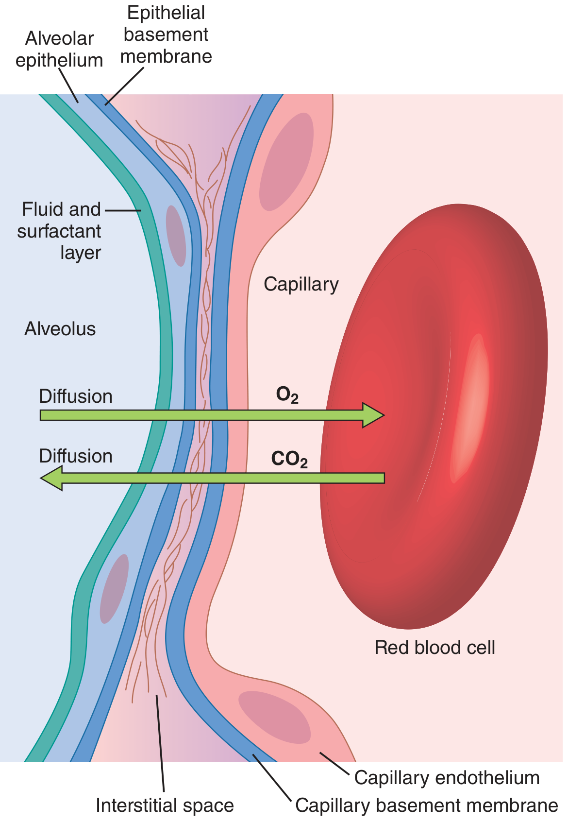
Definition
The 6 Layers (From Alveolus to Blood)
| Layer | Description |
|---|---|
| 1. Fluid + surfactant layer | Lines the alveolus; reduces surface tension of alveolar fluid |
| 2. Alveolar epithelium | Thin epithelial cells (Type I pneumocytes) forming the alveolar wall |
| 3. Epithelial basement membrane | Supports the alveolar epithelium |
| 4. Thin interstitial space | Between alveolar epithelium and capillary membrane |
| 5. Capillary basement membrane | Often fuses with the alveolar epithelial basement membrane, effectively eliminating the interstitial space in many areas |
| 6. Capillary endothelium | The inner lining of the pulmonary capillary |
- Guyton and Hall Textbook of Medical Physiology, p. 2394-2420
Key Physical Properties
| Property | Value |
|---|---|
| Minimum thickness | 0.2 µm |
| Average thickness | ~0.6 µm (except at cell nuclei) |
| Total surface area | ~70 m² (equivalent to a 25×30-foot room floor) |
| Pulmonary capillary blood volume | 60-140 mL at any given instant |
| Capillary diameter | ~5 µm (RBCs must squeeze through) |
Factors Affecting Rate of Gas Diffusion
- Thickness of the membrane - rate is inversely proportional; anything increasing thickness >2-3× normal significantly impairs exchange (e.g. pulmonary oedema, fibrosis)
- Surface area - directly proportional; emphysema can reduce surface area up to 5-fold by coalescence of alveolar walls; loss to 1/3-1/4 of normal substantially impairs exchange
- Diffusion coefficient - depends on gas solubility and inversely on √(molecular weight); CO2 diffuses ~20× faster than O2 through the membrane
- Partial pressure difference - driving force for diffusion (PO2 alveolus ~104 mmHg vs. capillary ~40 mmHg; PCO2 capillary ~45 mmHg vs. alveolus ~40 mmHg)
Clinical Relevance
| Disease | Effect on Respiratory Membrane |
|---|---|
| Pulmonary oedema | Edema fluid increases thickness - impairs diffusion |
| Pulmonary fibrosis | Fibrosis thickens membrane - reduces diffusion |
| Emphysema | Alveolar wall destruction reduces surface area up to 5-fold |
| Pneumonectomy | Halves total surface area |
| ARDS | Both surface area and thickness severely compromised |