
White colour object seeing in ear treatment
white object ear cholesteatoma otoscopy
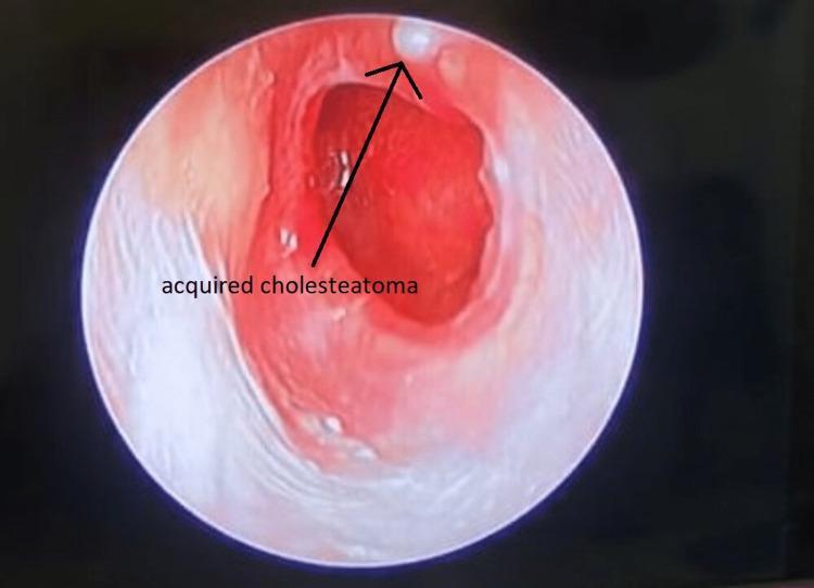
This clinical photograph, likely obtained via otoscopy or endoscopic ear examination, demonstrates an acquired cholesteatoma within the external auditory canal. The view reveals a significant area of pathological change characterized by intense erythema and inflammation of the canal wall and tympanic membrane region. A hallmark finding is the presence of a small, white, keratinous debris mass (cholesteatoma) located at the superior aspect of the canal, indicated by an arrow. The surrounding mucosal tissue appears thickened and irregular, suggesting chronic irritation or infection. The central portion of the field shows a deep red hue and compromised tissue integrity, typical of middle ear pathology extending into the canal. This image is an educational resource for identifying the visual hallmarks of primary acquired cholesteatoma, specifically the retraction pocket formation and the accumulation of squamous epithelial debris in the attic or pars flaccida region.
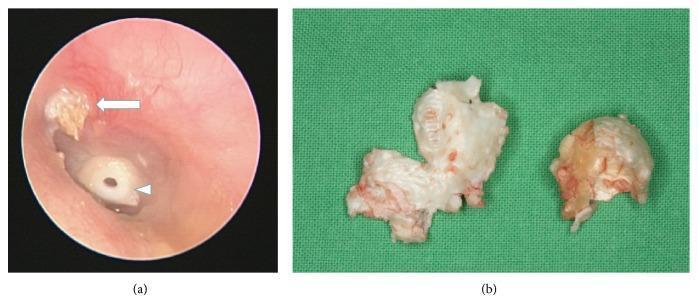
This composite image illustrates the clinical presentation and gross pathology of an acquired cholesteatoma. Image (a) is an otoscopic view of a left ear showing a keratinous debris accumulation in the attic region (white arrow), characteristic of a cholesteatoma originating from a retraction pocket in the pars flaccida. A tympanostomy tube or grommet (white arrowhead) is visible in the tympanic membrane, indicating a history of middle ear ventilation issues. Image (b) shows the macroscopic appearance of the dissected cholesteatoma tissue. The specimen exhibits a characteristic pearly-white, greasy-looking thin wall with a friable, pultaceous internal substance. This visual provides an educational comparison between the in situ endoscopic appearance of middle ear pathology and the gross morphological features of the excised squamous epithelial mass. It is relevant for otolaryngology training regarding chronic otitis media and surgical planning for mastoidectomy.
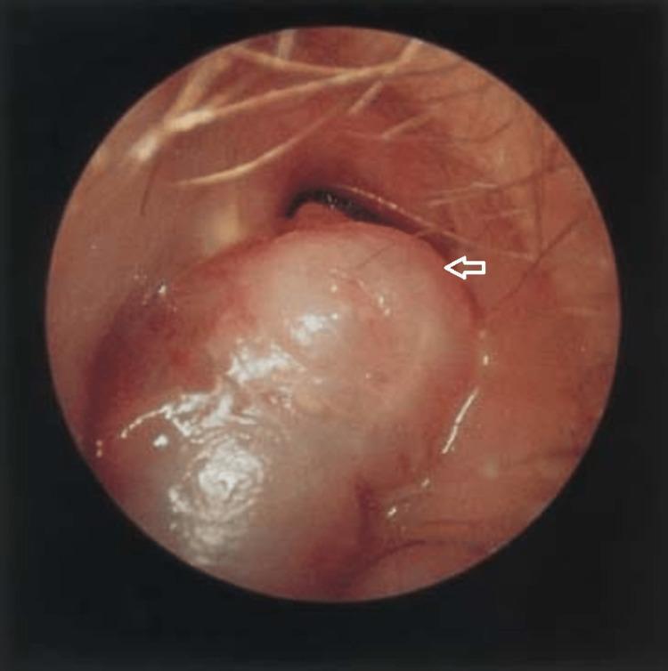
This clinical photograph, obtained through microscopic otoscopy, displays the right external auditory canal (EAC) of a patient. The central focus is a prominent, fleshy soft-tissue mass, indicated by a white arrow. The mass is pale pink to tan in color, with a smooth, glistening surface and a lobulated, irregular morphology. It occupies a significant portion of the canal lumen, originating from the posterosuperior wall. Fine hair follicles characteristic of the lateral EAC are visible in the foreground. This finding is clinically significant for evaluating chronic ear pathologies, such as aural polyps or extensive cholesteatoma, particularly when associated with chronic suppurative otitis media (CSOM). The image demonstrates how such a mass can obscure the view of the tympanic membrane and deeper middle ear structures. It serves as an educational example of otoscopic signs requiring further diagnostic imaging (CT or MRI) to assess for intracranial complications like mastoiditis or cerebral abscess.
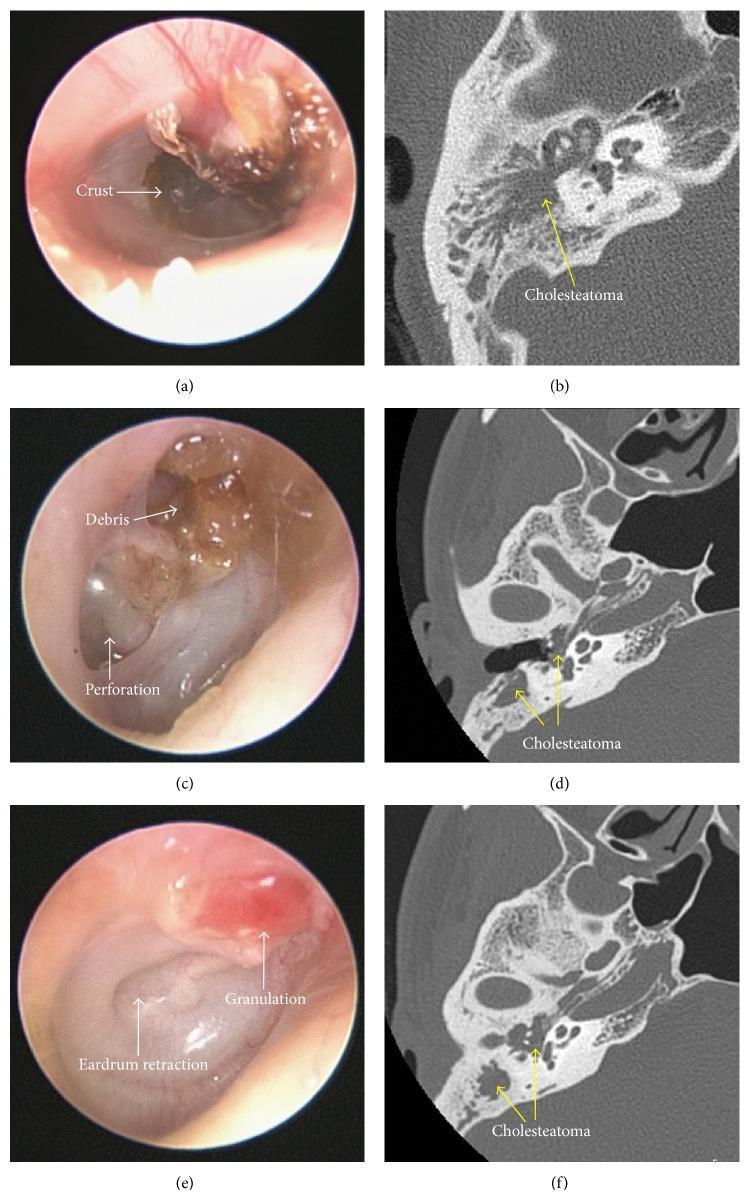
A six-panel clinical and diagnostic comparison illustrating the diagnostic challenges of identifying acquired cholesteatoma. The left column (a, c, e) contains otoscopic images showing common physical barriers to examination: (a) dark crusting along the superior canal wall, (c) brownish amorphous debris associated with a visible tympanic membrane perforation, and (e) erythematous granulation tissue with significant eardrum retraction. These external and middle ear findings often mask deeper pathology. The right column (b, d, f) provides corresponding high-resolution computed tomography (HRCT) axial scans of the temporal bone. The HRCT images clearly reveal soft-tissue opacification within the middle ear cleft, attic, and mastoid antrum, labeled as 'Cholesteatoma'. These diagnostic images demonstrate bony erosion and characteristic expansive growth that remained hidden or partially obscured during clinical otoscopy. This comparison emphasizes the clinical necessity of thorough canal debridement and the utility of HRCT in identifying cholesteatomas that are sequestered by superficial inflammatory changes, crust, or anatomical retractions.