
Gyi ve a detailed explanation about vescicular moles
hydatidiform mole vesicular mole pathology gross specimen
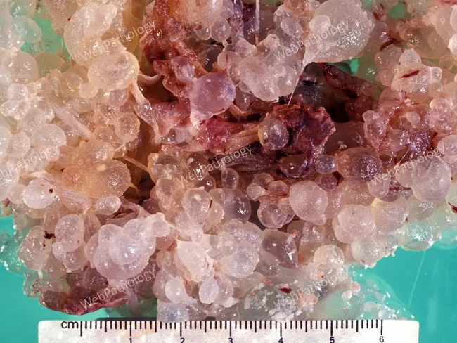
Gross pathology photograph of gestational tissue demonstrating complete hydatidiform mole. The specimen shows numerous swollen, vesicular, grape-like villous clusters with translucent to pale purple laminae and irregular surface architecture. No fetus or embryonic tissue is visible in the sample. A centimeter ruler is present for scale, confirming macroscopic vesicle diameters ranging from approximately 2-8 mm and overall tissue mass consistent with hydropic degeneration. The villous structures are edematous with cistern formation, and the tissue appears friable and bulky, representing diffuse trophoblastic involvement. The gross pattern is classic for a complete mole, characterized by diffuse villous edema, circumferential hydropic change, and lack of fetal tissue; the lesion may be accompanied by hemorrhagic foci and necrosis in larger specimens. This image is diagnostic when integrated with clinical data: markedly elevated beta-hCG, uterus enlargement, and ultrasound findings of a hollow, cystic placental mass. Diagnostic significance lies in identifying gestational trophoblastic disease early to guide uterine evacuation and obstetric management, while recognizing risk for persistent GTD and potential progression to choriocarcinoma. Potential clinical use cases include education on molar pregnancy morphology, gross pathology correlation with histology, and training for pathology residents in gross specimen handling and reporting.
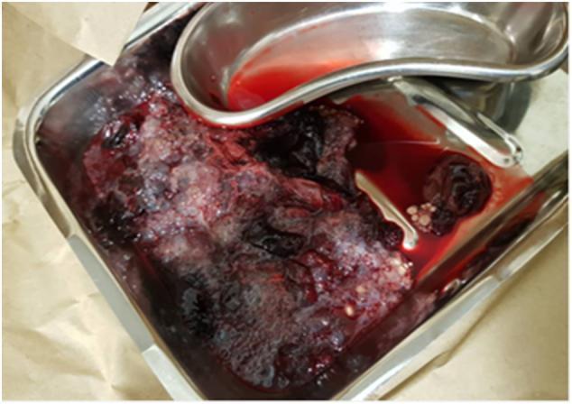
This clinical photograph shows a gross specimen of uterine contents expelled from a patient, presented in a stainless steel kidney dish and larger tray. The specimen demonstrates the classic gross appearance of a hydatidiform mole, a form of gestational trophoblastic disease. Visible pathology includes a large, heterogeneous mass composed of numerous small, translucent, grape-like vesicles (hydropic villi). These vesicles are intermixed with dark red-to-purple tissue and fresh blood. Small, dark fibrin clots are also visible within the fluid and adherent to the vesicular mass. The presence of these clusters of thin-walled cysts is a hallmark diagnostic feature often described as a 'bunch of grapes' appearance. This image illustrates the macro-pathology of molar pregnancy, highlighting the cystic swelling of the chorionic villi and associated intrauterine hemorrhage, which is clinically significant for the diagnosis and management of abnormal first-trimester vaginal bleeding.
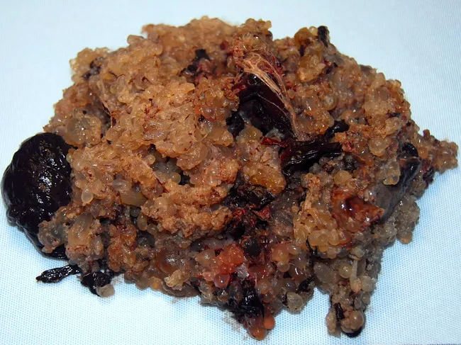
Gross pathology photograph of placental tissue containing hydropic vesicles consistent with a complete hydatidiform mole. This macroscopic specimen shows numerous variably sized, grape-like vesicular masses that replace normal placental architecture and lack detectable embryonic tissue. The vesicles are translucent to tan-brown, with sparse intervening hemorrhagic debris and dark, friable tissue components representing decidualized maternal tissue. The gross appearance corresponds to the pathognomonic feature of complete mole, which is characterized by diffuse trophoblastic proliferation and hydropic chorionic villi due to abnormal paternal genomic imprinting. Clinically, complete moles typically present with markedly elevated human chorionic gonadotropin (hCG), vaginal bleeding, and uterine enlargement disproportionate to gestational age; ultrasound in the modern era often reveals a heterogeneous, multicystic intrauterine mass. Histology (not shown in this image) would demonstrate diffuse villous edema with circumferential trophoblastic proliferation and absence of fetal tissue. The specimen’s appearance aids diagnosis, informs management (uterine evacuation and serial hCG monitoring), and raises consideration for persistent gestational trophoblastic disease. In differential terms, this contrasts with partial mole (embryo/fetus present with focal villous swelling) and with nonmolar miscarriage. This image is valuable for education, pathology training, and radiologic-pathologic correlation in obstetric medicine. This educational image supports recognition of mole pathology in clinical practice.
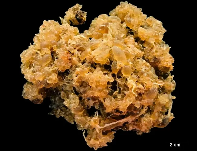
This is a gross pathology photograph of a complete hydatidiform mole derived from gestational tissue, illustrating numerous grape‑like vesicles consisting of hydropic chorionic villi without a fetal fetus. The specimen presents as an amorphous, multilobulated mass with tan‑yellow coloration and semitranslucent vesicles evident on the surface. A 2 cm scale bar is included for size reference, indicating a substantial vesicular conglomerate typical of molar tissue. Gross features include swollen villous cores with hydropic degeneration, numerous small vesicular cavities, and fibrous septa connecting some clusters. There is an absence of recognizable fetal parts or embryonic tissue, consistent with a complete mole in which paternal chromosomal duplication drives abnormal development. The image correlates with histologic patterns of diffuse villous edema and proliferation of trophoblastic tissue on microscopic examination, but the macroscopic morphology is diagnostic of gestational trophoblastic disease when correlated with clinical data. Clinically, complete moles can present with uterine enlargement, hyperemesis gravidarum, and markedly elevated β-hCG; management typically involves suction curettage with subsequent serial hCG monitoring to ensure resolution. This image, provided by Dr. Rosalinda Penaloza Ramirez, serves educational purposes in pathology teaching, differential diagnosis with partial mole and nonmolar abortion, and GTD management scenarios.
"hydatidiform mole" management diagnosis
Vesicular (Hydatidiform) Mole - Detailed Explanation
1. Classification
| Feature | Complete Mole | Partial Mole |
|---|---|---|
| Fetal/embryonic tissue | Absent | Present |
| Hydatidiform swelling | Diffuse | Focal |
| Trophoblastic hyperplasia | Diffuse | Focal |
| Karyotype | 46,XX or 46,XY (diploid, androgenetic) | 69,XXY or 69,XYY (triploid) |
| Karyotype origin | Entirely paternal | Two paternal + one maternal set |
| Risk of GTN | ~15-20% | ~1-4% |
2. Epidemiology
-
Extremes of reproductive age (very young and older patients - women >40 have a 37-60% risk of postmolar GTN)
-
Prior molar pregnancy
-
Low dietary intake of beta-carotene and animal fat
-
Rare: familial recurrent molar pregnancy (NLRP7 and KHDC3L gene mutations)
-
Berek & Novak's Gynecology, p. 2493
3. Pathogenesis and Cytogenetics
Complete Mole
- Monospermic mole (most common): A sperm fertilizes an empty oocyte (absent/inactive maternal pronucleus) and then duplicates → 46,XX (all paternal)
- Dispermic mole: Two sperms fertilize an empty oocyte → 46,XX or 46,XY
Partial Mole
- The Developing Human - Clinically Oriented Embryology, p. 223
4. Gross and Microscopic Pathology
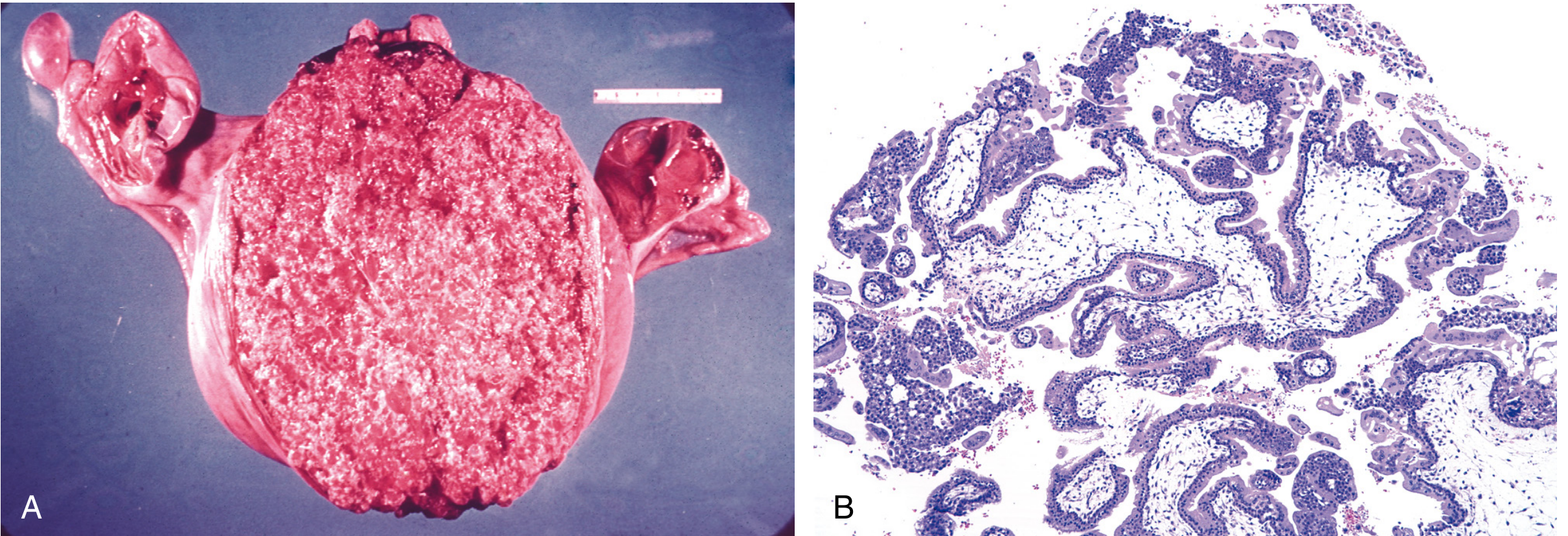
- Diffuse villous edema with cistern formation (central acellular lakes of fluid)
- Circumferential (360°) trophoblastic hyperplasia involving both cyto- and syncytiotrophoblast
- No fetal blood vessels
- Absence of embryonic/fetal tissue
- Focal villous swelling (only some villi are enlarged)
- Villi with irregular/scalloped outlines (like a jigsaw puzzle piece)
- Syncytiotrophoblast inclusions (pseudoinclusions invaginating into the villous stroma)
- Focal trophoblastic hyperplasia
- Fetal blood vessels and red blood cells may be present
5. Clinical Features
Complete Hydatidiform Mole
- Vaginal bleeding (most common symptom) - present in nearly all patients
- Uterine size large for dates (uterus enlarged out of proportion to gestational age)
- Markedly elevated beta-hCG - often >100,000 mIU/mL, higher than any normal pregnancy
- Hyperemesis gravidarum (due to very high hCG stimulating nausea)
- Preeclampsia before 20 weeks (pathognomonic - occurs in ~27% of complete moles; early-onset preeclampsia should always raise suspicion)
- Hyperthyroidism (hCG cross-reacts with TSH receptors): tachycardia, tremor, warm skin
- Theca lutein cysts of the ovaries (bilateral, multilocular - from ovarian hyperstimulation by high hCG); occur in ~25-30%
- Passage of vesicular tissue per vagina (classic but not always seen)
- No fetal heart sounds and no fetus on ultrasound
Partial Hydatidiform Mole
- Vaginal bleeding
- Uterus small or normal for dates (rarely large)
- hCG may be only mildly elevated
- Often no clinical features of a complete mole (no hyperthyroidism, no theca lutein cysts)
- Diagnosis is frequently made only on pathology after evacuation of what appeared to be a routine miscarriage
6. Diagnosis
Ultrasound
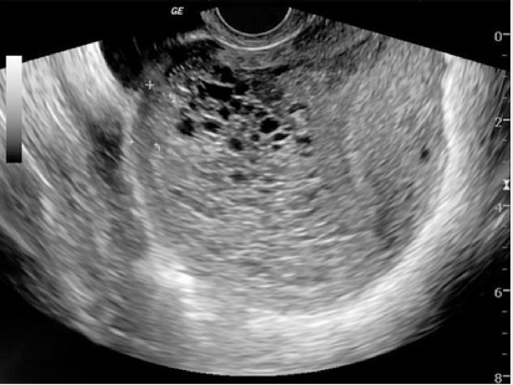
Beta-hCG
Histopathology
Additional workup at diagnosis:
- Chest X-ray (baseline; detect metastases)
- Complete blood count, liver and renal function tests
- Thyroid function (if clinical hyperthyroidism suspected)
- Blood group and crossmatch
7. Treatment
Suction Curettage (preferred)
- Start oxytocin infusion before anesthesia (reduces blood loss)
- Cervical dilation (increased bleeding is expected - do not stop)
- Suction curettage with a 12 mm cannula; one hand on the fundus if uterus >14 weeks to stimulate contractions and prevent perforation
- Sharp curettage to remove residual tissue
Hysterectomy
Prophylactic Chemotherapy
8. Post-Evacuation Follow-Up
hCG Monitoring Protocol
- Serial weekly serum beta-hCG until normal (three consecutive normal values)
- Then monthly for 6-12 months
Contraception
- Oral contraceptives are the preferred method during follow-up - they prevent a new pregnancy from confusing hCG surveillance and do NOT increase the risk of GTN
- Pregnancy should be avoided for 6-12 months after complete remission
Criteria for Diagnosing Persistent GTN (indicating need for chemotherapy)
- hCG plateau (4 values over 3 weeks, ±10%)
- hCG rise (3 values over 2 weeks showing >10% rise)
- hCG still detectable at 6 months after evacuation
- Histologic diagnosis of choriocarcinoma
9. Natural History and Complications
Complete Mole
- ~15% develop locally invasive GTN after evacuation
- ~4% develop metastatic GTN
- Risk is higher in older patients (>37% in women >40 years; up to 60% in women >50 years)
Partial Mole
- ~1-4% develop persistent (usually nonmetastatic) GTN requiring chemotherapy
- Patients who progress have no distinguishing clinical/pathologic characteristics at the time of initial diagnosis
Invasive Mole
Metastatic GTN
- Lungs: 80% (most common; "snowstorm," rounded densities, pleural effusion, or embolic patterns)
- Vagina: 30%
- Pelvis: 20%
- Liver: 10%
- Brain: 10%
10. Prognosis
Summary Table: Complete vs. Partial Mole
| Feature | Complete Mole | Partial Mole |
|---|---|---|
| Karyotype | 46,XX or 46,XY (diploid, androgenetic) | 69,XXY or 69,XYY (triploid) |
| Embryo/fetus | Absent | Present (abnormal) |
| Villous edema | Diffuse | Focal |
| Trophoblastic hyperplasia | Diffuse, circumferential | Focal, mild |
| hCG | Markedly elevated | Mildly elevated |
| Clinical presentation | Classic (large uterus, hyperemesis, preeclampsia <20 wks) | Mimics missed/incomplete abortion |
| Ultrasound | "Snowstorm" pattern | Focal cystic placental spaces |
| Risk of GTN | 15-20% | 1-4% |
| Risk of malignancy | Higher | Lower |
- Berek & Novak's Gynecology - Chapter 41, Gestational Trophoblastic Disease, pp. 2492-2510
- Robbins, Cotran & Kumar Pathologic Basis of Disease - Chapter 22, pp. 955-958
- The Developing Human - Clinically Oriented Embryology - Chapter 4, p. 223
- Recent evidence: A 2026 systematic review (PMID: 41066778) and a 2025 meta-analysis (PMID: 41155768) specifically address the rare scenario of complete hydatidiform mole coexisting with a normal twin fetus, confirming the management principles above still hold.
Normal labour, induction of labour and stages of labour
stages of labour cervical dilation fetal descent partograph
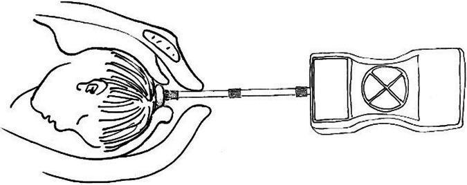
This schematic diagram illustrates a self-developed internal tocodynamometry machine used for monitoring fetal head descending thrust during labor. The illustration depicts three primary components: a pressure-sensitive sensor, a connecting rod, and an external monitoring unit. The sensor is shown in direct contact with the crown of a fetal head in the cephalic position, stabilized by a clinician's hand. A linear connecting rod, featuring three reinforcement bands, transmits the physical force from the sensor to the external device. The rectangular monitoring unit contains a display screen on the left and a circular dial with a cross-quadrant interface on the right. This diagnostic setup is designed to measure intrauterine pressure and fetal descent forces at specific stages of cervical dilation (3-5 cm, 5-8 cm, and 8-10 cm). The educational focus is on the instrumentation and clinical application of internal monitoring to assess labor progression and predict the necessity for emergency obstetric interventions.
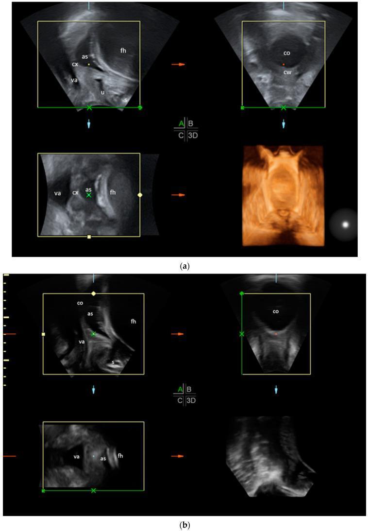
Transperineal ultrasound (TPU) compilation depicting two stages of labor progress in multiplanar views (sagittal, transverse, frontal, and 3D reconstruction). Figure (a) illustrates an earlier stage of labor where the cervix (cx) is relatively long and closed. The transverse plane clearly shows a thick cervical wall (cw) surrounding a small cervical opening (co). The sagittal view identifies the fetal head (fh), amniotic sac (as), vagina (va), urethra (u), and symphysis (s). Figure (b) demonstrates an advanced stage of labor characterized by significant cervical effacement and dilation. In the transverse plane, the cervical wall is minimally visible while the cervical opening (co) is widely dilated. The sagittal plane shows the descent of the fetal head (fh) and the amniotic sac (as) deeper into the birth canal, with the cervix (cx) appearing significantly shortened. This comparison serves as an educational tool for identifying anatomical landmarks and monitoring physiological changes such as cervical thinning and opening during the stages of delivery.
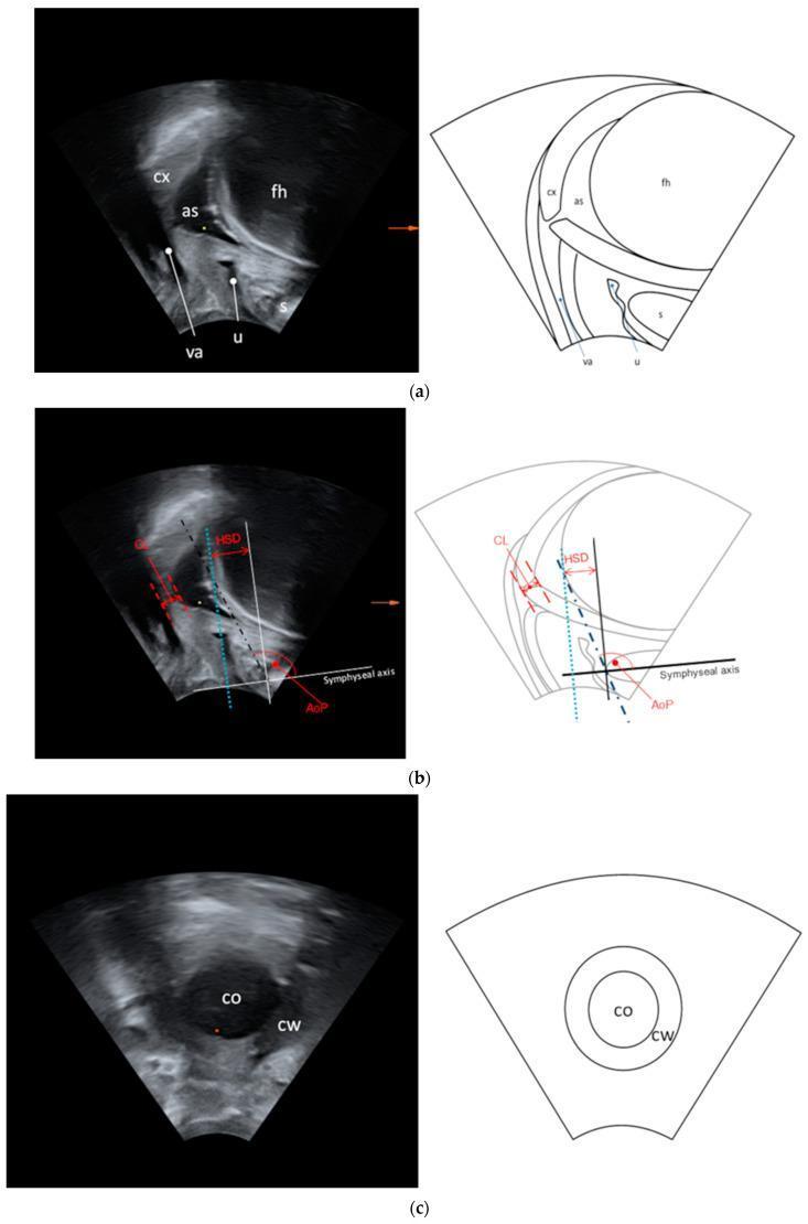
This composite educational resource demonstrates transperineal ultrasound (TPU) assessment during labor, featuring side-by-side sonographic images and corresponding schematic diagrams in sagittal and transverse planes. Section (a) illustrates maternal and fetal anatomy in the sagittal plane, identifying the symphysis (s), urethra (u), vagina (va), cervix (cx), amniotic sac (as), and fetal head (fh). Section (b) details objective labor progression measurements including the angle of progression (AoP), head-symphysis distance (HSD), and cervical length (CL), using the long symphyseal axis as a geometric reference. Section (c) provides a transverse view of the birth canal, clearly delineating the cervical opening (co) and the surrounding cervical wall (cw), which are essential for quantifying cervical dilation (CD) and wall thickness. This material serves as a clinical guide for using non-invasive ultrasound imaging to monitor fetal descent and cervical effacement during the first and second stages of labor.
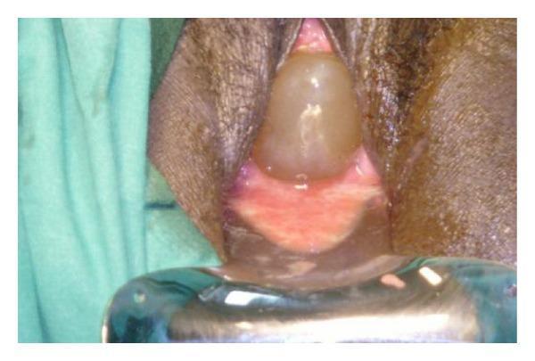
This clinical photograph illustrates a case of cervical insufficiency during pregnancy, specifically showing extensive bulging of the fetal membranes (hour-glassing) through the cervical os into the vaginal vault. The fetal membranes appear as a smooth, translucent, globular, and fluid-filled sac with a pale yellowish-tan hue. The surrounding cervical and vaginal tissues exhibit significant erythema and vascularity, indicative of inflammation. A metal vaginal speculum is positioned at the base of the image to provide exposure of the pelvic region. This visual is characteristic of second-trimester cervical dilation and prolapse of the amniotic sac, a critical clinical finding often necessitating emergent cerclage procedures such as the Shirodkar or McDonald technique. The image serves as an educational resource for identifying pre-viable or early viable pregnancy complications related to cervical incompetence.
Stages of labour