
Zenkers diverticulum refers to top 5 ent books and cover etiology pathogenesis clinical features treatment surgery steps
Reading File
Reading File
Finding Sources
Finding Sources
Searching PubMed
Zenker diverticulum AND treatment
Searching the Web
Zenker diverticulum guideline endoscopic surgical management 2025
Finding Sources
Finding Sources
Finding Sources
Reading Article
PMID: 40654274
Reading Web Page
https://pmc.ncbi.nlm.nih.gov/articles/PMC12429033
Reading File
Reading File
Reading File
Reading File
Reading File
Reading File
Reading File
Reading File
Reading File
Reading File
Zenker's diverticulum (pharyngeal pouch)
Definition: Zenker's diverticulum is an acquired false (pulsion) diverticulum of mucosa and submucosa arising posteriorly at the pharyngoesophageal junction through Killian's dehiscence, just above the cricopharyngeus muscle. It is not a true diverticulum because it lacks all layers of the pharyngeal wall.
Reference base: Cummings Otolaryngology, Scott-Brown's Otorhinolaryngology, K. J. Lee's Essential Otolaryngology, Bailey & Love, and Sabiston Textbook of Surgery.
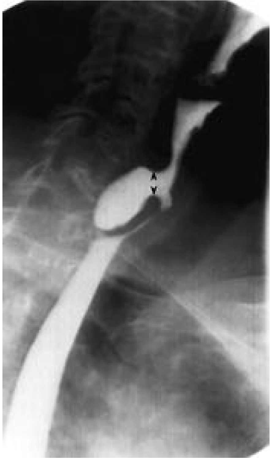
1. Anatomy and site
- It arises through Killian's dehiscence (Killian triangle), a weak posterior area between:
- Oblique fibers of the inferior pharyngeal constrictor superiorly
- Transverse fibers of the cricopharyngeus inferiorly
- The sac projects posteriorly, often toward the left, from the hypopharynx.
- It is a pharyngoesophageal diverticulum.
- Differentiate it from a Killian-Jamieson diverticulum, which arises below the cricopharyngeus and is anterolateral.
K. J. Lee's Essential Otolaryngology, p. 692; Bailey & Love's Short Practice of Surgery, p. 1148.
2. Etiology and pathogenesis
Etiology
Zenker's diverticulum is predominantly a disease of older people, usually in the seventh or eighth decade, and is more frequent in men.
Predisposing factors include:
- Age-related reduction in cricopharyngeal compliance
- Cricopharyngeal fibrosis or inflammation
- Failure of relaxation of the upper esophageal sphincter (UES)
- Impaired laryngeal elevation and impaired opening of the UES during swallowing
- Incoordination between pharyngeal contraction and cricopharyngeal relaxation
- Possible association with gastroesophageal reflux, though this does not by itself establish causation
Pathogenesis
The central abnormality is functional outflow obstruction at the UES.
- During swallowing, the pharyngeal constrictors contract.
- The cricopharyngeus fails to relax or open adequately.
- Intraluminal hypopharyngeal pressure rises.
- Mucosa and submucosa are forced posteriorly through Killian's dehiscence.
- Repeated pulsion produces an enlarging pouch.
- The pouch retains food and secretions, causing regurgitation, halitosis, and aspiration.
Exam sentence:
Zenker's diverticulum is an acquired pulsion pseudodiverticulum caused by impaired cricopharyngeal relaxation, producing high hypopharyngeal pressure and posterior mucosal herniation through Killian's dehiscence.
Sleisenger and Fordtran's Gastrointestinal and Liver Disease, p. 1688-1692; Fischer's Mastery of Surgery, p. 2811-2812.
3. Clinical features
Typical symptoms
| Feature | Explanation |
|---|---|
| Progressive oropharyngeal dysphagia | Usually starts with solids; later semisolids and liquids may be affected |
| Regurgitation of undigested food | Often occurs after meals, on bending, or when lying down |
| Halitosis | Food stagnation and decomposition within the pouch |
| Chronic cough and choking | Aspiration or laryngeal soiling |
| Aspiration pneumonia | Important complication, especially in older patients |
| Gurgling in neck | Cervical borborygmi due to fluid and food in the sac |
| Foreign-body or lump sensation | Food sticks at the level of the cricoid |
| Weight loss and malnutrition | In advanced dysphagia |
| Hoarseness or wet voice | Recurrent aspiration, laryngitis, or reflux |
| Neck swelling | Rare, usually left sided and compressible in a large pouch |
Clinical signs
- Thin or malnourished older patient
- Occasionally a soft compressible left anterior neck swelling
- Palpation may produce gurgling, termed Boyce sign
- Pooling of saliva in the hypopharynx may be seen on endoscopic examination
- A "rising tide" phenomenon can be seen during swallow assessment: retained fluid returns upward from the postcricoid region after swallowing.
Red flag: odynophagia is unusual. New pain, bleeding, rapid weight loss, or progressive worsening warrants evaluation for malignancy or another obstructing lesion.
Scott-Brown's Otorhinolaryngology Head & Neck Surgery, p. 855-856; Fischer's Mastery of Surgery, p. 2812.
4. Diagnosis
Investigation of choice: barium swallow with videofluoroscopy
Findings:
- Posterior contrast-filled pouch at the pharyngoesophageal junction
- Best seen in the lateral projection
- Demonstrates sac size, neck, retention, emptying, aspiration, and the functional swallowing abnormality
- Large pouches may compress and displace the cervical esophagus.
Cummings Otolaryngology Head and Neck Surgery, p. 1880; K. J. Lee's Essential Otolaryngology, p. 692.
Other investigations
- Flexible endoscopy or upper GI endoscopy: evaluates mucosa, clears retained food, excludes tumor or other esophageal pathology. It must be performed carefully because the scope may enter the pouch and cause perforation.
- FEES or modified barium swallow: useful where aspiration and pharyngeal dysphagia need functional assessment.
- Manometry: not routinely required. It can be difficult to perform and may not alter management.
- CT neck/chest: reserved for diagnostic uncertainty, complications, or concern for malignancy.
5. Treatment
Indications for intervention
Treat symptomatic Zenker's diverticulum, particularly if there is:
- Significant dysphagia
- Regurgitation
- Weight loss or malnutrition
- Aspiration, recurrent chest infections, or aspiration pneumonia
- Large or progressively enlarging sac
- Impaired quality of life
Small asymptomatic diverticula can usually be observed with dietary advice, aspiration precautions, and nutritional monitoring.
Principle of definitive treatment
The essential treatment is division of the cricopharyngeal muscle.
Simply excising the sac without correcting UES obstruction risks persistence or recurrence of symptoms.
Treatment options
| Method | Main role |
|---|---|
| Open transcervical cricopharyngeal myotomy with diverticulectomy | Large pouch, unfavorable endoscopic exposure, recurrent or complex disease, concern about pathology |
| Open diverticulopexy with CP myotomy | Selected patients, often frail patients or when avoiding a pharyngeal suture line is desirable |
| Open CP myotomy alone | Very small pouches |
| Rigid endoscopic stapled diverticulotomy | Common ENT approach when neck extension and exposure are adequate |
| Rigid endoscopic CO₂ laser diverticulotomy | Alternative to stapling |
| Flexible endoscopic septotomy | Minimally invasive approach, useful for older or high-risk patients |
| Z-POEM | Submucosal tunnel approach enabling a complete cricopharyngeal myotomy in experienced centers |
Recent evidence is evolving. A 2025 systematic review of seven comparative studies found higher clinical success with Z-POEM than alternative endoscopic approaches, with comparable adverse-event and reintervention rates, but the authors rated the underlying evidence as low quality (2025 systematic review).
6. Open transcervical surgery: operative steps
This is an exam-oriented outline, not a substitute for supervised operative training.
A. Preparation
- Confirm anatomy and pouch size on contrast swallow.
- Correct dehydration, malnutrition, and pulmonary infection if present.
- General anesthesia with careful airway planning because the pouch may contain retained material and aspiration is a risk.
- Position supine with shoulder roll and neck extension.
- Consider endoscopic evacuation of retained debris before dissection.
B. Exposure
- Make a left-sided transverse cervical incision, generally along a skin crease at the level of the cricoid.
- Divide platysma and enter the plane anterior to the sternocleidomastoid muscle.
- Retract carotid sheath laterally and thyroid/trachea medially as needed.
- Expose the cervical esophagus and identify the diverticular sac.
- Mobilize the sac gently, preserving the recurrent laryngeal nerve and surrounding vascular structures.
- Identify the neck of the pouch and the cricopharyngeus. A bougie may be passed in the esophagus to define the lumen and facilitate safe resection.
C. Cricopharyngeal myotomy
- Perform a longitudinal myotomy through the cricopharyngeus.
- Extend the division adequately across the dysfunctional cricopharyngeal segment and onto the proximal esophageal muscle as required.
- Confirm that the restrictive muscle has been fully divided.
Key point: cricopharyngeal myotomy is the essential component because it removes the functional obstruction that caused the pouch.
D. Management of the sac
Option 1: Diverticulectomy
Used most commonly for a large pouch.
- Apply a linear stapler or carefully close the pouch neck after placing a bougie within the esophageal lumen.
- Excise the diverticulum.
- Inspect the staple or suture line for integrity.
- Perform a leak test where appropriate.
Option 2: Diverticulopexy
Useful in selected patients, particularly where avoidance of a pharyngeal suture line is preferred.
- Mobilize the pouch.
- Suspend the sac superiorly by suturing its apex to prevertebral fascia or nearby cervical musculature.
- This keeps the pouch above the food stream, reducing filling.
- Complete the cricopharyngeal myotomy.
Option 3: Myotomy alone
- Consider for a very small pouch, where correction of the outflow obstruction may relieve symptoms and prevent progression.
E. Closure and postoperative care
- Secure hemostasis and place a drain selectively.
- Close the wound in layers.
- Keep the patient nil by mouth initially.
- Perform a contrast swallow according to local protocol before oral intake, particularly after diverticulectomy.
- Start liquids if there is no leak, then advance to a soft diet.
- Monitor for aspiration, neck swelling, fever, dysphonia, leak, and mediastinitis.
Bailey & Love's Short Practice of Surgery, p. 1148; Sabiston Textbook of Surgery, p. 200; Fischer's Mastery of Surgery, p. 2812-2813.
7. Rigid endoscopic stapled diverticulotomy: key steps
This is usually the classic ENT minimally invasive operation.
- General anesthesia and supine positioning with neck extension.
- Insert a rigid diverticuloscope to expose the common septum between the esophageal lumen and diverticulum.
- One blade of the endoscopic stapler enters the esophagus and the other enters the pouch.
- Fire the stapler across the septum.
- This divides the septum and simultaneously creates:
- A common channel between pouch and esophagus
- A cricopharyngeal myotomy
- A stapled sealed edge
- Inspect for adequate division, hemostasis, and mucosal integrity.
- Contrast swallow and diet advancement follow institutional practice.
Advantages: no neck incision, shorter recovery, lower morbidity in suitable patients.
Limitations: difficult exposure in patients with short neck, restricted neck extension, obesity, poor dentition, cervical spine disease, or a small pouch.
Limitations: difficult exposure in patients with short neck, restricted neck extension, obesity, poor dentition, cervical spine disease, or a small pouch.
K. J. Lee's Essential Otolaryngology, p. 692; Bailey & Love's Short Practice of Surgery, p. 1148; Sabiston Textbook of Surgery, p. 200.
8. Flexible endoscopic septotomy / Z-POEM: essential concept
Flexible endoscopic septotomy
- The septum between the pouch and esophagus is divided with an endoscopic knife.
- Division includes the cricopharyngeal muscle.
- The aim is to form a common cavity so retained food drains into the esophagus.
Z-POEM
- Inject submucosa proximal to the septum.
- Make a mucosal entry and create a submucosal tunnel on both sides of the septum.
- Expose the cricopharyngeal muscle.
- Divide the muscle completely along the septum.
- Achieve hemostasis.
- Close the mucosal entry with endoscopic clips or suturing.
Sabiston describes postoperative overnight fasting, contrast swallow on postoperative day 1, then gradual progression from liquid to soft diet if no leak or obstruction is found. Sabiston Textbook of Surgery, p. 200.
9. Complications of treatment
- Cervical esophageal or pharyngeal leak
- Pharyngocutaneous fistula
- Mediastinitis
- Retropharyngeal or parapharyngeal abscess
- Recurrent laryngeal nerve injury and vocal cord paralysis
- Bleeding
- Perforation
- Subcutaneous emphysema
- Aspiration
- Persistent dysphagia or recurrence from incomplete myotomy
- Residual large pouch after endoscopic treatment
Short exam answer
Zenker's diverticulum is an acquired false pulsion diverticulum through Killian's dehiscence, above the cricopharyngeus. Cricopharyngeal dysfunction causes raised hypopharyngeal pressure, resulting in posterior herniation of mucosa and submucosa. It presents in elderly patients with progressive dysphagia, regurgitation of undigested food, halitosis, cough, aspiration, weight loss, and cervical borborygmi. Barium swallow with lateral videofluoroscopy confirms the diagnosis. Symptomatic disease is treated by cricopharyngeal myotomy with diverticulectomy, diverticulopexy, or endoscopic stapled/laser/flexible endoscopic septotomy.
This is a shared conversation. Sign in to Orris to start your own chat.