
A 2-year-old girl is brought to the physician because of episodes of turning blue while playing over the past 6 months; the episodes have been increasing in frequency over the past month and occur most often when she is agitated. She was born at term and weighed 3317 g (7 lb 5 oz). She has a normal diet for her age. She is at the 25th percentile for height and weight. Her temperature is 37°C (98.6°F), pulse is 100/min, respirations are 26/min, and blood pressure is 80/46 mm Hg. Pulse oximetry on room air shows an oxygen saturation of 87%. The lungs are clear to auscultation. There is a heave at the left sternal border. A grade 3/6, harsh systolic murmur is heard at the left sternal border with radiation to the axilla. An x-ray of the chest shows decreased pulmonary vascularity and a prominent right ventricle
Tetralogy of Fallot chest x-ray boot shaped heart decreased pulmonary vascularity
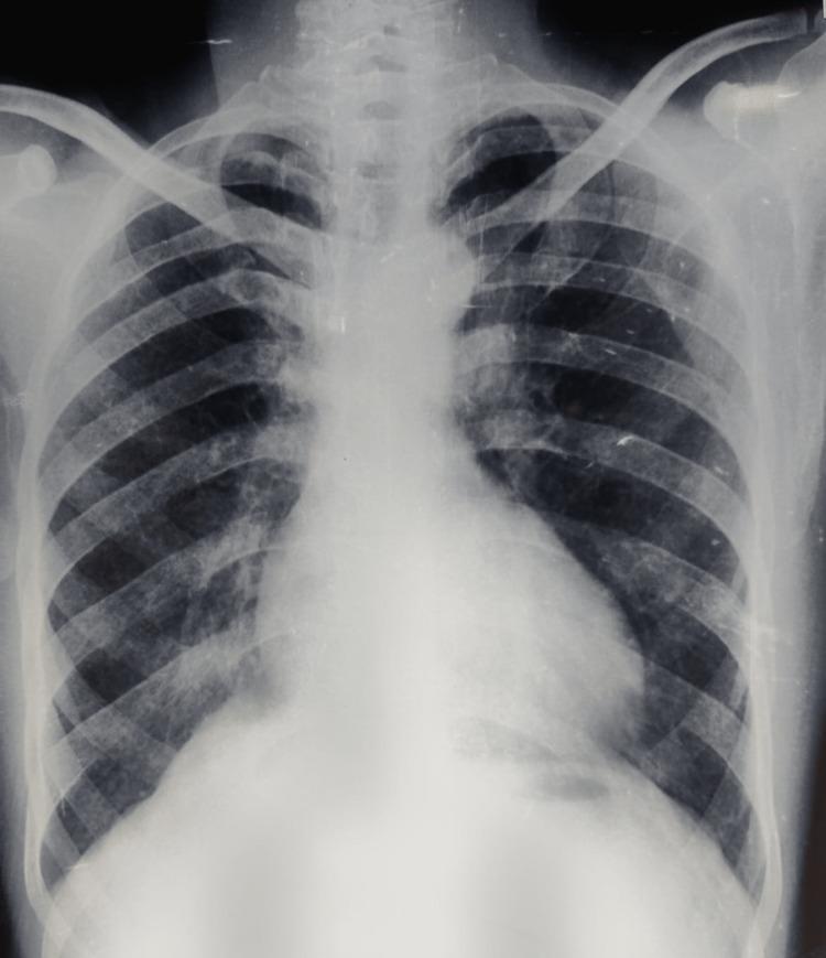
A posteroanterior (PA) chest X-ray of an adult male demonstrating classic radiological features associated with Tetralogy of Fallot (TOF). The cardiac silhouette exhibits mild cardiomegaly and a characteristic 'boot-shaped' heart (coeur en sabot). This morphology is defined by an upturned, rounded cardiac apex, indicating right ventricular hypertrophy, and a concave pulmonary artery segment (pulmonary conus). The lung fields appear relatively oligemic, reflecting decreased pulmonary blood flow typical of right-to-left shunting or pulmonary stenosis. No acute pulmonary infiltrates or pleural effusions are visible. The costophrenic angles are clear. This diagnostic image is intended for medical students and clinicians to identify visual markers of cyanotic congenital heart disease and right ventricular pressure overload in an adult clinical presentation.
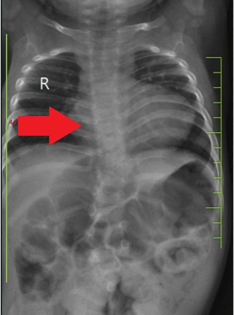
This diagnostic image is a posteroanterior (PA) chest X-ray of a pediatric patient demonstrating classic radiographic signs of Tetralogy of Fallot (TOF). The most prominent finding is a 'boot-shaped' heart (coeur en sabot), characterized by an upturned cardiac apex due to right ventricular hypertrophy and a concave pulmonary artery segment (pulmonary conus). The lung fields appear relatively oligemic, consistent with decreased pulmonary blood flow. The mediastinum is centrally located, and the diaphragm is well-defined, with the right hemidiaphragm slightly superior to the left. The skeletal structures show visible ribs and a vertebral column that appears to have mild alignment irregularities, potentially correlating with lumbar kyphoscoliosis mentioned in clinical context. The lower portion of the radiograph captures the upper abdomen, showing normal bowel gas patterns. This imaging is a classic educational example for pediatric cardiology and radiology, illustrating the morphological changes in the heart and great vessels associated with cyanotic congenital heart disease.
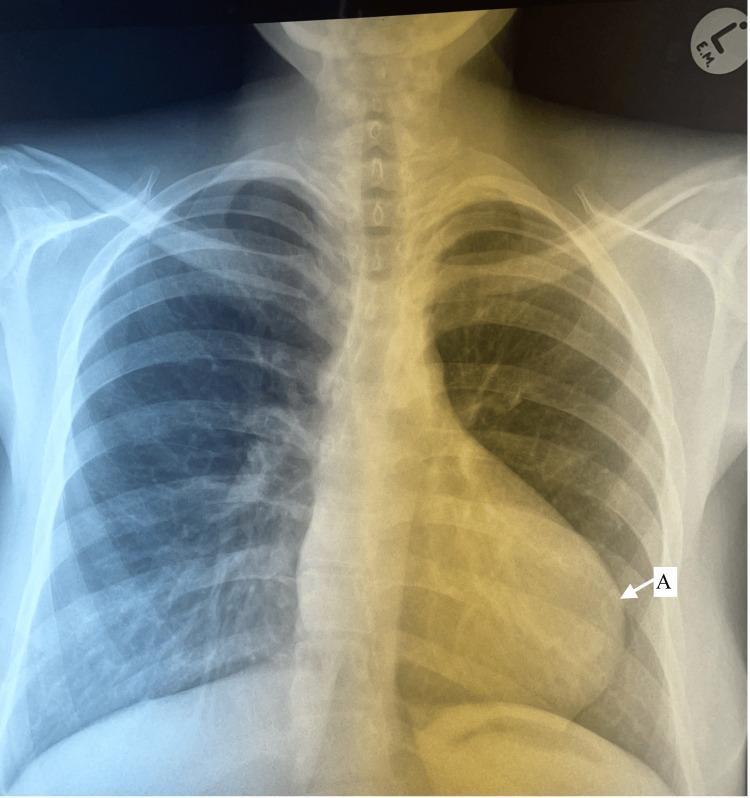
This diagnostic image is a posteroanterior (PA) chest X-ray of a pediatric patient, demonstrating classic radiographic findings of Tetralogy of Fallot. The most prominent feature is a 'boot-shaped' cardiac silhouette (coeur en sabot), characterized by an upturned apex (labeled 'A') due to right ventricular hypertrophy and a concave pulmonary artery segment (the 'waist' of the heart). The lungs exhibit generalized hyperlucency with significantly diminished pulmonary vascular markings (oligemia), suggesting decreased pulmonary blood flow secondary to right ventricular outflow tract obstruction or pulmonary stenosis. The mediastinum is narrow, and the aortic arch position is visible. Bony structures, including the ribs, clavicles, and spine, appear developmentally normal for the age group without acute pathology. This visual serves as a hallmark educational example for identifying cyanotic congenital heart disease and understanding the anatomical correlations of Tetralogy of Fallot in a clinical setting.
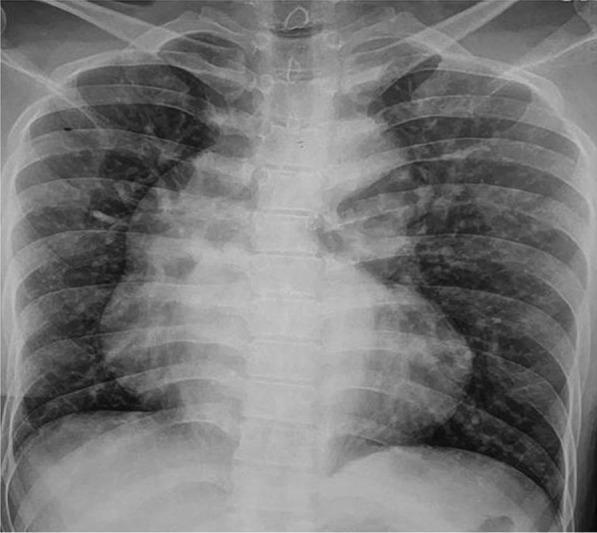
This diagnostic image is a posteroanterior (PA) chest X-ray demonstrating classic radiographic signs associated with cyanotic congenital heart disease, specifically Tetralogy of Fallot (TOF). The most prominent feature is the 'boot-shaped' heart (coeur en sabot), characterized by an upturned cardiac apex due to right ventricular hypertrophy and a concave pulmonary artery segment. The mediastinal shadow shows a significantly dilated ascending aorta, which is a common compensatory finding in TOF or pulmonary atresia. The lung fields appear remarkably clear, indicating decreased pulmonary vascular markings (oligemia) rather than pulmonary congestion, consistent with reduced pulmonary blood flow. This visual representation serves as a key educational tool for identifying the anatomical distortions caused by right-to-left shunting and right ventricular outflow tract obstruction in pediatric and adult congenital cardiology.
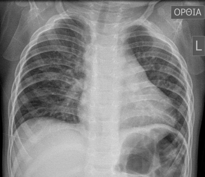
This diagnostic image is an anteroposterior (AP) chest X-ray of a pediatric patient, specifically a 16-month-old male. The image demonstrates a classic 'boot-shaped' heart (coeur en sabot), a hallmark radiographic sign of Tetralogy of Fallot. The cardiac silhouette shows an upturned apex due to right ventricular hypertrophy and a concave main pulmonary artery segment. The lung fields exhibit increased, patchy opacities and markings, particularly prominent in the right lung, which may suggest compensatory pulmonary blood flow or associated bronchial anomalies. The mediastinum appears slightly shifted, and the overall lung volume is clear enough to evaluate the bronchovascular markings. This radiograph serves as a primary educational example of congenital heart disease (CHD) manifestations in pediatric radiology, illustrating the morphological changes to the heart and the secondary effects on pulmonary vascular distribution.
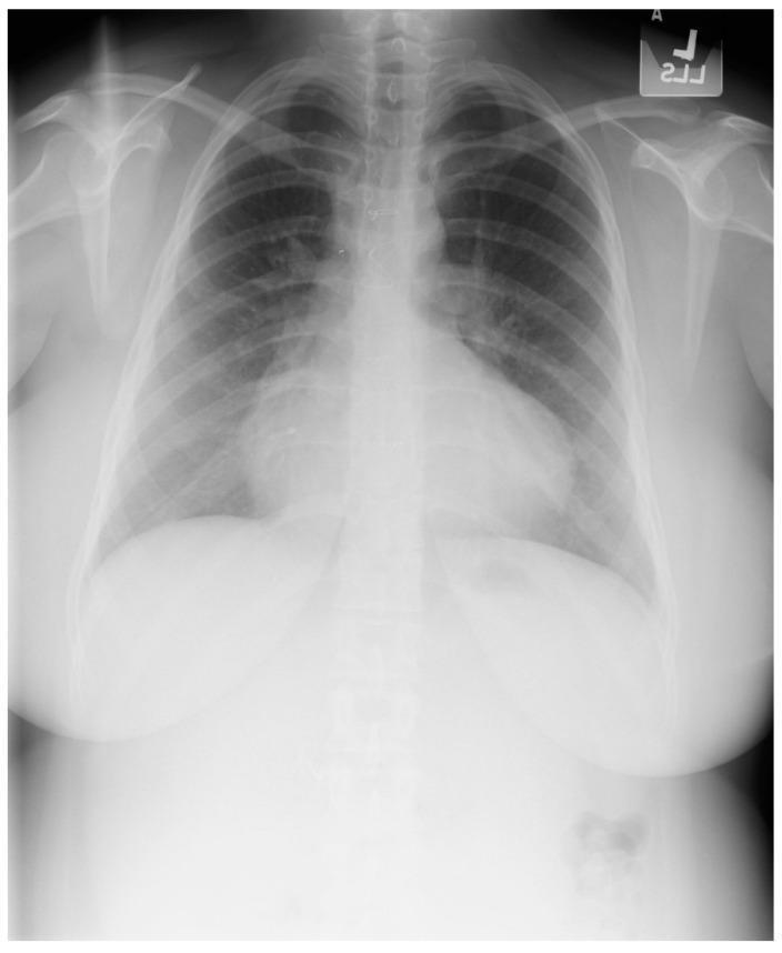
This diagnostic posteroanterior (PA) chest X-ray demonstrates the characteristic findings of Tetralogy of Fallot in a 21-year-old female patient. The image shows a well-centered view with adequate inspiratory effort. The primary finding is an increased cardiothoracic ratio with a distinctive 'boot-shaped' heart, also known as 'coeur en sabot.' This morphology is characterized by an upturned cardiac apex, resulting from significant right ventricular hypertrophy, and a concave pulmonary artery segment. The right heart border is notably prominent, indicating right atrial enlargement. Despite the history of surgical repair (including VSD closure and RV outflow tract reconstruction), these structural changes remain visible. The pulmonary vasculature appears normal, and the lung fields are clear without evidence of opacities, focal lesions, or pleural effusions. Sternal wires and surgical markers from prior cardiac procedures may be visible in the midline. The image serves as a classic educational example of chronic cardiac remodeling in congenital heart disease.
Tetralogy of Fallot four components anatomy diagram VSD overriding aorta pulmonary stenosis right ventricular hypertrophy
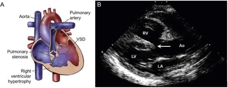
This composite educational graphic illustrates the pathophysiology and diagnostic imaging of Tetralogy of Fallot (TOF). Panel A is an anatomical diagram depicting the four hallmark features of TOF: a large ventricular septal defect (VSD), pulmonary stenosis (subvalvular and valvular), right ventricular hypertrophy with a thickened muscular wall, and an overriding aorta that sits above the septal defect, receiving blood from both ventricles. Panel B is a diagnostic 2D echocardiogram in a parasternal long-axis view. It confirms the clinical findings illustrated in Panel A, showing the right ventricle (RV) situated superiorly and the left ventricle (LV) inferiorly. A prominent white arrow points to the large malaligned VSD. The aorta (Ao) is clearly visualized overriding the interventricular septum, demonstrating its communication with both the LV and RV. The left atrium (LA) is seen posterior to the LV. The image serves as a clinical correlation between conceptual cardiac defects and their real-time ultrasound presentation.
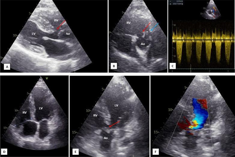
This composite of transthoracic echocardiographic (TTE) images displays key diagnostic features of Tetralogy of Fallot (TOF). The 2D grayscale images (A, B, E) identify a large malalignment-type ventricular septal defect (VSD, red arrows) and an overriding aorta (Ao) positioned over the interventricular septum. Image B, a parasternal short-axis view, further highlights a hypoplastic main pulmonary artery (blue arrow). Image D, an apical four-chamber view, shows a moderately dilated and hypertrophied right ventricle (RV) compared to the left ventricle (LV). Functional assessment via Continuous Wave Doppler (C) demonstrates severe right ventricular outflow tract (RVOT) obstruction with a peak velocity (Vmax) of 6.3 m/s, indicating a high pressure gradient. Image F uses Color Doppler to visualize bidirectional shunt flow across the VSD. This set illustrates the classic anatomical components of TOF: VSD, overriding aorta, RVOT stenosis/pulmonary hypoplasia, and subsequent RV hypertrophy, providing a comprehensive pedagogical overview of cyanotic congenital heart disease imaging.
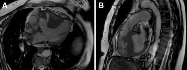
This diagnostic image consists of two panels (A and B) showing Cardiac Magnetic Resonance Imaging (MRI) in a patient with Tetralogy of Fallot. Panel A presents an axial view demonstrating a large ventricular septal defect (VSD) and an overriding aorta, where the aortic root is positioned directly above the septal defect rather than solely over the left ventricle. Panel B provides a sagittal/oblique view highlighting significant right ventricular hypertrophy, characterized by thickened muscular walls of the right ventricle. Additionally, this view illustrates subpulmonary stenosis, visible as an anatomical narrowing of the right ventricular outflow tract (RVOT) below the pulmonary valve. These findings represent the classic morphological components of Tetralogy of Fallot, a cyanotic congenital heart disease. The images serve as an educational example of how cardiac MRI can definitively characterize complex intracardiac shunts, valvular obstructions, and ventricular remodeling for surgical planning.
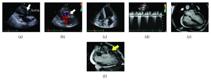
This composite figure presents multi-modal cardiac imaging of a patient with Tetralogy of Fallot. Panels (a) through (d) utilize transthoracic echocardiography: (a) Parasternal long-axis view showing a large subaortic ventricular septal defect (VSD, white arrow) with an overriding aorta. (b) Color Doppler confirms bidirectional shunting across the VSD. (c) Apical four-chamber view demonstrating marked right ventricular (RV) hypertrophy. (d) Spectral Doppler showing significant RV outflow tract (RVOT) obstruction with a high-velocity jet of 4.9 m/s. Panels (e) and (f) display Cardiac Magnetic Resonance Imaging (CMRI): (e) 4-chamber cine image used for volumetric analysis showing RV enlargement. (f) Detailed CMRI of the RVOT (yellow arrow) identifying severe infundibular (subvalvular) pulmonary stenosis. Collectively, these images illustrate the classic anatomical components of Tetralogy of Fallot including VSD, overriding aorta, RV hypertrophy, and RVOT obstruction, emphasizing the importance of multimodality imaging in assessing congenital heart disease.
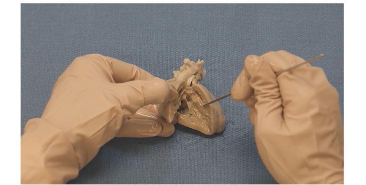
Anatomical Diagram/Clinical Photograph: This image depicts a gross anatomical dissection of a human heart specimen, specifically prepared to demonstrate the pathophysiology of Tetralogy of Fallot (TOF). The cadaveric specimen exhibits a preserved, tan, and fibrous texture, having been bisected to reveal the internal cardiac chambers. Two gloved hands are shown; one stabilizes the specimen while the other utilizes a metal surgical probe to indicate specific structural defects. The educational focus is on visualizing the four key components of TOF: ventricular septal defect (VSD), pulmonary stenosis, right ventricular hypertrophy, and the overriding aorta. The clinical significance of this specimen lies in its use for pediatric cardiology education, providing a three-dimensional perspective on congenital heart disease (CHD) morphology. The image is intended for intermediate to advanced medical learners to understand the spatial relationships between the malaligned interventricular septum and the great vessels.
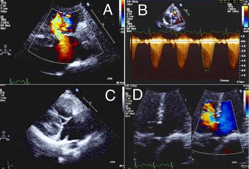
A composite of four echocardiographic images (A-D) demonstrating clinical findings consistent with Tetralogy of Fallot in an adult. (A) Color Flow Doppler imaging showing a membranous ventricular septal defect (VSD) with a significant right-to-left shunt, visualized as a turbulent, multicolored mosaic jet. (B) Spectral Continuous-Wave (CW) Doppler across the pulmonary valve, revealing a high-velocity systolic jet (reaching approximately -7.0 m/s), indicating severe pulmonic valve stenosis with dynamic obstruction. (C) Two-dimensional grayscale transthoracic echocardiogram in a four-chamber view illustrating marked right ventricular hypertrophy (RVH), characterized by thickened ventricular walls and altered chamber morphology. (D) Dual-panel view with and without Color Doppler demonstrating an overriding aorta, where the aortic root is positioned directly over the ventricular septal defect, receiving blood from both ventricles. This educational image illustrates key pathophysiological components of cyanotic congenital heart disease, relevant for cardiology and radiology training regarding anatomical abnormalities and hemodynamic shunt patterns.
Tetralogy of Fallot (ToF) - Complete Clinical Analysis
Diagnosis: Tetralogy of Fallot
The Four Cardinal Features
| # | Feature | This Patient |
|---|---|---|
| 1 | VSD (large, unrestrictive, malaligned) | Harsh grade 3/6 systolic murmur at LSB |
| 2 | Right ventricular outflow tract (RVOT) obstruction - subpulmonic/infundibular stenosis | Decreased pulmonary vascularity on CXR |
| 3 | Overriding aorta (straddles the VSD) | Murmur radiates to axilla (aortic flow) |
| 4 | Right ventricular hypertrophy (secondary to RVOT pressure) | Left sternal border heave; prominent RV on CXR |
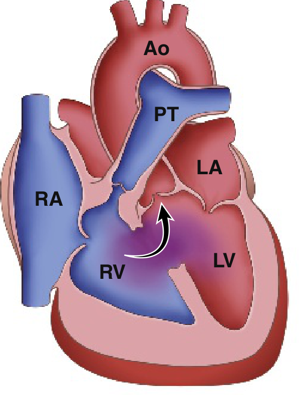
Mapping the Clinical Clues
- Episodes of turning blue during agitation - classic tet spells triggered by crying/agitation (lowers SVR, worsens right-to-left shunt)
- Peak incidence of tet spells: 2-4 months, but episodes at 2 years reflect worsening RVOT obstruction as the child grows
- Born at term, normal early life - consistent with a duct-dependent or milder form that decompensated over time
- SpO2 87% - confirms significant right-to-left shunting
- Heave at left sternal border - right ventricular hypertrophy
- Grade 3/6 harsh systolic murmur at LSB - from RVOT obstruction (NOT the VSD itself, as the VSD is large and non-restrictive)
- Radiation to axilla - suggests significant aortic flow / overriding aorta
- Decreased pulmonary vascularity - reduced pulmonary blood flow from RVOT obstruction
- Prominent right ventricle - the "boot-shaped heart" (coeur en sabot) due to RVH with upturned apex and concave pulmonary artery segment
Pathophysiology of Tet Spells
- SVR suddenly drops (crying, physical activity, defecation, fever) or HR rises (tachycardia)
- Right-to-left shunt across the VSD increases markedly
- Deoxygenated blood bypasses lungs → PaO2 falls, PaCO2 rises, pH falls
- Metabolic acidosis stimulates respiratory center → hyperpnea (deep, rapid breathing)
- Increased negative intrathoracic pressure pulls more venous blood into the RV
- More blood shunted right-to-left → further hypoxia → self-perpetuating cycle
- Rosen's Emergency Medicine, p. 3210
Why This Isn't the Other Diagnoses
| Condition | Why Excluded |
|---|---|
| VSD alone | Would show increased pulmonary vascularity (left-to-right shunt) and no cyanosis |
| Transposition of the Great Arteries | Presents in neonates, not a 2-year-old; no tet spells, "egg-on-a-string" CXR |
| Truncus arteriosus | Single vessel, very early cyanosis, increased pulmonary flow |
| Pulmonary stenosis alone | No VSD, murmur would be isolated; no shunt/cyanosis |
| Eisenmenger syndrome | Develops over years after a large L→R shunt; not at age 2 |
Immediate Management of a Tet Spell
- Knee-to-chest position (squatting equivalent) - increases SVR, reduces right-to-left shunt
- Supplemental oxygen (limited value alone, but used)
- Morphine 0.1-0.2 mg/kg IV/IM - reduces hyperpnea, decreases SVR by venodilation, decreases RVOT spasm
- Sodium bicarbonate 1 mEq/kg IV - corrects metabolic acidosis
- Propranolol 0.1-0.2 mg/kg - reduces heart rate and RVOT spasm
- Phenylephrine 0.01-0.02 mg/kg IV - raises SVR directly, decreases R-to-L shunt
- Ketamine 1-2 mg/kg IV - increases SVR, provides sedation
- IV fluids - treat hypovolemia which precipitates spells
Note: Squatting (knee-to-chest in infants) is the classic maneuver because it both increases venous return AND raises SVR by compressing femoral arteries, reducing the right-to-left shunt from both ends.
Definitive Treatment
- Total corrective repair: Patch closure of the VSD + RVOT reconstruction (resection of infundibular muscle, pulmonary valvuloplasty or transannular patch)
- Typically performed in the first 6-12 months of life
- Palliative Blalock-Taussig (BT) shunt (subclavian to pulmonary artery) may be performed first in very small infants to increase pulmonary blood flow, followed by complete repair later
Key Associations to Remember
- Most common cyanotic congenital heart disease beyond infancy (accounts for ~5% of all congenital cardiac defects)
- Right-sided aortic arch in ~25% of cases
- Associated with DiGeorge syndrome (22q11 deletion), Down syndrome, VACTERL
- Chronic hypoxemia leads to compensatory polycythemia and digital clubbing
- Without repair, some patients survive to adulthood; prognosis is excellent with surgical correction
- Robbins & Kumar Basic Pathology, p. 271-283 (ToF morphology and clinical features)
- Rosen's Emergency Medicine, p. 3210 (ToF clinical features, tet spell management)