
Sweetish smale having Glossitis in a patient with immune compromised situation .what is tha possibilitys
oral candidiasis glossitis immunocompromised tongue
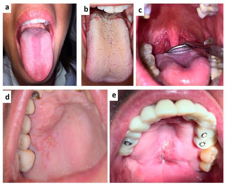
This composite of clinical intraoral photographs illustrates various oral manifestations associated with HIV infection and immunocompromised states. (a) Displays median rhomboid glossitis, appearing as a depapillated, erythematous longitudinal band on the dorsal midline of the tongue. (b) Shows pseudomembranous candidiasis (thrush) characterized by a thick, confluent, creamy-white coating across the dorsal tongue surface. (c) Depicts oropharyngeal candidiasis and herpetic-like ulcerations on the soft palate and tonsillar pillars, with visible dental prosthetics. (d) Shows an irregular herpetic lesion on the hard palate presenting as grouped erosions and erythematous patches. (e) Exhibits a large, ulcerated, and necrotizing lesion on the hard palate consistent with non-Hodgkin lymphoma in an HIV-positive patient; the maxillary arch shows extensive dental rehabilitation, including a bridge and several crown restorations. These images serve as educational markers for opportunistic infections and malignancies that frequently act as early clinical indicators of systemic immunosuppression.
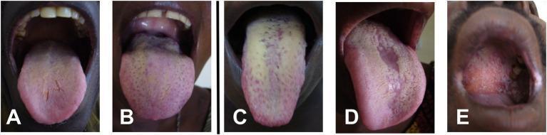
A compilation of five clinical intraoral photographs (A–E) illustrating various presentations of oral candidiasis. Images A and B demonstrate mild involvement, characterized by thin yellowish-white coatings on the dorsal tongue covering less than 50% of the surface area. Image C displays a more severe case of pseudomembranous candidiasis, with thick, confluent whitish-yellow plaques covering over 50% of the tongue's dorsal surface. Image D shows median rhomboid glossitis, appearing as a well-defined, erythematous, atrophic area along the midline of the tongue, where filiform papillae are absent, surrounded by light peripheral plaques. Image E depicts palatal involvement, showing a diffuse erythematous area with granular texture and small inflammatory lesions on the hard and soft palate. This series is used for the clinical classification of oral fungal infections, often seen in immunocompromised patients, such as those with HIV/AIDS, and demonstrates the spectrum from mild yeast colonization to severe acute thrush and atrophic mucosal changes.
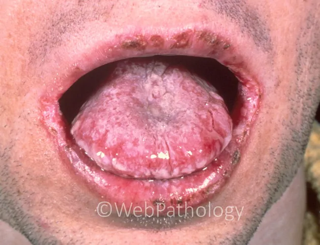
This is an intraoral clinical photograph of a patient with oral candidiasis (thrush). An adherent, white, curd-like pseudomembrane extensively coats the tongue surface (dorsum and lateral margins) and is removable with gentle scraping, exposing erythematous, friable mucosa beneath. The tissue appears inflamed with mucosal glossitis and reduced turgor at the tongue base; the white plaques may extend to the buccal mucosa. In addition, the lips show areas of erosion and crusting consistent with traumatic or infectious mucosal damage that can accompany candidal infection or secondary bacterial superinfection. The image demonstrates characteristic features of pseudomembranous candidiasis: cottage-cheese-like plaques, positive scraping test, and mucosal erythema. Clinical significance: this presentation is typical in immunocompromised individuals, diabetics, antibiotic or inhaled corticosteroid users, the elderly, or patients with HIV/AIDS; suggests need for antifungal therapy (topical nystatin/clotrimazole or systemic fluconazole) and evaluation of underlying risk factors. Potential differential diagnoses include oral leukoplakia (non-scrapable white patch), erythematous candidiasis, aphthous ulcers, or herpes stomatitis; correlation with systemic symptoms and immune status is advised. This image is useful for educational illustration of thrush morphology, scraping test, and mucosal involvement in the oral cavity. Documented features support prompt clinical management and targeted laboratory evaluation as indicated for patient safety and care.
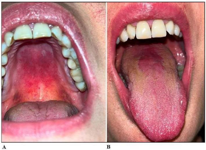
Two-panel clinical photograph displaying intraoral manifestations of erythematous oral candidiasis. Panel A shows the maxillary arch and palate, characterized by diffuse erythema and focal erosive areas across both the hard and soft palate mucosal surfaces. Panel B illustrates the lingual surface, demonstrating erythematous-atrophic glossitis. The dorsal tongue exhibits a central zone of depapillation and atrophy, resulting in a smooth, red appearance, contrasted with surrounding areas that show a brownish-tan discoloration and altered texture. These clinical features are characteristic of atrophic candidiasis in a patient with chronic symptoms. This image is an educational resource for dental and medical students to identify fungal infections of the oral cavity and distinguish between palatal erythema and atrophic glossitis.
Sweet Smell + Glossitis in an Immunocompromised Patient - Differential Possibilities
Understanding the Key Clue: Sweet/Fruity Oral Odor
| Sweet Odor Type | Associated Cause |
|---|---|
| Sweet/yeasty/bread-like | Candida infection (acetaldehyde byproducts) |
| Fruity/acetone-like | Diabetic ketoacidosis (DKA) - often co-exists with immunosuppression |
| Grape-like/fruity | Pseudomonas aeruginosa infection |
| Sweet putrid | Anaerobic/mixed infection (necrotizing stomatitis) |
Top Differential Diagnoses
1. Oral Candidiasis (MOST LIKELY) - "Thrush"
- In adults, the tongue may appear atrophic with a smooth, glazed, bright red surface (erythematous/atrophic candidiasis = a form of glossitis), or there may be grayish-white pseudomembranous plaques on the tongue and buccal mucosa
- Candida produces volatile organic compounds including acetaldehyde and ethanol, which give a sweet/yeasty smell
- It is often the first clinical sign of AIDS and is present in almost all untreated HIV patients
- Candida species beyond C. albicans (e.g. C. glabrata, C. krusei) are more prevalent in HIV disease
- Pseudomembranous (white curd-like plaques that scrape off, leaving red mucosa)
- Erythematous/atrophic (smooth, red, depapillated tongue = glossitis)
- Median rhomboid glossitis (depapillated erythematous patch at dorsal tongue midline)
- Andrews' Diseases of the Skin, block 4 - "The papillae of the tongue may appear atrophic, with the surface smooth, glazed, and bright red. It is often the first manifestation of AIDS."
- Scott-Brown's Otorhinolaryngology - "Oral candidiasis has been reported as the most prevalent oral lesion [in HIV disease]."
2. Herpetic Geometric Glossitis (HSV Reactivation)
- Characterized by painful, geometric, linear fissures on the dorsal tongue surface, making eating difficult
- Almost exclusively seen in immunosuppressed patients (HSV-1 reactivation), including those with HIV, leukemia, or neutropenia
- Can produce a sweetish/musty odor from ulcerated tissue
- Responds to oral acyclovir
"Herpetic geometric glossitis...tends to present in immunosuppressed patients as a reactivation of chronic oral herpes simplex infection." - Sleisenger & Fordtran's GI and Liver Disease
"Although the glossitis usually affects only immunocompromised patients...linear longitudinal, cross-hatched, or branching fissures of the dorsal tongue...may be quite painful and may limit oral intake." - Andrews' Diseases of the Skin
3. Diabetic Ketoacidosis (DKA) / Uncontrolled Diabetes
- Diabetes is both a cause of immunocompromise and produces a fruity/sweet acetone breath (ketone bodies - acetone)
- Glossitis in DKA patients is also driven by secondary candidiasis (diabetes predisposes strongly)
- A dry mouth from hyperglycemia further impairs salivary innate immunity, promoting Candida overgrowth
- Consider this if the patient has diabetes or if immunosuppression is from diabetic complications
4. Pseudomonas aeruginosa Infection
- Pseudomonas has a characteristic grape-like/sweet fruity smell (from 2-aminoacetophenone)
- In neutropenic/immunocompromised patients, Pseudomonas can cause oral/tongue ulcers and necrotizing stomatitis
- Less common cause of glossitis but important in severely neutropenic patients (e.g., post-chemotherapy)
5. Hairy Leukoplakia (EBV)
- White corrugated patches on lateral tongue borders, caused by Epstein-Barr virus reactivation in immunocompromised (especially HIV)
- Not primarily sweet-smelling but can co-exist with Candida (which provides the odor)
- Unlike candidiasis, these plaques cannot be scraped off
6. Acute Necrotizing Ulcerative Gingivitis/Stomatitis (ANUG/NOMA)
- In severely immunocompromised patients, anaerobic bacterial infection can produce necrotic, sweet-putrid smelling oral lesions with tongue involvement
- Particularly relevant in HIV patients with very low CD4 counts
Quick Diagnostic Approach
| Step | Action |
|---|---|
| 1. Scrape the white patches | If they come off - Candida (thrush). If not - consider hairy leukoplakia or dysplasia |
| 2. Check blood glucose & ketones | Rule out DKA |
| 3. Check CD4 count / HIV status | CD4 <200: all opportunistic infections more likely |
| 4. KOH preparation / culture | Confirm Candida; identify species for azole resistance |
| 5. Swab for HSV PCR | If painful fissures/geometric pattern on tongue |
| 6. Check for Pseudomonas | In neutropenic patients with necrotic lesions |
Summary Priority List
- Oral Candidiasis (erythematous/atrophic form = glossitis) - most likely by far
- Concurrent DKA/hyperglycemia driving both immunosuppression and sweet breath
- Herpetic Geometric Glossitis (HSV reactivation) - if painful linear fissures
- Pseudomonas stomatitis - if neutropenic with necrotic lesions
- Mixed/anaerobic infection - if severe necrotizing picture