
Mechanism of wound healing in detail for 20 marks from Scott brown Cummings Zakir Hussain Dhingra Hazarika and related articles in detail
Reading File
Finding Sources
Finding Sources
Finding Sources
Reading File
Reading File
Finding Sources
Reading File
Reading File
Finding Sources
Reading File
Reading File
Finding Sources
Reading File
Reading File
Reading File
Reading File
Searching PubMed
wound healing phases mechanisms review
Reading File
Reading File
Loading Image
Loading Image
Reading File
Reading File
Reading File
Reading File
I now have comprehensive content from all the key textbooks. Let me compile the complete, detailed 20-mark answer.
Mechanism of Wound Healing - Detailed Answer (20 Marks)
Sources: Cummings Otolaryngology Head & Neck Surgery, Scott-Brown's Otorhinolaryngology Head & Neck Surgery (Vol 1), Schwartz's Principles of Surgery (11e), Bailey & Love's Short Practice of Surgery (28e)
INTRODUCTION
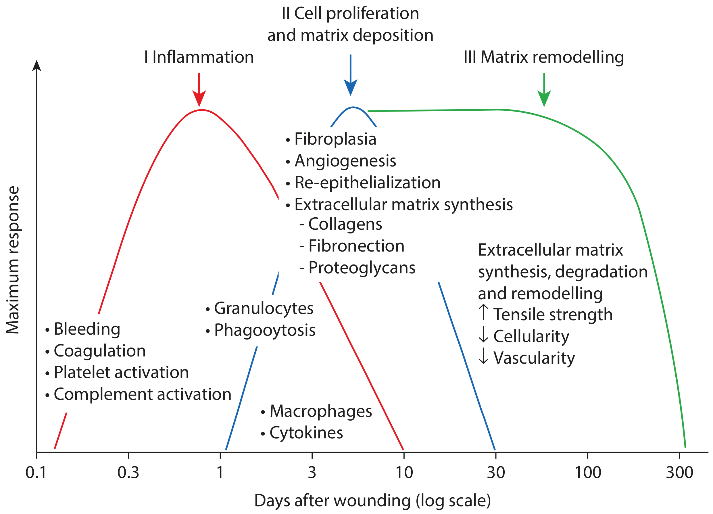
Wound healing is a complex, dynamic, and coordinated cellular and biochemical cascade that leads to restitution of tissue integrity and function following injury. Regardless of the causative agent, the events associated with wound repair are relatively consistent. The process has classically been divided into three (or four, if haemostasis is separated) overlapping phases:
- Haemostasis (immediate, minutes to hours)
- Inflammation (days 1-7)
- Proliferation / Fibroplasia (day 3 to week 4)
- Remodelling / Maturation (week 1 onward, up to 2 years)
(Cummings Otolaryngology)
TYPES OF WOUND HEALING
Before describing the mechanism, it is important to classify how wounds heal:
1. Primary Intention (Healing by First Intention)
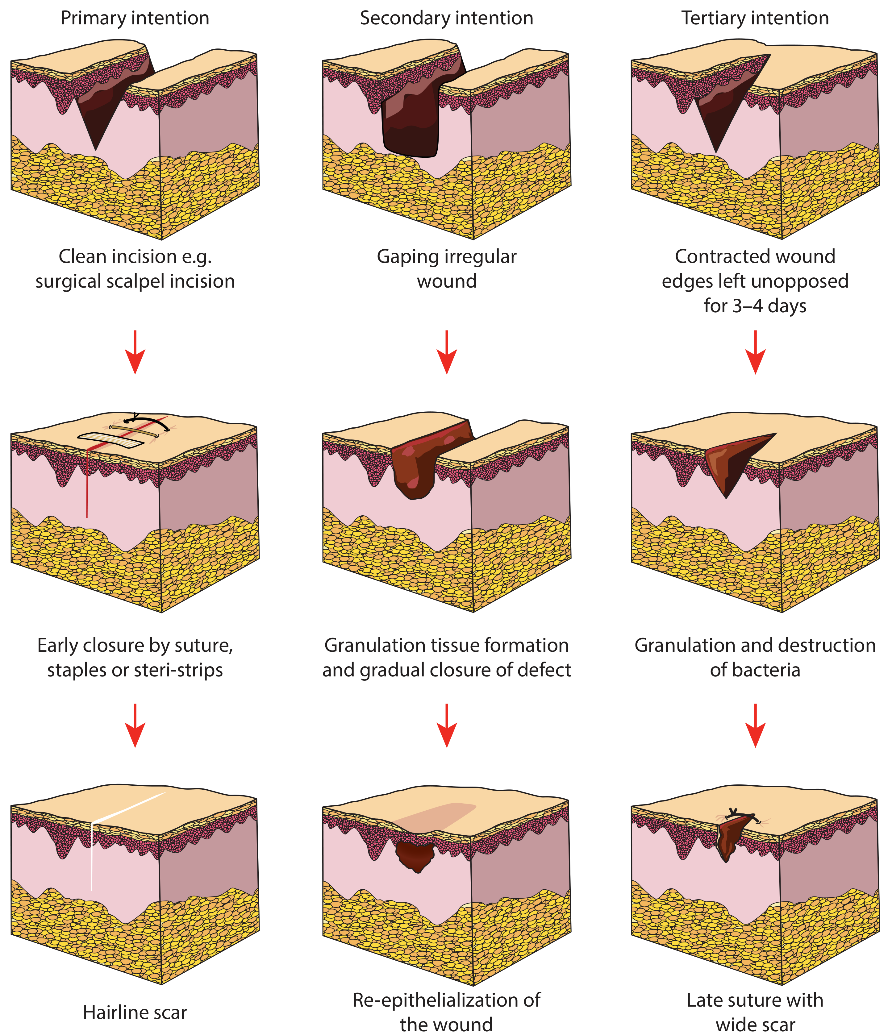
Occurs when wound edges are cleanly apposed within 12-24 hours - e.g., surgical incisions closed with sutures, skin glue, or steri-strips. Apposition reduces the distance for epidermal proliferation. Results in a thin, hairline scar. (Scott-Brown's)
2. Secondary Intention (Healing by Second Intention)
Occurs in large, gaping, or infected wounds where edges cannot be apposed. Epithelial cells must grow both downwards and across the wound bed. Granulation tissue fills the defect; wound contraction and subsequent epithelialization close the wound. Myofibroblasts appear around day 3. The process is slower and may result in contracture. (Scott-Brown's)
3. Tertiary Intention (Delayed Primary Closure)
Occurs in contaminated wounds (bites, peritoneal soiling). Wound edges are intentionally left unopposed for 3-4 days, during which host phagocytes debride the wound and destroy bacteria. Edges are then approximated. (Scott-Brown's)
PHASE 1: HAEMOSTASIS
Timeline: Immediate (minutes)
Tissue injury disrupts vascular endothelium, causes extravasation of blood constituents, and exposes basal lamina. The vascular response consists of:
- Vasoconstriction lasting 10-15 minutes (mediated by catecholamines and prostaglandins)
- Platelet activation: Exposure of sub-endothelial collagen triggers platelet adhesion, aggregation, and degranulation
- Coagulation cascade (extrinsic pathway) is activated, resulting in fibrin deposition
- A clot forms composed of collagen, platelets, thrombin, and fibronectin
This clot:
- Covers and protects the wound from contamination
- Provides an initial scaffold for cell attachment and migration
- Acts as a reservoir of growth factors
Growth factors released from platelet α-granules:
| Growth Factor | Source | Function |
|---|---|---|
| PDGF (Platelet-Derived GF) | Platelets, macrophages, endothelial cells | Chemoattractant for macrophages; collagen synthesis; fibroblast stimulation |
| TGF-β (Transforming GF-β) | Platelets, lymphocytes, macrophages, keratinocytes | Chemoattractant for monocytes, macrophages, neutrophils, fibroblasts; stimulates angiogenesis and fibrosis |
| EGF (Epidermal GF) | Platelets, macrophages | Keratinocyte migration; stimulates fibroblasts and collagen synthesis |
| VEGF | Macrophages | Promotes angiogenesis |
| PAF (Platelet Activating Factor) | Platelets | Initiates inflammation |
| Serotonin | Platelets | Vasoconstriction |
(Cummings; Scott-Brown's; Schwartz's)
Vasodilatation sets in after ~10-15 minutes due to release of leukotrienes, prostaglandins, and histamine from endothelial and mast cells, along with thrombin and kinins - facilitating extravascular migration of inflammatory cells. (Cummings)
PHASE 2: INFLAMMATION
Timeline: Days 1-7; lasts 4-5 days in a sutured wound, 7-10 days in an open wound
This phase is divided into early (days 1-2) and late (days 2-3) sub-phases.
Early Inflammation - Neutrophil Phase (Days 1-2)
Activation of classical and alternative complement cascades leads to neutrophil infiltration. Chemoattractants include:
- TGF-β, complement constituents (C3a, C5a)
- Bacterial peptide products
- Fragments of ECM proteins, IL-1, TNF-α, platelet factor 4
Steps of neutrophil migration:
- Margination - neutrophils adhere to endothelial cells of surrounding blood vessels
- Diapedesis - movement through the vessel wall into tissue
- Chemotaxis - directed migration toward the wound
Neutrophil functions:
- Phagocytosis and intracellular destruction of bacteria
- Release of proteolytic enzymes (collagenases) to digest devitalized tissue and bacteria
- Formation of oxygen-free radicals via the myeloperoxidase pathway to sterilize the wound
- Release of IL-1α, IL-1β, and TNF-α - early activators of growth factor expression in macrophages, keratinocytes, and fibroblasts
Neutrophils constitute ~50% of all cells at the wound site after day 1. They decline within days through apoptosis and phagocytosis by macrophages.
Note: Neutrophils are not essential for healing in a sterile wound - their primary role is infection control. (Schwartz's)
Late Inflammation - Macrophage Phase (Days 2-3)
Monocytes are attracted to the wound by cytokines, complement components, and breakdown products of collagen and elastin. They arrive 48-72 hours post-injury and differentiate into macrophages at the wound site - becoming the most important cell of the late inflammatory stage and the "master regulators" of wound healing.
Macrophage activities:
| Activity | Mediators |
|---|---|
| Phagocytosis | Reactive oxygen species, nitric oxide |
| Debridement | Collagenase, elastase |
| Cell recruitment & activation | PDGF, TGF-β, EGF, IGF, TNF-α, IL-1, IL-6 |
| Matrix synthesis | TGF-β, EGF, PDGF, prostaglandins |
| Angiogenesis | FGF, VEGF, TNF-α, nitric oxide |
(Schwartz's Table 9-1; Scott-Brown's)
Macrophages are the bridge between inflammation and proliferation - without them, healing fails.
T-lymphocytes (Days 5-7)
T-lymphocytes infiltrate the wound peaking at ~1 week and bridge the inflammatory and proliferative phases. They are less numerous than macrophages but have important regulatory roles:
- CD8+ suppressor T cells: depletion enhances wound healing
- CD4+ helper T cells: depletion has no effect
- Lymphocytes exert a downregulating effect on fibroblast collagen synthesis via IFN-γ, TNF-α, and IL-1 through direct cell-cell contact - suggesting extracellular matrix synthesis is regulated not only by soluble factors but also by direct cell-cell contact (Schwartz's)
PHASE 3: PROLIFERATION (FIBROPLASIA)
Timeline: Day 3 to week 4 (overlaps with inflammation)
The proliferative phase begins on day 3, lasts about 2-4 weeks, and consists of three key processes: re-epithelialization, granulation tissue formation (fibroplasia + angiogenesis), and wound contraction.
3A. Re-epithelialization
Attempts to restore the protective barrier of the skin. Begins within hours of injury.
Process:
- Increased mitotic activity within basal cells of wound edges begins within 12 hours
- Keratinocytes migrate - as a sheet, extending lamellipodia across the defect, migrating between the fibrin clot and the collagenous dermis
- "Epiboly" - creation of a delicate epithelial covering over the raw area
- Migration is guided by integrin receptor-ECM interactions
- Keratinocytes produce plasminogen activators which activate plasmin and collagenase-1 (MMP-1) to dissolve fibrin and type I collagen for migration
- Contact inhibition stops migration once epithelial sheets meet
- Keratinocytes return to normal differentiation; epithelial stratification is restored
Keratinocyte proliferation begins 1-2 days post-injury. In partial-thickness wounds, skin appendages (hair follicles, sebaceous glands) serve as an additional source of epithelial cells. (Cummings; Scott-Brown's)
Migration is most rapid in hyperbaric/moist conditions and dependent on oxygen tension. (Scott-Brown's)
Growth factors driving re-epithelialization: EGF, TGF-α, KGF (keratinocyte growth factor from fibroblasts), IGF-1, IL-8
3B. Granulation Tissue Formation
At approximately day 3-4, granulation tissue begins to replace the fibrin clot. It consists of:
- New blood vessels (angiogenesis)
- Fibroblasts producing ECM
- Loose collagenous matrix (pink, granular appearance on examination)
Fibroplasia
The fibroblast is the most important cell in granulation tissue production. Fibroblasts are attracted 2-4 days after injury by PDGF and TGF-β.
Steps:
- Fibroblasts proliferate and migrate into the wound
- They produce: fibronectin, hyaluronan, collagen (types I and III), elastin, and proteoglycans
- Initial matrix: fibrin and fibronectin → gradually replaced by collagen and glycoproteins
- Lactate (accumulates ~10 mmol in wound) is a potent regulator of collagen synthesis via adenosine-related mechanisms
Collagen synthesis (detailed):
- Fibroblasts synthesize procollagen chains (protocollagen, ~1000 amino acids), each with glycine in every third position + proline or lysine
- In the endoplasmic reticulum: hydroxylation (prolyl hydroxylase requiring O₂, iron, α-ketoglutarate, vitamin C as cofactors) + glycosylation → forces α-helical configuration
- Three α-helical chains entwine to form procollagen (right-handed superhelix with registration peptides)
- Extracellularly: registration peptides are cleaved by procollagen peptidase → collagen monomer
- Further polymerization and cross-linking via intra/intermolecular covalent bonds → mature collagen fibrils (Schwartz's)
Collagen types in wound healing:
- Type III collagen predominates early (scar collagen) - more flexible, less strong
- Type I collagen predominates in mature scar - thicker fibers, greater tensile strength
- Aberrant collagen deposition → keloid (excess) or poor wound healing (deficit)
Proteoglycan synthesis: Glycosaminoglycans (repeating disaccharide units) couple with proteins → proteoglycans, forming ground substance of granulation tissue.
Angiogenesis
New blood vessel formation begins 2-3 days after wounding - essential for metabolic support of the healing wound.
Steps of angiogenesis:
- Degradation of basement membrane of parent vessel
- Endothelial cell migration toward angiogenic stimuli
- Endothelial cell proliferation
- Formation and maturation into capillary tubes
- Invasion creates a microvascular network
Key inducers:
- VEGF (vascular endothelial growth factor) - produced by macrophages and keratinocytes in response to hypoxia
- FGF-1 and FGF-2 (acidic and basic fibroblast growth factor) - released from disrupted cells; most potent angiogenic factors
- PDGF, TGF-β
- Hypoxia is the strongest physiologic inducer of angiogenesis
Chronic wounds with insufficient vascular supply show delayed healing.
3C. Wound Contraction
Begins 7 days after wounding; peaks at 2 weeks. Reduces size of defect at a rate of 0.6-0.7 mm/day. (Scott-Brown's)
Mechanism:
- Fibroblasts differentiate into myofibroblasts (contain smooth muscle actin - α-SMA)
- Myofibroblasts attach to the ECM and to each other
- Through contractile activity, they pull wound edges together
- Driven by TGF-β
Significance:
- Reduces the amount of scar tissue needed
- In humans, skin is relatively immobile - contraction alone rarely closes a wound completely
- Excessive contraction → contracture (impairs function, especially at mobile sites like the neck, joints)
- Insufficient contraction → delayed wound closure, bleeding, infection
PHASE 4: REMODELLING AND SCAR MATURATION
Timeline: Week 1 onward, continuing for up to 1-2 years
This is the longest phase of wound healing and determines the final wound appearance.
Key events:
- Equilibrium is reached between collagen formation and degradation around day 21
- Matrix metalloproteinases (MMPs) - collagenases, gelatinases, stromelysins - synthesized by fibroblasts, neutrophils, and macrophages degrade excess collagen
- TIMPs (tissue inhibitors of metalloproteinases) regulate MMP activity
- Type III collagen is progressively replaced by type I collagen
- Collagen fibers reorganize from a disorganized array of fine fibers → thicker fibers aligned parallel to skin stress lines (Langer's lines)
- Myofibroblasts and vascular cells undergo apoptosis → granulation tissue transforms into scar
- Reduced: cellularity, vascularity, macrophage activity, capillary outgrowth
Tensile strength progression (Cummings):
- At 1 week: 3% of normal dermis
- At 3 weeks: 20% of normal dermis
- At 3 months: 80% of normal dermis
- The scar never fully regains normal dermal strength (maximum ~80%)
Lymphocytes are the most common leukocyte subset in human skin wounds during this phase. (Cummings)
GROWTH FACTORS AND CYTOKINES IN WOUND HEALING (SUMMARY TABLE)
| Cytokine/GF | Origin | Key Role |
|---|---|---|
| TGF-β | Platelets, lymphocytes, macrophages, keratinocytes | Chemoattractant (monocytes, fibroblasts); stimulates angiogenesis, fibrosis, ECM deposition |
| PDGF | Platelets, macrophages, endothelial cells | Chemoattractant for macrophages; fibroblast stimulation; collagen synthesis |
| VEGF | Macrophages | Angiogenesis |
| FGF (acidic & basic) | Macrophages, lymphocytes, endothelial cells | Potent angiogenesis; stimulates fibroblasts and keratinocytes |
| EGF | Platelets, macrophages | Keratinocyte migration; collagen synthesis |
| TGF-α | Macrophages, keratinocytes | Stimulates fibroblast and keratinocyte migration |
| IGF-1 | Keratinocytes, fibroblasts, platelets | Re-epithelialization; granulation tissue formation |
| KGF | Fibroblasts | Keratinocyte migration, differentiation, proliferation |
| IL-8 | Macrophages, keratinocytes | Keratinocyte proliferation; neutrophil chemotaxis |
| TNF-α | Lymphocytes, macrophages | Matrix formation; stimulates fibroblasts; early activator of healing |
(Cummings Table 77.3)
FACTORS AFFECTING WOUND HEALING
(Scott-Brown's, Cummings)
Local Factors
- Infection (most common cause of delayed healing)
- Foreign bodies, devitalized tissue
- Ischemia / poor blood supply
- Mechanical trauma, excessive tension
- Radiation damage
- Wound type and size
Systemic Factors
- Age: Elderly patients show delayed epithelialization, altered inflammatory response, reduced collagen content, impaired angiogenesis
- Nutritional status: Protein-calorie malnutrition delays healing; key nutrients include arginine, vitamins A and C, and zinc. Serum albumin <30 g/L predicts delayed healing
- Diabetes: Immune dysfunction, slower collagen synthesis, decreased angiogenesis, poor tensile strength - multifactorial
- Malnutrition: Decreased fibroblast proliferation, decreased proteoglycan and collagen synthesis
- Medications: Corticosteroids, immunosuppressants, and chemotherapy agents alter the healing environment in the first 3 days - critical molecular modulators are released during this window
- Smoking: Reduced oxygenation, vasoconstriction, impairs all phases
- Immunosuppression: Failure of inflammatory phase, absent molecular modulators
- Weight loss >10% (common in cancer patients) correlates with decreased wound healing
EXCESSIVE / ABNORMAL WOUND HEALING
Hypertrophic Scars
- Excess collagen deposition within the original wound boundaries
- Elevated, red, firm scar
- Common at mobile sites (neck, joints)
- Management: topical silicone, intralesional corticosteroid, compression therapy, surgical excision (Bailey & Love)
Keloid Scars
- Extend beyond original wound borders - locally destructive
- Occur more commonly in darker skin types (African, Asian)
- Most commonly affect: face, earlobes, deltoid area, presternal region
- Appear months after injury
- More resistant to treatment; may require repeated excision + adjuvant radiotherapy (Bailey & Love)
Chronic Wounds
- Healing delayed >3 weeks
- Deficient in growth factors; decreased keratinocyte and fibroblast migration
- Increased reactive oxygen species and tissue proteases
- Microbial contamination; fibroblasts become senescent
RECENT EVIDENCE (PubMed, 2022-2024)
- Peña & Martin, 2024 (Nat Rev Mol Cell Biol) - "Cellular and molecular mechanisms of skin wound healing" - updated review confirming the four-phase model with emphasis on immune cell crosstalk and stem cell roles (PMID: 38528155)
- Sorg & Sorg, 2023 (Eur Surg Res) - "Skin Wound Healing: Of Players, Patterns, and Processes" - comprehensive review of cellular players (PMID: 36417847)
SUMMARY
Wound healing is a coordinated four-phase process:
| Phase | Timing | Key Cells | Key Events |
|---|---|---|---|
| Haemostasis | Minutes | Platelets | Vasoconstriction, clot, growth factor release |
| Inflammation | Days 1-7 | Neutrophils (1-2 days), Macrophages (2-3 days), T-lymphocytes (5-7 days) | Debridement, phagocytosis, chemokine release |
| Proliferation | Days 3-28 | Keratinocytes, Fibroblasts, Endothelial cells | Re-epithelialization, granulation tissue, angiogenesis, wound contraction |
| Remodelling | Week 1 to 2 years | Fibroblasts, MMPs | Collagen reorganization, scar maturation, tensile strength gain |
All phases overlap in time. Any disruption to this cascade leads to delayed healing or chronic wounds. The scar tissue never fully recaptures the tensile strength of normal dermis (maximum ~80% at 3 months).
References: Cummings Otolaryngology Head & Neck Surgery (9780323612173) | Scott-Brown's Otorhinolaryngology Head & Neck Surgery Vol 1 (9781138094611) | Schwartz's Principles of Surgery 11e (9781259835346) | Bailey & Love's Short Practice of Surgery 28e (9780367548117)
This is a shared conversation. Sign in to Orris to start your own chat.