Tuberculous lymphadenitis

Reading File
Finding Sources
Finding Sources
Searching PubMed

"tuberculous lymphadenitis"

Searching the Web

WHO tuberculosis treatment extrapulmonary lymph node tuberculosis guideline adults 2025

Reading File
Reading File
Reading File
Reading Web Page

https://tbksp.who.int/ar/node/3024

Reading File
Reading File
Searching Images

tuberculous cervical lymphadenitis cold abscess clinical photograph
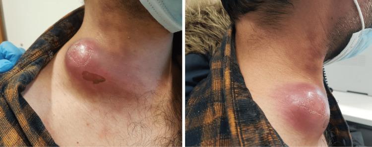
This clinical photograph consists of two panels showing the right lateral and anterolateral views of a patient's neck. The primary finding is a large, prominent, circumscribed mass approximately 6 cm in diameter located in the right cervical region. The overlying skin exhibits significant inflammatory changes, including intense erythema and a shiny, taut texture suggesting edema and underlying tension. There are areas of subtle desquamation and a small, darker, well-demarcated area that may represent a focal scab or localized skin necrosis. The mass appears fluctuant, which is characteristic of a cold abscess or severe lymphadenitis. Clinically, this presentation is highly suggestive of tuberculous lymphadenitis (scrofula) or a severe bacterial cervical lymphadenitis. The presence of a surgical mask on the patient and a blue-gloved hand in the frame indicates a clinical examination setting. This visual material is used to teach the physical signs of extrapulmonary tuberculosis and the differential diagnosis of unilateral cervical masses in adults.
This clinical photograph consists of two panels showing the right lateral and anterolateral views of a patient's neck. The primary finding is a large, prominent, circumscribed mass approximately 6 cm in diameter located in the right cervical region. The overlying skin exhibits significant inflammatory changes, including intense erythema and a shiny, taut texture suggesting edema and underlying tension. There are areas of subtle desquamation and a small, darker, well-demarcated area that may represent a focal scab or localized skin necrosis. The mass appears fluctuant, which is characteristic of a cold abscess or severe lymphadenitis. Clinically, this presentation is highly suggestive of tuberculous lymphadenitis (scrofula) or a severe bacterial cervical lymphadenitis. The presence of a surgical mask on the patient and a blue-gloved hand in the frame indicates a clinical examination setting. This visual material is used to teach the physical signs of extrapulmonary tuberculosis and the differential diagnosis of unilateral cervical masses in adults.

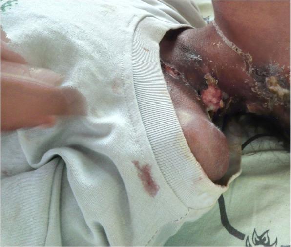
Clinical photograph of the left lateral neck and supraclavicular region of a patient, demonstrating classic features of scrofuloderma (cutaneous tuberculosis). The image shows multiple matted, enlarged cervical lymph nodes that have progressed to involve the overlying skin. Key visible findings include irregular skin ulcerations with undermined borders, friable granulation tissue, and active purulent drainage of a yellowish-white consistency. Distinctive draining sinus tracts are visible, along with thick, dark-colored crusts and scabs adjacent to the open lesions. The surrounding skin exhibits inflammatory changes, including erythema and hyperpigmentation. This presentation illustrates the progression of tuberculous lymphadenitis (scrofula) where the infection extends from deeper lymphatic structures to the cutaneous surface, resulting in chronic ulceration and cold abscess formation.

Clinical photograph of the left lateral neck and supraclavicular region of a patient, demonstrating classic features of scrofuloderma (cutaneous tuberculosis). The image shows multiple matted, enlarged cervical lymph nodes that have progressed to involve the overlying skin. Key visible findings include irregular skin ulcerations with undermined borders, friable granulation tissue, and active purulent drainage of a yellowish-white consistency. Distinctive draining sinus tracts are visible, along with thick, dark-colored crusts and scabs adjacent to the open lesions. The surrounding skin exhibits inflammatory changes, including erythema and hyperpigmentation. This presentation illustrates the progression of tuberculous lymphadenitis (scrofula) where the infection extends from deeper lymphatic structures to the cutaneous surface, resulting in chronic ulceration and cold abscess formation.

Tuberculous lymphadenitis

Tuberculous lymphadenitis is tuberculosis involving lymph nodes. It is one of the most common forms of extrapulmonary TB and most often affects the cervical lymph nodes. The classic term is scrofula.
Tuberculous cervical lymphadenitis with cold abscess

Typical presentation

  • Gradually enlarging, usually painless lymph nodes, commonly posterior cervical or supraclavicular
  • Nodes may be firm and matted
  • Low-grade fever, night sweats, fatigue, weight loss, or cough may occur, but may be absent
  • With progression: caseation leads to a painless cold abscess
  • A cold abscess may form a draining sinus or ulcer, often with undermined edges
Most patients with isolated lymph-node TB do not have obvious active pulmonary TB, though pulmonary involvement should still be assessed. Goldman-Cecil Medicine, p. 2912-16; Bailey and Love's Short Practice of Surgery, p. 108.

Diagnosis

Diagnosis should be microbiologically or histologically confirmed whenever possible.
  1. Ultrasound of the node can identify necrosis/abscess and guide sampling.
  2. Fine-needle aspiration cytology (FNAC) is usually first-line:
    • Cytology for granulomatous inflammation/caseation
    • Acid-fast bacilli smear
    • Mycobacterial culture with drug-susceptibility testing
    • Rapid molecular testing such as Xpert MTB/RIF, where available
  3. Core or excision biopsy if FNAC is non-diagnostic or lymphoma/malignancy remains a concern.
  4. Evaluate for other TB sites:
    • Chest radiograph and sputum testing if respiratory symptoms or abnormal imaging
    • HIV testing is recommended as it changes evaluation and treatment considerations.
  5. Tuberculin skin test or IGRA supports evidence of TB infection but does not independently establish that the lymph node disease is active TB.
Important differentials: bacterial lymphadenitis, lymphoma, metastatic head-and-neck cancer, toxoplasmosis, and non-tuberculous mycobacterial infection.

Treatment

For drug-susceptible lymph-node TB in adults, the usual regimen is 6 months of combination anti-TB therapy:
  • 2 months: isoniazid + rifampicin + pyrazinamide + ethambutol (2HRZE)
  • 4 months: isoniazid + rifampicin (4HR)
Drug doses, pyridoxine use with isoniazid, monitoring, and regimen selection should be prescribed through a TB program or clinician, especially with liver disease, pregnancy, HIV, or possible drug resistance. WHO guidance states that adults with extrapulmonary TB, including lymph-node disease, generally receive the 6-month 2HRZE/4HR regimen. WHO extrapulmonary TB guidance.

Important treatment point: nodes can enlarge initially

A node can enlarge, become more tender, or develop a sinus after starting effective treatment. This may be a paradoxical reaction, rather than drug failure. It occurs in over 20% of lymph-node TB cases in one respiratory-medicine reference. However, clinicians should reassess adherence, culture/drug susceptibility, alternative diagnoses, and possible resistance before assuming it is paradoxical. Murray & Nadel's Textbook of Respiratory Medicine, p. 2434.

Role of procedures

  • Needle aspiration may help a tense or fluctuant abscess and provides material for testing.
  • Routine incision and drainage of cervical nodes is generally avoided when possible because it can cause persistent discharge and scarring.
  • Excision is mainly for diagnostic uncertainty, persistent troublesome nodes, or selected complications.

Seek urgent assessment if

  • Trouble breathing or swallowing, voice change, rapidly expanding neck mass
  • High fever or severe redness/pain
  • Neurologic symptoms, severe headache, confusion
  • Significant immune suppression, including untreated HIV
  • A new neck lump in an adult, since cancer and lymphoma also need exclusion.
A recent PubMed search did not identify a directly relevant 2024-26 systematic review that would change the standard approach above.

Potts disease

Reading File
Finding Sources
Finding Sources
Searching PubMed

"spinal tuberculosis" AND treatment

Searching the Web

WHO spinal tuberculosis extrapulmonary TB treatment guidance 2025

Reading File
Reading File
Finding Sources
Searching Images

spinal tuberculosis Pott disease MRI vertebral destruction paraspinal abscess
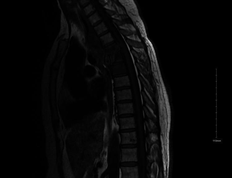
This diagnostic image is a T1-weighted sagittal MRI of the thoracic spine demonstrating classic radiological features of tuberculous spondylitis (Pott disease). The image shows significant pathology concentrated at the T4, T5, and T6 levels, characterized by altered marrow signal intensity within the vertebral bodies. There is evidence of endplate destruction and narrowing of the T4-T5 intervertebral disc space, indicating discitis-osteomyelitis. A large, multiloculated prevertebral and paraspinal soft tissue mass is visible, representing a tuberculous 'cold abscess' in the posterior mediastinum. This collection appears to exert a mass effect on adjacent anterior structures, such as the esophagus. The spinal canal shows potential for impingement due to the localized inflammation and bony destruction. This image serves as a key educational example of extrapulmonary tuberculosis presenting as spinal infection with associated paraspinal abscess formation.
This diagnostic image is a T1-weighted sagittal MRI of the thoracic spine demonstrating classic radiological features of tuberculous spondylitis (Pott disease). The image shows significant pathology concentrated at the T4, T5, and T6 levels, characterized by altered marrow signal intensity within the vertebral bodies. There is evidence of endplate destruction and narrowing of the T4-T5 intervertebral disc space, indicating discitis-osteomyelitis. A large, multiloculated prevertebral and paraspinal soft tissue mass is visible, representing a tuberculous 'cold abscess' in the posterior mediastinum. This collection appears to exert a mass effect on adjacent anterior structures, such as the esophagus. The spinal canal shows potential for impingement due to the localized inflammation and bony destruction. This image serves as a key educational example of extrapulmonary tuberculosis presenting as spinal infection with associated paraspinal abscess formation.

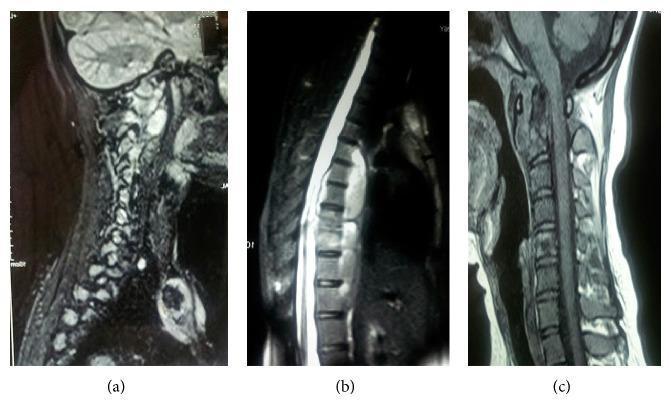
This composite diagnostic image consists of three sagittal MRI views (a, b, and c) of the spinal column demonstrating multisegmental tuberculous spondylitis (Pott disease). Panel (a) is a sagittal T2-weighted sequence showing the cervical spine with signal hyperintensity and irregularities at the craniovertebral junction and upper cervical levels, suggestive of inflammatory involvement and prevertebral edema. Panel (b) shows the thoracic spine with significant pathology, including destruction and collapse of mid-thoracic vertebral bodies (D7-D8 region) and a large, hyperintense anterior fluid collection (cold abscess) extending from D5 to D8. This collection causes mass effect and compression of the spinal cord. Panel (c) is a T1-weighted sagittal view of the cervical spine further detailing the loss of normal vertebral architecture and prevertebral soft tissue thickening. Collectively, these images illustrate characteristic findings of spinal tuberculosis: spondylodiscitis, vertebral destruction, and paraspinal abscess formation across non-contiguous spinal levels.

This composite diagnostic image consists of three sagittal MRI views (a, b, and c) of the spinal column demonstrating multisegmental tuberculous spondylitis (Pott disease). Panel (a) is a sagittal T2-weighted sequence showing the cervical spine with signal hyperintensity and irregularities at the craniovertebral junction and upper cervical levels, suggestive of inflammatory involvement and prevertebral edema. Panel (b) shows the thoracic spine with significant pathology, including destruction and collapse of mid-thoracic vertebral bodies (D7-D8 region) and a large, hyperintense anterior fluid collection (cold abscess) extending from D5 to D8. This collection causes mass effect and compression of the spinal cord. Panel (c) is a T1-weighted sagittal view of the cervical spine further detailing the loss of normal vertebral architecture and prevertebral soft tissue thickening. Collectively, these images illustrate characteristic findings of spinal tuberculosis: spondylodiscitis, vertebral destruction, and paraspinal abscess formation across non-contiguous spinal levels.

Reading Web Page

https://tbksp.who.int/ar/node/3024

Pott disease

Pott disease, also called tuberculous spondylitis or spinal tuberculosis, is infection of the vertebrae by Mycobacterium tuberculosis. It is a form of skeletal TB that can cause vertebral destruction, spinal deformity, paraspinal abscesses, and spinal cord compression.
MRI showing thoracic spinal tuberculosis with vertebral destruction and paraspinal cold abscess

Pathology and common sites

TB usually reaches the spine by hematogenous spread and often begins in the anterior vertebral body. It may spread beneath the anterior longitudinal ligament to adjacent levels.
  • Thoracic and thoracolumbar regions are frequently affected.
  • Adjacent vertebrae may be involved, though non-contiguous "skip" lesions can occur.
  • Vertebral collapse produces angular kyphosis, called a gibbus deformity.
  • Extension into soft tissues can form a paraspinal or psoas cold abscess. Miller's Review of Orthopaedics, p. 787.

Clinical features

  • Chronic, progressive localized back pain and stiffness
  • Constitutional symptoms may be mild or absent: fever, night sweats, weight loss, fatigue
  • Local tenderness, restricted movements, kyphotic deformity
  • Paraspinal swelling or psoas abscess
  • Neurologic complications: radicular pain, weakness, sensory loss, bladder/bowel disturbance, or Pott paraplegia due to cord compression from abscess, granulation tissue, deformity, or bony fragments.

Diagnosis

MRI with contrast is the preferred imaging test to define vertebral involvement, abscesses, epidural extension, and neural compression. Typical findings include vertebral-body/endplate destruction, subligamentous spread, deformity, and large paraspinal collections.
Confirmation should be sought through:
  • CT-guided aspiration or biopsy of vertebral/paraspinal material
  • Acid-fast bacilli smear, mycobacterial culture with drug-susceptibility testing
  • Molecular tests such as Xpert MTB/RIF, where available
  • Histology, often showing granulomatous inflammation with caseation
  • Chest imaging, sputum testing when indicated, and HIV testing
Differentials include pyogenic vertebral osteomyelitis, brucellosis, fungal infection, metastatic disease, lymphoma, and inflammatory spondyloarthropathy.

Treatment

Treatment requires a TB specialist plus spine/orthopedic or neurosurgical assessment.
For drug-susceptible disease, treatment uses multidrug anti-TB therapy. The core adult regimen is commonly:
  • 2 months: isoniazid, rifampicin, pyrazinamide, ethambutol
  • Continuation: isoniazid plus rifampicin
Duration for spinal TB varies by national program, disease extent, microbiology, and response. WHO notes that although 6 months is the core regimen for extrapulmonary TB, many experts use 9-12 months for bone and joint TB, including spinal disease, because assessment of healing is difficult. WHO extrapulmonary TB guidance.
Supportive care may include analgesia, nutritional support, bracing in selected cases, and rehabilitation. Drug-resistant TB requires an individualized regimen based on susceptibility results.

When surgery is considered

Anti-TB chemotherapy is primary treatment, but surgery may be necessary for:
  • Progressive or significant neurologic deficit/spinal cord compression
  • Spinal instability or severe progressive kyphosis
  • Large abscess needing drainage or diagnostic sampling
  • Persistent pain, deformity, or failure to improve despite appropriate treatment
  • Uncertain diagnosis, including possible tumor
Recent evidence on surgical strategy remains heterogeneous. A 2024 systematic review and meta-analysis assessed surgical outcomes, but does not replace individualized spine-specialist decision-making.

Red flags: urgent same-day hospital assessment

Seek urgent care for back pain with any of:
  • New leg weakness, numbness, difficulty walking
  • Urinary retention, incontinence, or bowel dysfunction
  • Fever/systemic illness with severe back pain
  • Rapidly worsening spinal deformity
  • Known TB with new neurologic symptoms
Sources: Miller's Review of Orthopaedics, p. 787; WHO extrapulmonary TB guidance.

Love message for girl friend

Sweet message:
My love, every day with you feels more beautiful than the last. Your smile gives me peace, your voice makes my day better, and your love means more to me than I can ever explain. I am truly grateful to have you in my life. No matter what happens, I want you to know that I will always care for you, support you, and choose you. I love you so much. ❤️
Short version:
You are my favorite person, my happiness, and my peace. I am so lucky to have you in my life. I love you more than words can say. ❤️

Birthday message for girldfriend

Happy Birthday, my love! ❤️
You make my life brighter, happier, and more meaningful just by being in it. I am so grateful for every smile, every conversation, and every memory we share. You deserve all the love, happiness, success, and beautiful moments in the world today and always.
May this new year of your life bring you everything your heart wishes for. I promise to keep supporting you, caring for you, and making more wonderful memories with you.
I love
!

peer closed connection without sending complete message body (incomplete chunked read)

We encountered an error while processing your request. Please try again or reach out to us for help.

Cold abscess

Reading File
Finding Sources
Searching PubMed

"cold abscess" AND tuberculosis

Searching the Web

cold abscess tuberculosis clinical diagnosis treatment CDC WHO

Cold abscess

A cold abscess is a chronic collection of pus that develops with little or no acute inflammation. Unlike a typical pyogenic abscess, it is usually:
  • Painless or only mildly painful
  • Not hot or markedly red
  • Soft/fluctuant and slowly enlarging
  • Associated with little fever or systemic toxicity
It is called “cold” because the usual inflammatory signs of a hot abscess, such as warmth, redness, tenderness, and edema, are absent or mild.

Common causes

The classic cause is tuberculosis, particularly:
  • Tuberculous cervical lymphadenitis
  • Spinal TB (Pott disease), with paraspinal or psoas abscess
  • TB of bones and joints
Less commonly, chronic infections such as actinomycosis or non-tuberculous mycobacterial infection can cause a similar presentation.

What happens in TB

Tuberculous inflammation causes tissue necrosis, often called caseation, which liquefies to form an abscess. It may track along tissue planes and later rupture through skin, producing a chronic draining sinus.

Diagnosis

Assessment commonly includes:
  • Ultrasound, CT, or MRI depending on the location
  • Needle aspiration or tissue biopsy
  • Microscopy for acid-fast bacilli, mycobacterial culture, and molecular testing such as Xpert MTB/RIF
  • Histopathology for granulomas/caseation
  • Evaluation for pulmonary TB and HIV where appropriate
A TB skin test or IGRA can support TB infection but cannot alone prove that the mass is active TB disease. The CDC diagnostic guidance emphasizes that TB infection tests must be interpreted alongside clinical evaluation and microbiologic testing.

Treatment

Treatment is directed at the cause. For a tuberculous cold abscess, this usually means a full multidrug anti-TB regimen prescribed through a TB specialist or national TB program. Needle aspiration or image-guided drainage may be needed for a large, tense, symptomatic, or diagnostically uncertain collection. Routine open incision and drainage can lead to persistent sinus formation and scarring in some TB lesions.
Seek urgent assessment if there is fever, rapidly worsening swelling, severe pain, neurological weakness, difficulty breathing or swallowing, or a neck/spinal mass.
Bailey and Love's Short Practice of Surgery, p. 108.

Milliary TB

Reading File
Finding Sources
Finding Sources
Searching PubMed

"miliary tuberculosis"

Searching the Web

WHO miliary tuberculosis diagnosis treatment extrapulmonary 2025

Reading File
Reading File
Searching Images

miliary tuberculosis chest x ray diffuse 1 to 3 mm nodules
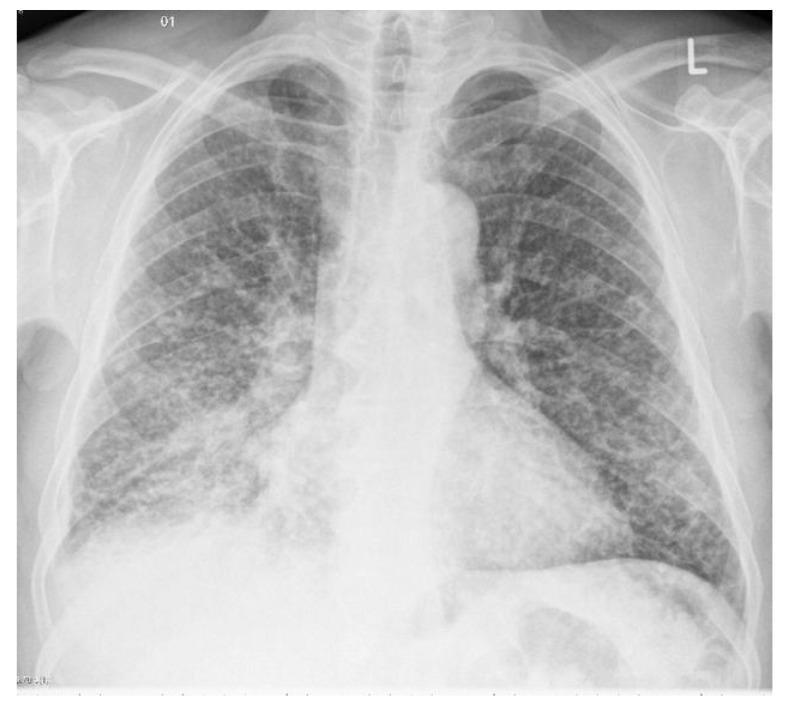
This diagnostic image is a posterior-anterior (PA) chest X-ray demonstrating miliary tuberculosis. The primary radiographic finding is a diffuse, bilateral, and uniform distribution of fine pulmonary nodules throughout both lung fields. These nodules are approximately 1–3 mm in diameter and are spread evenly from the apices to the lung bases, creating the characteristic 'miliary' (millet seed-like) pattern. The distribution involves all lobes without significant sparing, indicating hematogenous dissemination of Mycobacterium tuberculosis. The lung parenchyma shows a granular texture due to the density of these nodules. Cardiac silhouette and mediastinal contours appear within normal limits in this view, and no obvious pleural effusions are noted. This image serves as a classic educational example of miliary pulmonary involvement, emphasizing the importance of recognizing diffuse micronodular patterns in infectious disease diagnostics.
This diagnostic image is a posterior-anterior (PA) chest X-ray demonstrating miliary tuberculosis. The primary radiographic finding is a diffuse, bilateral, and uniform distribution of fine pulmonary nodules throughout both lung fields. These nodules are approximately 1–3 mm in diameter and are spread evenly from the apices to the lung bases, creating the characteristic 'miliary' (millet seed-like) pattern. The distribution involves all lobes without significant sparing, indicating hematogenous dissemination of Mycobacterium tuberculosis. The lung parenchyma shows a granular texture due to the density of these nodules. Cardiac silhouette and mediastinal contours appear within normal limits in this view, and no obvious pleural effusions are noted. This image serves as a classic educational example of miliary pulmonary involvement, emphasizing the importance of recognizing diffuse micronodular patterns in infectious disease diagnostics.

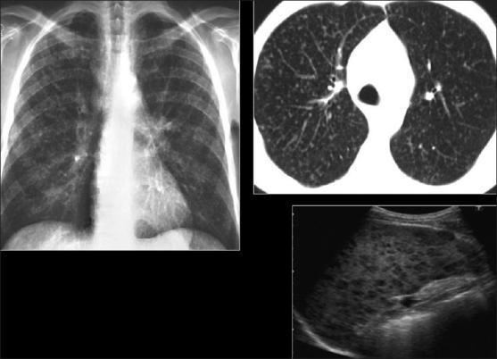
This composite of diagnostic images illustrates a case of disseminated miliary Mycobacterium tuberculosis (MTB) involving the pulmonary and lymphatic systems. The top-left panel is a frontal chest X-ray (CXR) demonstrating diffuse, symmetric interstitial opacities throughout both lung fields. The top-right panel shows high-resolution computed tomography (HRCT) of the lungs, providing a clearer visualization of a classic miliary pattern consisting of tiny (1-3 mm), randomly distributed, well-defined nodules with no predilection for specific lung zones. The bottom-right panel displays a grayscale ultrasound of the spleen, revealing splenomegaly and a heterogeneous parenchymal texture characterized by multiple small, hypoechoic nodules. These splenic findings represent MTB granulomas or microabscesses. Collectively, these imaging modalities depict the typical appearance of 'primary' miliary tuberculosis in an immunocompromised context, highlighting the hematogenous spread of the infection to both pulmonary and extrapulmonary organs.

This composite of diagnostic images illustrates a case of disseminated miliary Mycobacterium tuberculosis (MTB) involving the pulmonary and lymphatic systems. The top-left panel is a frontal chest X-ray (CXR) demonstrating diffuse, symmetric interstitial opacities throughout both lung fields. The top-right panel shows high-resolution computed tomography (HRCT) of the lungs, providing a clearer visualization of a classic miliary pattern consisting of tiny (1-3 mm), randomly distributed, well-defined nodules with no predilection for specific lung zones. The bottom-right panel displays a grayscale ultrasound of the spleen, revealing splenomegaly and a heterogeneous parenchymal texture characterized by multiple small, hypoechoic nodules. These splenic findings represent MTB granulomas or microabscesses. Collectively, these imaging modalities depict the typical appearance of 'primary' miliary tuberculosis in an immunocompromised context, highlighting the hematogenous spread of the infection to both pulmonary and extrapulmonary organs.

Searching the Web

site:tbksp.who.int miliary tuberculosis treatment disseminated tuberculosis

Miliary tuberculosis

Miliary TB is a severe form of disseminated tuberculosis in which Mycobacterium tuberculosis spreads through the bloodstream to the lungs and often to multiple other organs. It is named for the many tiny, millet seed-like lesions it can produce.
Chest X-ray: diffuse 1-3 mm miliary nodules

Who is at greater risk?

  • Infants and young children
  • Older adults
  • People living with HIV or other immune suppression
  • Malnutrition, diabetes, renal disease, or treatment with immunosuppressive medicines
  • Recent primary TB infection or reactivation of latent TB

Clinical features

Symptoms may be gradual and nonspecific:
  • Persistent fever and night sweats
  • Weight loss, loss of appetite, fatigue
  • Cough and breathlessness
  • Enlarged liver, spleen, or lymph nodes
  • Headache, confusion, neck stiffness, or seizures if TB meningitis occurs
  • Abdominal symptoms, anemia, low blood counts, or jaundice from multi-organ involvement
Acute miliary TB can cause acute respiratory distress syndrome (ARDS), shock, multi-organ failure, and death, so it needs urgent evaluation. Tintinalli's Emergency Medicine, section “Miliary Tuberculosis.”

Diagnosis

  • Chest X-ray: numerous diffuse, evenly distributed 1-3 mm nodules, the classic miliary pattern. This pattern may be absent early in disease.
  • High-resolution CT chest: more sensitive for tiny random nodules.
  • Samples for TB testing: sputum, induced sputum, bronchoalveolar lavage, lymph-node/other tissue biopsy, blood, bone marrow, or fluid from involved organs.
  • Tests include acid-fast bacilli smear, mycobacterial culture, molecular testing such as Xpert MTB/RIF, and drug-susceptibility testing.
  • Evaluate for TB meningitis, liver involvement, kidney disease, adrenal insufficiency, and HIV.
A miliary pattern is not exclusive to TB. Fungal infections, metastatic cancer, sarcoidosis, and pneumoconiosis can look similar, so microbiologic confirmation is important.

Treatment

Miliary TB is treated as active TB with multidrug anti-TB therapy under a TB specialist or national TB program. For drug-susceptible TB, the common initial regimen includes:
  • Isoniazid
  • Rifampicin
  • Pyrazinamide
  • Ethambutol
Treatment duration depends on drug susceptibility and organs involved. If there is TB meningitis, treatment is longer and corticosteroids may be indicated. Drug-resistant TB requires a regimen based on susceptibility results.

Urgent warning signs

Go to emergency care immediately for:
  • Severe shortness of breath, blue lips, or low oxygen
  • Confusion, severe headache, stiff neck, seizures, or weakness
  • Fainting, very low blood pressure, persistent vomiting
  • High fever with marked drowsiness or rapid deterioration
Miliary TB commonly affects the liver, spleen, bone marrow, adrenals, kidneys, and meninges in addition to lungs. Robbins, Cotran & Kumar Pathologic Basis of Disease, section “Morphology.”
This is a shared conversation. Sign in to Orris to start your own chat.