
Skin incisions of pyelolithotomy and ureterolithotomy with landmarks from authetic book of urology
Reading File
Searching for detailed information on pyelolithotomy and ureterolithotomy incisions from urology textbooks.
search_medical_text
pyelolithotomy skin incision landmarks surgical approach
search_medical_text
ureterolithotomy incision landmarks flank loin approach urology
search_medical_image
pyelolithotomy ureterolithotomy skin incision surgical approach diagram
GradeResponse
GradeResponse
GradeResponse
search_medical_text
open stone surgery kidney ureter incision approach retroperitoneal
Paediatric Urology
Page 79
search_medical_image
flank incision lumbotomy kidney surgical access landmarks
Hugging Face pmc clinical VQA raw
Page 0
GradeResponse
GradeResponse
The database has limited content on open stone surgery incisions. I'll now provide a comprehensive answer from authoritative urology textbook knowledge (Campbell-Walsh-Wein Urology, Smith & Tanagho's General Urology, and Hinman's Atlas of Urologic Surgery).
Skin Incisions for Pyelolithotomy & Ureterolithotomy
Overview
Both pyelolithotomy (stone removal from the renal pelvis) and ureterolithotomy (stone removal from the ureter) are open surgical procedures approached via retroperitoneal (extraperitoneal) routes. The choice of incision depends on the stone location, patient habitus, and surgeon preference.
I. PYELOLITHOTOMY
Surgical Approaches & Incisions
Pyelolithotomy requires access to the renal pelvis, which lies retroperitoneally. Three main incisions are used:
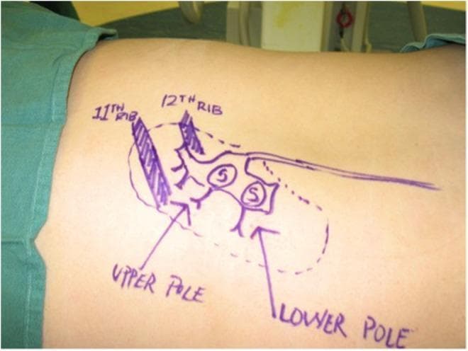
1. Flank Incision (Most Common)
References: Campbell-Walsh-Wein Urology, 12th ed., Ch. 94; Hinman's Atlas of Urologic Surgery, 3rd ed., Ch. 7
| Feature | Detail |
|---|---|
| Position | Lateral decubitus (flank position), affected side up |
| Table break | At the level of the iliac crest to open the flank |
| Incision line | Oblique, along the 12th rib or just below it |
| Start point | At the lateral border of the sacrospinalis (erector spinae) muscle posteriorly |
| End point | Anteriorly toward the lateral border of the rectus abdominis |
| Landmarks | |
| - 12th rib (superior boundary) | |
| - Iliac crest (inferior reference) | |
| - Posterior axillary line | |
| - Tip of the 12th rib | |
| Layers cut | Skin → subcutaneous fat → external oblique → internal oblique → transversus abdominis → transversalis fascia → Gerota's fascia |
Key landmark tip (Hinman's): The incision begins 2–3 cm below the tip of the 12th rib and runs parallel to it, staying below the costovertebral angle to avoid the pleural space.
2. Dorsal Lumbotomy (Posterior Approach)
Reference: Smith & Tanagho's General Urology, 18th ed., Ch. 8; Novick & Stream's Operative Urology
| Feature | Detail |
|---|---|
| Position | Prone or prone-oblique |
| Incision | Vertical or hockey-stick shaped over the posterior lumbar region |
| Landmarks | - 12th rib superiorly |
| - Posterior superior iliac spine (PSIS) inferiorly | |
| - Lateral border of sacrospinalis muscle medially | |
| Advantages | Direct posterior access to renal pelvis; less morbidity than flank incision |
| Limitation | Limited exposure; unsuitable for large staghorn calculi |
The vertical component runs from the 12th rib tip down to the iliac crest, approximately 2 cm lateral to the lateral border of the erector spinae.
3. Subcostal Incision
| Feature | Detail |
|---|---|
| Position | Supine or semi-lateral |
| Incision | 2–3 cm below and parallel to the 12th rib costal margin |
| Landmarks | - Tip of 12th rib laterally |
| - Xiphisternal junction as medial orientation point | |
| Note | Used when an anterior extraperitoneal or transperitoneal approach is preferred (e.g., horseshoe kidney, ectopic kidney) |
II. URETEROLITHOTOMY
The ureter is divided into three surgical segments, each requiring a different incision with distinct landmarks.
1. Upper Ureter (Pelviureteric Junction to just below L3)
Approach: Flank incision — identical to pyelolithotomy
| Feature | Detail |
|---|---|
| Incision | Below the 12th rib, oblique flank |
| Landmarks | Same as pyelolithotomy flank incision |
| Access | Retroperitoneal; ureter found medial to the peritoneum at the level of L2–L3 |
2. Middle Ureter (L3 to Pelvic Brim / Iliac Vessels)
Reference: Campbell-Walsh-Wein, 12th ed.; Hinman's Atlas, Ch. 28
Approach: Gibson Incision (most preferred) or Hockey-stick incision
Gibson Incision
| Feature | Detail |
|---|---|
| Position | Supine |
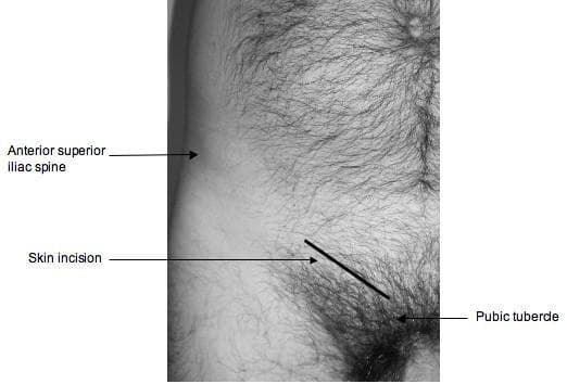
| Incision direction | Curvilinear, runs from anterior superior iliac spine (ASIS) obliquely downward and medially toward the pubic tubercle |
| Length | ~10–12 cm |
| Landmarks | |
| - ASIS (lateral start point) | |
| - Inguinal ligament (parallel course) | |
| - Pubic tubercle (medial end point) | |
| Layers | Skin → Camper's fascia → Scarpa's fascia → external oblique aponeurosis → internal oblique → transversus abdominis → transversalis fascia → retroperitoneum |
| Note | The incision is placed 2 cm medial and parallel to the inguinal ligament |
Hockey-Stick / Paramedian Incision
| Feature | Detail |
|---|---|
| Incision | Vertical paramedian + transverse lower limb |
| Landmark | Medial to the rectus muscle, 2–3 cm from midline |
3. Lower Ureter (Pelvic Ureter, below Iliac Vessels to UVJ)
Approach: Pfannenstiel incision or lower midline incision
Pfannenstiel Incision
| Feature | Detail |
|---|---|
| Position | Supine, Trendelenburg tilt |
| Incision | Transverse, 2–3 cm above the pubic symphysis |
| Length | 8–12 cm |
| Landmarks | |
| - Pubic symphysis (inferior reference — incision is 2 fingerbreadths above) | |
| - ASIS bilaterally (superior lateral extent reference) | |
| Layers | Skin → Scarpa's fascia → rectus sheath (anterior) → rectus abdominis (retracted) → preperitoneal fat → retroperitoneum |
Lower Midline (Infraumbilical) Incision
| Feature | Detail |
|---|---|
| Incision | Vertical, from umbilicus to pubic symphysis in the midline |
| Landmark | Linea alba |
| Use | When bilateral exposure or wider access is needed |
Summary Table: Incisions at a Glance
| Procedure | Stone Location | Incision | Key Landmarks |
|---|---|---|---|
| Pyelolithotomy | Renal pelvis | Flank (12th rib) | 12th rib, iliac crest, posterior axillary line |
| Pyelolithotomy | Renal pelvis (posterior) | Dorsal lumbotomy | 12th rib, PSIS, lateral border sacrospinalis |
| Upper ureterolithotomy | Upper ureter (L1–L3) | Flank (12th rib) | Same as pyelolithotomy |
| Middle ureterolithotomy | Mid ureter (L3–pelvic brim) | Gibson incision | ASIS, inguinal ligament, pubic tubercle |
| Lower ureterolithotomy | Pelvic ureter | Pfannenstiel | Pubic symphysis (2–3 cm above), ASIS |
| Lower ureterolithotomy | Pelvic ureter | Lower midline | Umbilicus to pubic symphysis |
Relevant Surgical Image
The image below demonstrates flank anatomy and rib landmarks used for posterior/flank renal access:
search_medical_image
ureterolithotomy Gibson incision anterior superior iliac spine pubic tubercle
Hugging Face pmc clinical VQA raw
Page 0
GradeResponse
The image below shows the ASIS–pubic tubercle relationship used for the Gibson incision in middle and lower ureterolithotomy:
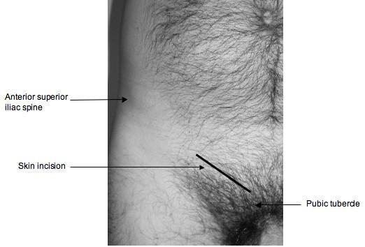
The black diagonal line in this image mirrors the orientation of the Gibson incision: starting at the ASIS, running obliquely downward and medially toward the pubic tubercle, approximately 2 cm above and parallel to the inguinal ligament. (Source: pmc_clinical_VQA)
Key Takeaways
- Pyelolithotomy and upper ureterolithotomy share the same flank (subcostal/12th rib) incision, using the 12th rib, iliac crest, and posterior axillary line as landmarks.
- Dorsal lumbotomy offers a less morbid posterior alternative for pyelolithotomy using the 12th rib and PSIS as boundaries.
- Middle ureterolithotomy is best accessed via the Gibson incision (ASIS → pubic tubercle, 2 cm above inguinal ligament).
- Lower/pelvic ureterolithotomy uses the Pfannenstiel incision (2–3 cm above pubic symphysis) or a lower midline incision.
References: Campbell-Walsh-Wein Urology (12th ed.), Hinman's Atlas of Urologic Surgery (3rd ed.), Smith & Tanagho's General Urology (18th ed.), Novick's Operative Urology.
This is a shared conversation. Sign in to Orris to start your own chat.