
Hidden spaces of middle ear
Reading File
Finding Sources
Finding Sources
Searching PubMed
"middle ear"[MeSH Terms] AND "anatomy"
Reading File
Reading File
Searching Images
sinus tympani facial recess retrotympanum middle ear anatomy
Reading File
query_images_db
Hidden spaces of the middle ear
"Hidden" spaces are the recesses that are hard to see with the operating microscope. Cholesteatoma can sit in them and be left behind. The Scott-Brown chapter on endoscopy lists the main ones: the sinus tympani, the anterior epitympanum, the protympanic (supratubal) recess and the facial recess (Scott-Brown's Otorhinolaryngology Head & Neck Surgery, Vol 2, Otoendoscopy versus the microscope).
1. Retrotympanum (posterior mesotympanum)
This region has two main recesses, separated by the pyramidal eminence.
Sinus tympani
- It lies medial to the facial nerve and behind the promontory.
- Its boundaries are the promontory anteriorly, the facial canal superiorly, and the pyramidal eminence laterally. The subiculum lies beneath it and the ponticulus above it (K J Lee's Essential Otolaryngology, p. 496 area).
- It is the most difficult recess to reach. Cummings classifies it as type A (small), type B or type C (deeper, extending behind the facial nerve), and the type determines whether an endoscopic approach is enough.
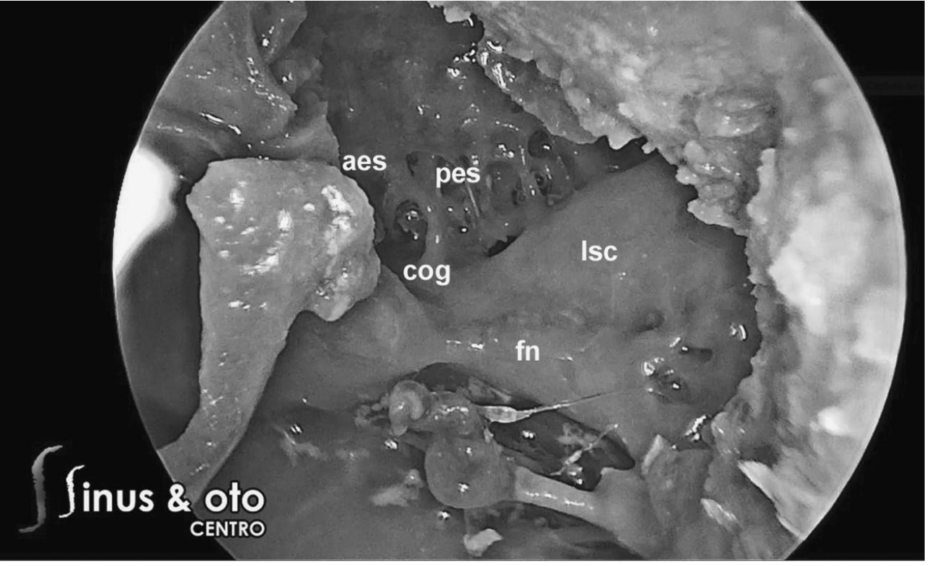
- Endoscopic anatomy describes a ridge called the "finiculus" (Scott-Brown Vol 2).
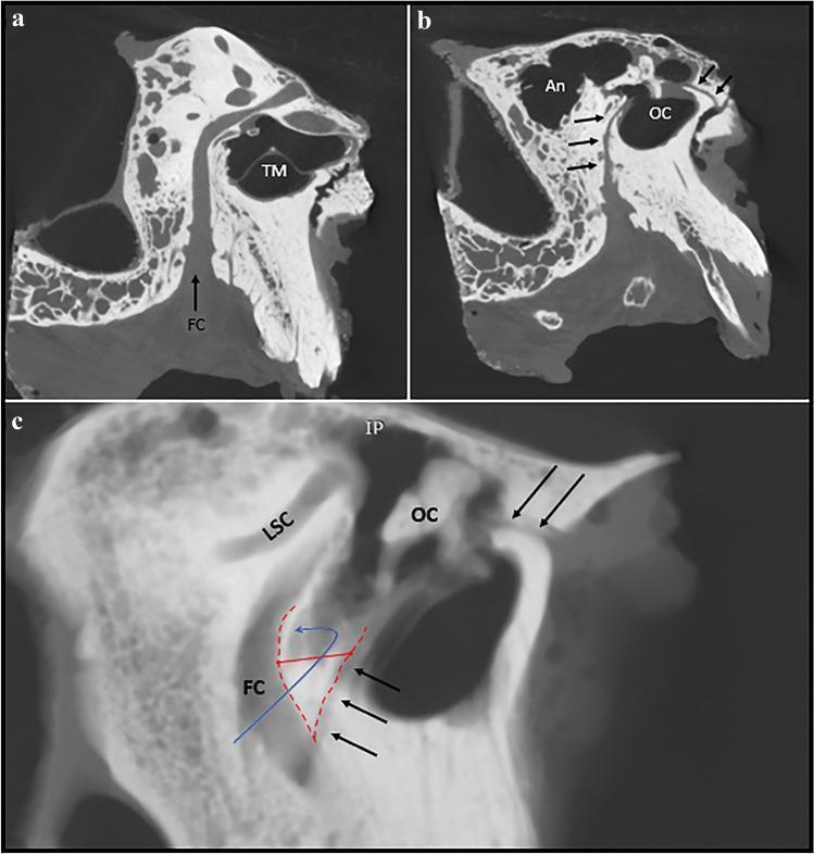
Facial recess
- It is bounded by the facial nerve posteriorly, the chorda tympani anteriorly, and the incudal buttress superiorly (K J Lee's Essential Otolaryngology).
- It is the surgical window for posterior tympanotomy, used in cochlear implantation and in cholesteatoma surgery.
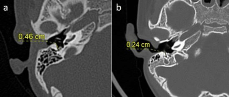
- A wide recess is about 4.6 mm and a narrow one about 2.4 mm on HRCT. A narrow recess limits access.
2. Epitympanum (attic) and its pockets
- Limits:
- Superior: tegmen tympani.
- Medial: facial canal and lateral semicircular canal.
- Lateral: pars flaccida and scutum.
- Anterior: zygomatic root.
- Posterior: incudal fossa and aditus ad antrum (K J Lee's Essential Otolaryngology).
- Prussak space is the commonest site of attic cholesteatoma.
- Lateral wall: pars flaccida.
- Medial wall: malleus neck.
- Roof: lateral malleal fold.
- Floor: short process of the malleus.
- Boundaries per the textbook: scutum, lateral malleolar ligament, neck of malleus, anterior malleolar ligament, pars flaccida.
- Anterior epitympanic space and supratubal recess: the cog (a bony septum) can separate them. They are hidden from the microscope.
- Posterior epitympanum: lies behind the superior malleal fold.
- Tympanic isthmus: a roughly 2.5 mm opening in the tympanic diaphragm that ventilates the whole attic. The anterior isthmus lies between the incudostapedial joint and the tensor tympani tendon. Blockage by granulation tissue or webs causes selective attic dysventilation, retraction pockets and cholesteatoma.
3. Other spaces (from general anatomy, not retrieved from the library)
- Protympanum: the anterior part of the middle ear, around the eustachian tube opening.
- Hypotympanum: the part below the level of the tympanic annulus.
- Round window niche: the recess in front of the round window membrane.
- Von Tröltsch, anterior and posterior pouches: these lie between the tympanic membrane and the malleal folds.
Why this matters clinically
- The microscope gives poor control of these areas, so cholesteatoma can persist or recur. Endoscopy or second-look surgery is often used to check them. Shambaugh notes that second-look mastoidectomy allows a panoramic view of the sinus tympani, facial recess, epitympanum and supratubal recess.
- Cummings describes endoscopic, facial recess or wide canaloplasty routes for sinus tympani cholesteatoma.
Recent evidence check: A quick PubMed search for recent systematic reviews on middle ear anatomy found nothing that changes this anatomical picture. A 2024 meta-analysis (PMID 38722318) compared endoscopic and microscopic resection of temporal bone paraganglioma, which is related to endoscopic access but not to the anatomy itself.
If you want, I can go into the sinus tympani classification or how to remember these spaces for exams.
This is a shared conversation. Sign in to Orris to start your own chat.