
c/o pain hip , no trauma
Reading File
search_medical_image
hip X-ray avascular necrosis femoral head atraumatic
search_medical_text
avascular necrosis femoral head radiographic findings staging management
GradeResponse
GradeResponse
search_medical_text
avascular necrosis femoral head Ficat Arlet staging X-ray crescent sign subchondral collapse
search_medical_text
atraumatic hip pain 56 year female osteoarthritis AVN differential diagnosis causes risk factors
GradeResponse
Radiograph Analysis — V. Aruna Kumari, 56F | Hip AP/Lateral
Key Radiographic Findings
Looking at both the AP and lateral views of the right hip (marked "R"):
| Finding | Observation |
|---|---|
| Femoral head contour | Loss of normal spherical shape; flattening of the superior weight-bearing surface |
| Subchondral region | Increased sclerosis with areas of mixed lucency — suggestive of crescent sign (subchondral fracture) |
| Joint space | Relatively preserved at this stage |
| Femoral neck | Intact, no fracture line |
| Acetabulum | No significant degenerative change |
| Bone density | Patchy sclerosis in the femoral head consistent with ischemic necrosis |
Primary Diagnosis
Avascular Necrosis (AVN) / Osteonecrosis of the Femoral Head — Right Hip
Ficat & Arlet Stage II–III (sclerosis + early subchondral collapse without significant joint space loss)
Staging (Ficat & Arlet)
| Stage | X-ray Finding | This Patient |
|---|---|---|
| I | Normal X-ray | No |
| II | Sclerosis/cysts, preserved head shape | Borderline |
| III | Crescent sign, subchondral collapse, head flattening | Likely |
| IV | Joint space narrowing, acetabular involvement | Not yet |
Etiology to Investigate (No Trauma)
In a 56-year-old female with no trauma, consider:
- Corticosteroid use (most common secondary cause)
- Alcohol use
- Systemic lupus erythematosus (SLE)
- Hypothyroidism / dyslipidemia
- Sickle cell disease
- Gaucher's disease
- Idiopathic (primary AVN)
Per Bailey & Love's 28th Edition (p. 572): "AVN can be primary (idiopathic) or secondary to other pathology" — interruption of blood supply leads to bone death, femoral head collapse, and eventual secondary OA.
Recommended Next Steps
Investigations:
- MRI both hips — gold standard; detects early AVN, assesses extent of necrosis, checks contralateral hip (bilateral in ~50–80%)
- CBC, ESR, CRP, ANA, lipid profile, HbA1c
- History: steroid use, alcohol intake, autoimmune disease
Management based on stage:
- Stage I–II (pre-collapse): Core decompression ± bone grafting; bisphosphonates (adjunct); protected weight-bearing
- Stage III (early collapse): Core decompression with structural bone graft, or consider rotational osteotomy
- Stage IV: Total Hip Arthroplasty (THA)
Reference Image (AVN Femoral Head — Bilateral for comparison)
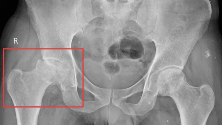
Bottom line: This is right hip AVN, approximately Ficat Stage II-III, presenting as atraumatic hip pain in a 56-year-old female. MRI is urgently indicated to confirm staging, assess viability, and screen the contralateral hip before irreversible collapse occurs.
This is a shared conversation. Sign in to Orris to start your own chat.