
Management of acute intestinal obstruction
"intestinal obstruction" AND management
acute intestinal obstruction abdominal X-ray dilated bowel loops
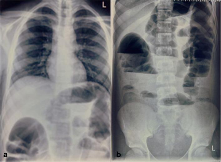
Two diagnostic X-ray images demonstrating clinical features of intestinal obstruction. Image (a) is a posteroanterior chest X-ray that shows clear lung fields and a normal cardiomediastinal silhouette, but notably reveals dilated bowel loops containing gas beneath the diaphragmatic plane. Image (b) is an anteroposterior erect abdominal X-ray, which is diagnostic for bowel obstruction. It displays multiple markedly dilated loops of bowel distributed throughout the central and peripheral abdominal cavity. Pathognomonic 'string of pearls' or 'stepladder' appearances are indicated by numerous air-fluid levels visible in the erect position. These findings are consistent with an acute mechanical intestinal obstruction or adynamic ileus, requiring further clinical correlation with the patient's history of rectal pathology and metastatic workup. The spinal column and pelvic structures appear unremarkable, focusing the clinical relevance on the abnormal bowel gas pattern.
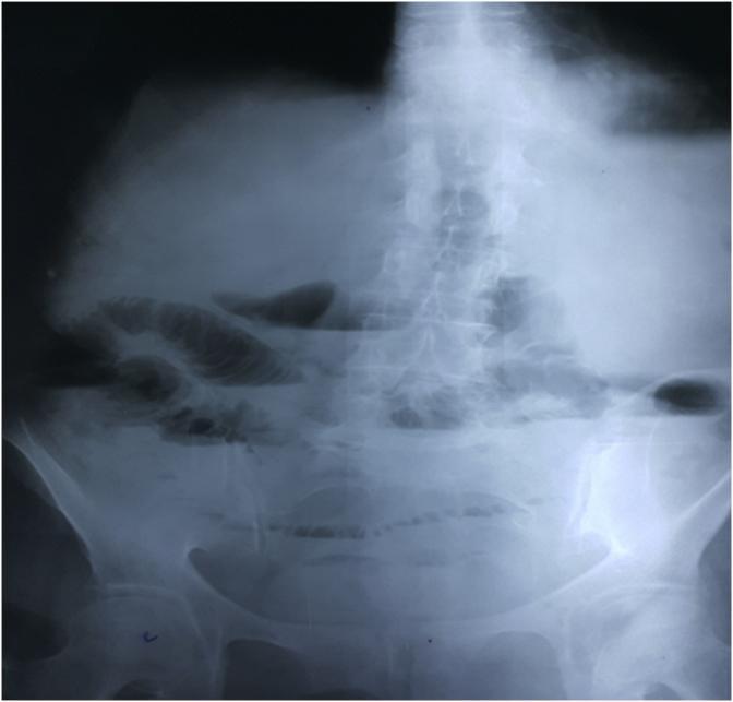
This diagnostic image is a plain erect abdominal X-ray (radiography) demonstrating classic radiographic features of an intestinal obstruction. The most prominent finding is the presence of multiple dilated loops of small bowel containing horizontal air-fluid levels, concentrated in the central and upper abdominal regions. The dilated loops exhibit visible valvulae conniventes (plicae circulares), which are thin mucosal folds that span the entire diameter of the bowel lumen, identifying these as small intestinal loops. A significant clinical sign is the complete absence of gas within the large bowel and rectum, indicating a high-grade or complete mechanical obstruction proximal to the colon. The lumbar spine and pelvic bones are visible, providing anatomical context for the bowel loop distribution. This image is a primary educational resource for recognizing the 'step-ladder' pattern of small bowel obstruction and differentiating small bowel from large bowel dilation in an acute clinical setting.
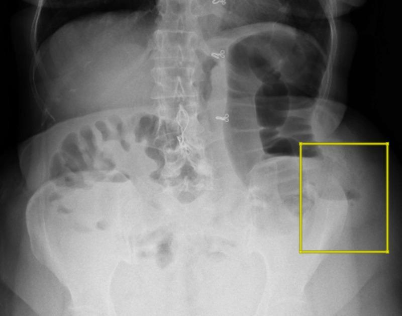
This diagnostic image is a plain erect abdominal radiograph (X-ray) demonstrating signs of intestinal obstruction. The radiograph reveals multiple significantly dilated loops of small and large bowel distributed across the abdominal cavity. Characteristic multiple air-fluid levels are visible, representing the gravitational separation of liquid and gas within the distended lumen. A yellow square highlights the left lower quadrant (left iliac region), where focal dilated bowel loops and prominent air-fluid levels are concentrated near the iliac crest. These findings are clinically significant as they indicate a mechanical bowel obstruction or a localized paralytic ileus. The image serves as a teaching tool for identifying radiological hallmarks of intestinal distension and fluid stasis in acute surgical abdominal presentations, specifically correlating with bowel herniation through an iliac crest defect.
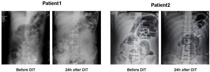
This diagnostic imaging panel consists of four anteroposterior abdominal X-ray plain films comparing two patients (Patient 1 and Patient 2) with acute intestinal obstruction before and 24 hours after a deeper intubation technique (DIT). In the 'Before DIT' images for both patients, there are classic signs of small bowel obstruction, including significant gaseous distension of bowel loops and visible air-fluid levels. In Patient 2, the 'Before DIT' film particularly highlights multiple dilated small bowel loops in a step-ladder pattern. The '24h after DIT' images demonstrate a marked reduction in intestinal gas and decompression of the previously dilated bowel loops. Notably, in the post-procedure film for Patient 2, a radio-opaque intestinal decompression tube is clearly visible, extending through the stomach and deep into the intestinal tract with the distal tip positioned in the lower abdominal quadrant. These images illustrate the radiological progression of bowel decompression and the effective placement of deeper intubation for managing obstructive pathologies.
Management of Acute Intestinal Obstruction
1. Classification - Essential First Step
| Feature | Small Bowel Obstruction (SBO) | Large Bowel Obstruction (LBO) |
|---|---|---|
| Common causes | Adhesions (most common), hernias, Crohn's disease, intussusception | Colorectal carcinoma (~50%), diverticular stricture (~17%), volvulus (5-10%) |
| Vomiting | Early, frequent, bilious | Late or absent |
| Distension | Central | Peripheral/generalized |
| Pain | Colicky, central, high-pitched bowel sounds | Lower abdominal colic |
| X-ray | Valvulae conniventes - folds traverse full width, central | Haustra - partial folds, peripheral |
2. Initial Assessment and Resuscitation ("Drip & Suck")
A. Intravenous Fluid Resuscitation
- SBO leads to loss of up to 6 litres/day of salt-rich fluid by vomiting or sequestration into the bowel lumen
- Aggressive IV crystalloid replacement (normal saline or Hartmann's/Ringer's lactate)
- Correct electrolyte imbalances - monitor K+, Na+, Cl-
- Insert urinary catheter for accurate fluid balance
B. Nasogastric Decompression
- NG tube suction empties the stomach, reduces the risk of pulmonary aspiration and limits further distension from swallowed air
- Long intestinal tubes (Cantor or Baker tube) offer no significant advantage over standard NG tubes - prospective RCTs show no difference in decompression achieved, success of non-operative treatment, or post-operative morbidity. They are associated with longer hospital stay and longer post-operative ileus - Sabiston Textbook of Surgery
- Use should be determined on a case-by-case basis (evidence remains inconclusive for universal use)
C. Monitoring and Work-up
- IV access (large bore x2), blood for FBC, U&E, LFTs, amylase, group & save
- Erect chest X-ray (to exclude pneumoperitoneum)
- Erect and supine plain abdominal X-ray as initial imaging
- CT scan (with IV and oral contrast) - now standard for confirming diagnosis, identifying the level and cause, and detecting strangulation/ischemia
3. Radiological Diagnosis
Plain Abdominal X-Ray
- Multiple air-fluid levels on the erect film ("step-ladder" or "string of pearls" pattern)
- Dilated loops with valvulae conniventes (SBO) vs. haustra (LBO)
- Absence of colonic gas suggests complete SBO
CT Abdomen (Gold Standard)
- Identifies the exact transition point, cause (adhesion, hernia, mass), and level
- Detects early features of strangulation: mesenteric edema, free fluid, pneumatosis intestinalis, portal venous gas
- Differentiates benign from malignant causes: malignant obstruction - mass at transition zone, adenopathy, abrupt cutoff, irregular thickening; benign - smooth transition zone, mesenteric vascular changes
- Acute cecal dilatation >12-14 cm is a surgical emergency due to high risk of rupture - Harrison's Principles of Internal Medicine, 22nd Ed.
4. Identifying Strangulation - The Critical Decision Point
| Clinical Signs | Investigations |
|---|---|
| Constant (not colicky) pain | Leukocytosis (WBC >15,000) |
| Fever (>38°C) | Raised serum lactate |
| Tachycardia | CT - pneumatosis, mesenteric edema, free fluid |
| Peritonism/rebound tenderness | Loss of enhancement of bowel wall on CT |
| Ominously quiet bowel sounds |
"Local tenderness over part of the abdomen may suggest developing strangulation... with developing peritoneal irritation due to strangulation the bowel may become atonic and the bowel sounds ominously quiet." - Pye's Surgical Handicraft
5. Non-Operative (Conservative) Management
Indications
- Partial SBO (up to 85% of partial obstructions resolve without surgery)
- Adhesive SBO without strangulation signs
- Post-operative ileus
- First episode after known pelvic/abdominal malignancy (trial of conservative)
- Crohn's disease-related acute obstruction (often resolves with conservative care + steroids)
Components
- NG decompression + bowel rest
- IV fluid and electrolyte replacement
- Analgesia
- Regular clinical reassessment (vital signs, abdominal exam, urine output)
- Serial abdominal X-rays
Water-Soluble Contrast Challenge (Gastrografin)
- 100 mL of water-soluble contrast given via NG tube or orally, with follow-up X-rays at 8 and 24 hours
- If contrast has not reached the colon at 24 hours - conservative management is unlikely to succeed and surgery is indicated
- Evidence supports its use in adhesive SBO specifically; not established for non-adhesive obstruction - Sabiston Textbook of Surgery
Failure of Conservative Management
- Clinical deterioration (increasing pain, fever, tachycardia)
- Increasing distension on serial X-rays
- Failure of contrast to pass at 24 hours
- No improvement after 48-72 hours
- New signs of strangulation at any point
6. Operative Management
Indications for Urgent/Emergency Surgery
- Strangulation or vascular compromise (closed-loop obstruction, volvulus, hernia with vascular compromise)
- Complete obstruction with failure of conservative management
- Peritonitis / perforation (pneumoperitoneum on CXR)
- Signs of strangulation on CT
- Cecal dilatation >12 cm (LBO)
Approach by Etiology
| Cause | Surgical Approach |
|---|---|
| Adhesion band | Laparoscopy/laparotomy: lysis of adhesions. Gentle bowel handling to minimize serosal trauma |
| Incarcerated hernia | Manual reduction of herniated bowel + hernia defect repair |
| Colonic carcinoma (LBO) | Hartmann's procedure; or resection with primary anastomosis +/- defunctioning stoma; SEMS as bridge to surgery |
| Sigmoid volvulus | Endoscopic detorsion (sigmoidoscopy) as first-line; elective sigmoid resection after |
| Cecal volvulus | Surgery required (right hemicolectomy) |
| Crohn's fibrotic stricture | Resection or strictureplasty |
| Radiation enteropathy (chronic) | Resection or bypass of irradiated segment |
| Malignant obstruction / terminal | Bypass, stenting, or venting gastrostomy; avoid major resections in poor prognosis |
| Intra-abdominal abscess | Percutaneous/laparoscopic drainage; laparotomy for large abscesses |
Laparoscopic vs. Open
- Laparoscopic approach for SBO - multiple studies confirm lower morbidity and mortality, shorter length of stay
- Conversion to open if extensive adhesions, ischemic bowel, or dense inflammation
- Sabiston Textbook of Surgery
Assessing Bowel Viability Intraoperatively
- Wrap segment in warm saline-moistened sponges for 15-20 minutes and re-examine
- If normal color returns and peristalsis resumes - safe to retain
- Doppler flow probe - identifies mesenteric flow but adds little to surgeon's clinical judgment
- Fluorescein or indocyanine green (ICG) fluorescence - useful in difficult borderline cases
- If viability still uncertain - temporary abdominal closure with second-look laparotomy at 18-24 hours
- Sabiston Textbook of Surgery
7. Large Bowel Obstruction - Special Considerations
SEMS (Self-Expanding Metal Stents)
- Endoscopic colonic stenting increasingly used in malignant LBO
- "Bridge to surgery": converts emergency to elective operation, allowing bowel preparation and reduction of stoma rate
- Definitive palliation in patients with advanced/metastatic disease
- Fischer's Mastery of Surgery, 8th Ed.
Colonic Pseudo-Obstruction (Ogilvie's Syndrome)
- Functional obstruction - treat the underlying cause (stop opioids, correct electrolytes, mobilize)
- Neostigmine 2 mg IV - effective first-line pharmacological treatment
- Colonoscopic decompression for cecal diameter >10-12 cm or failure of neostigmine
- Surgery only if perforation or failure of all other measures
- Current Surgical Therapy, 14th Ed.
Sigmoid Volvulus
- Flexible sigmoidoscopy / rigid sigmoidoscopy + rectal tube: first-line decompression (>90% success)
- High risk of recurrence (~40-60%) - elective sigmoid resection after recovery
- Emergency surgery if signs of gangrene/perforation or failed endoscopic decompression
8. Malignant Bowel Obstruction (Palliative Setting)
- Median survival with malignant obstruction is only 3-4 months
- ~25-30% have a benign cause (adhesions, drug-induced ileus) - exclude before declaring palignant
- Conservative approach preferred for known advanced disease: NG decompression, IV fluids, antiemetics, antispasmodics, analgesia
- Octreotide - reduces GI secretions, relieves obstructive symptoms
- Glucocorticoids - anti-inflammatory, may reduce edema at obstruction site
- Venting gastrostomy (PEG) - palliates nausea/vomiting without major surgery
- SEMS for accessible colonic/duodenal obstruction
- Harrison's Principles of Internal Medicine, 22nd Ed.
9. Summary Algorithm
Acute Intestinal Obstruction
        |
        ├── Resuscitation (IV fluids, NG tube, catheter, monitoring)
        ├── Investigations (AXR, CT scan, bloods)
        |
        ├── SIGNS OF STRANGULATION / PERITONITIS / PERFORATION?
        |       YES → Emergency laparotomy / laparoscopy
        |
        └── NO → Conservative Management (48-72 hrs)
                    |
                    ├── SBO
                    |    ├── Partial → 85% resolve; water-soluble contrast challenge at 24-48h
                    |    └── Complete → Careful trial; contrast challenge; operate if no resolution at 24-48h
                    |
                    └── LBO
                         ├── Malignancy → SEMS (bridge/palliation) or urgent resection
                         ├── Volvulus → Endoscopic detorsion (sigmoid) / surgery (cecal)
                         └── Pseudo-obstruction → Neostigmine / colonoscopic decompression
- Sabiston Textbook of Surgery: The Biological Basis of Modern Surgical Practice - Operative and non-operative management
- Pye's Surgical Handicraft, 22nd Ed. - Clinical features and initial assessment
- Current Surgical Therapy, 14th Ed. - LBO etiology and management
- Harrison's Principles of Internal Medicine, 22nd Ed. - Malignant obstruction
- Fischer's Mastery of Surgery, 8th Ed. - SEMS in LBO