
Congestive cardiac failure treatment
"heart failure" AND treatment
congestive heart failure treatment algorithm management
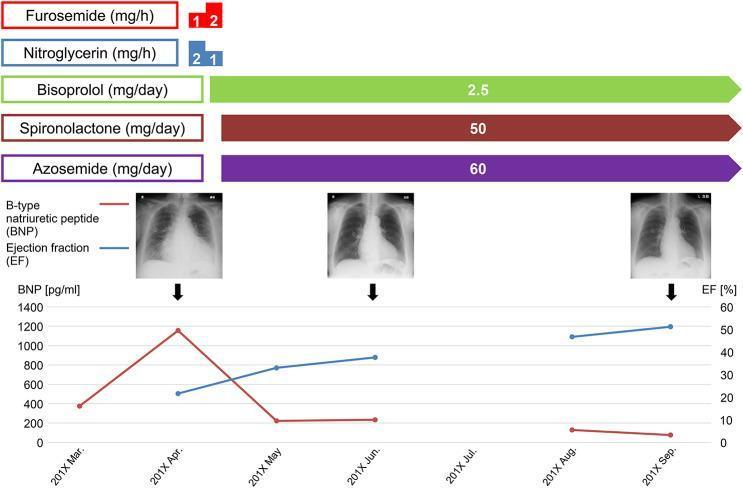
This infographic illustrates the clinical timeline and treatment progression of a patient with congestive heart failure. The visual combines pharmacological management, serial diagnostic imaging, and biochemical/functional markers. The top section displays a medication timeline featuring intravenous diuretics (furosemide) and vasodilators (nitroglycerin) in the acute phase, followed by long-term oral maintenance with bisoprolol (2.5 mg/day), spironolactone (50 mg/day), and azosemide (60 mg/day). The middle section shows three serial posterior-anterior (PA) chest radiographs from March, June, and September, demonstrating a progressive reduction in cardiomegaly and clearance of pulmonary congestion. The bottom dual-axis line graph tracks B-type natriuretic peptide (BNP) levels (red line) and Ejection Fraction (EF) percentages (blue line). It reveals a sharp peak in BNP (~1200 pg/ml) in April corresponding with acute decompensation, followed by a significant decline to <200 pg/ml. Simultaneously, the EF shows a steady improvement from approximately 25% to 52% by September, correlating with clinical recovery and the effectiveness of the heart failure medication regimen.
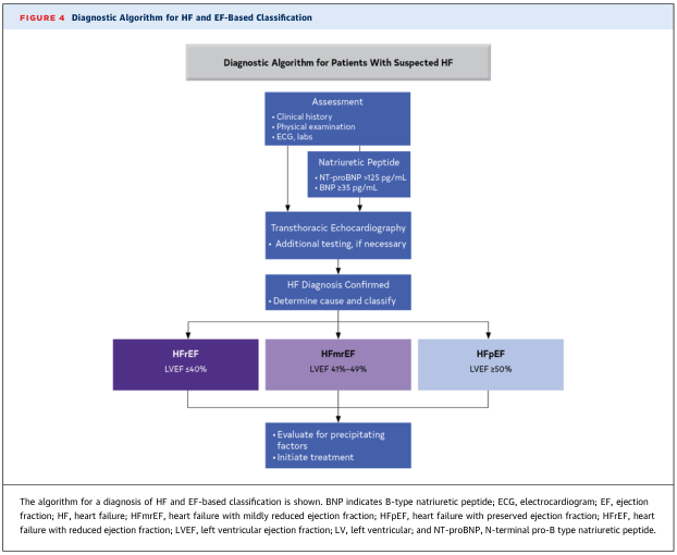
Summary : This figure presents a diagnostic algorithm for heart failure (HF) and its classification based on ejection fraction (EF), guiding clinicians through assessment, testing, and classification steps for patients with suspected HF. flowchart: # Nodes : • Diagnostic Algorithm for Patients With Suspected HF (rectangular header) • Assessment (rectangle): Clinical history, Physical examination, ECG, labs • Natriuretic Peptide (rectangle): NT-proBNP >125 pg/mL, BNP ≥35 pg/mL • Transthoracic Echocardiography (rectangle): Additional testing, if necessary • HF Diagnosis Confirmed (rectangle): Determine cause and classify • HFrEF (rectangle): LVEF ≤40% • HFmrEF (rectangle): LVEF 41%-49% • HFpEF (rectangle): LVEF ≥50% • Evaluate for precipitating factors, Initiate treatment (rectangle under all three EF categories) # Connectors : • Top-down arrows connect each step sequentially: Assessment → Natriuretic Peptide → Transthoracic Echocardiography → HF Diagnosis Confirmed. • From "HF Diagnosis Confirmed," three branches lead to HFrEF, HFmrEF, and HFpEF. • Each EF category leads to a shared final step: Evaluate for precipitating factors, Initiate treatment. # Layout : • Vertical flow from top to bottom, with a split into three parallel branches for EF-based classification. • All three EF branches converge to a common final action box. # Analysis : • The algorithm provides a clear, stepwise approach for diagnosing HF, starting with clinical assessment and progressing through biomarker testing and echocardiography. • Classification is based on left ventricular ejection fraction (LVEF): reduced (≤40%), mildly reduced (41%-49%), and preserved (≥50%). • The final step emphasizes evaluating causes and initiating treatment regardless of EF category, supporting a systematic and comprehensive management strategy for HF.
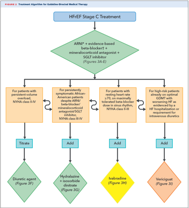
Summary : This figure presents a treatment algorithm flowchart for guideline-directed medical therapy in patients with Heart Failure with Reduced Ejection Fraction (HFrEF) Stage C, outlining medication choices and escalation steps based on patient characteristics. flowchart: # Nodes : • Start (rounded rectangle): "HFrEF Stage C Treatment" • Main therapy (diamond): "ARNI* + evidence-based beta-blocker† + mineralocorticoid antagonist + SGLT inhibitor (Figures 3A-E)" • Four decision nodes (rectangles, left to right): – "For patients with persistent volume overload, NYHA class II-IV" – "For persistently symptomatic African-American patients despite ARNI/beta-blocker/mineralocorticoid antagonist/SGLT inhibitor, NYHA class III-IV" – "For patients with resting heart rate ≥70, on maximally tolerated beta-blocker dose in sinus rhythm, NYHA class II-III" – "For high-risk patients already on optimal GDMT with worsening HF as evidenced by a HF hospitalization or requirement for intravenous diuretics" • Four action nodes (diamonds, left to right): – "Diuretic agent (Figure 3F)" – "Hydralazine + isosorbide dinitrate (Figure 3G)" – "Ivabradine (Figure 3H)" – "Vericiguat (Figure 3I)" # Connectors : • Downward arrows from "HFrEF Stage C Treatment" to "ARNI* + evidence-based beta-blocker† + mineralocorticoid antagonist + SGLT inhibitor" • Four downward arrows from main therapy node to each decision node • Each decision node connects downward to its respective action node: – "Titrate" arrow to "Diuretic agent" – "Add" arrow to "Hydralazine + isosorbide dinitrate" – "Add" arrow to "Ivabradine" – "Add" arrow to "Vericiguat" # Layout : • Vertical flow from top to bottom • After main therapy, splits into four parallel branches, each with a decision node and corresponding action node • Colour coding: main therapy node (green diamond), decision nodes (orange rectangles), action nodes (green or orange diamonds), connectors (black arrows) # Analysis : • The flowchart emphasizes a foundational quadruple therapy for all HFrEF Stage C patients, then stratifies further medication choices based on specific clinical scenarios (volume overload, persistent symptoms in African-American patients, elevated heart rate, or high-risk status). • Each branch provides a clear escalation or addition of therapy tailored to patient phenotype, supporting individualized care. • The structure allows for rapid identification of next steps in management depending on patient response and risk factors.
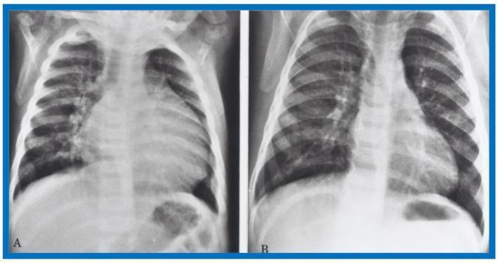
This Comparison Chart displays two side-by-side antero-posterior (AP) chest X-rays of an infant, demonstrating the radiographic progression of congestive heart failure management. Panel A (pre-therapy) shows significant cardiomegaly with an increased cardiothoracic ratio and prominence of the left heart border. The lung fields exhibit increased perihilar opacities and blurred vascular markings, characteristic of pulmonary venous congestion. Panel B (post-therapy, following hydralazine administration) demonstrates a remarkable improvement in the clinical state. There is a visible reduction in the heart silhouette size, suggesting a decrease in moderate cardiomegaly. The lungs appear significantly clearer with increased radiolucency and sharper definition of vascular markings and rib outlines, indicating the resolution of pulmonary venous congestion. These diagnostic images are essential for teaching pediatric cardiology concepts, specifically the radiographic manifestations of left ventricular dysfunction and the effectiveness of vasodilator therapy in improving cardiac output and reducing pulmonary backup in infants.
Congestive Cardiac Failure (Heart Failure) - Treatment
Classification by Ejection Fraction
| Type | LVEF | Evidence for Treatment |
|---|---|---|
| HFrEF (Heart Failure with reduced EF) | ≤40% | Strong RCT evidence |
| HFmrEF (mildly reduced EF) | 41-49% | Moderate evidence |
| HFpEF (preserved EF) | ≥50% | Limited specific evidence |
| HFimpEF (improved EF) | Previously ≤40%, now >40% | Continue GDMT |
Guideline-Directed Medical Therapy (GDMT) - Stage C HFrEF

1. RAAS Blockade (ACEi / ARB / ARNI)
- ACE Inhibitors (e.g., enalapril, lisinopril, ramipril): First-line for HFrEF. Reduce mortality and hospitalization. Also used in asymptomatic LV dysfunction (Stage B).
- ARBs (e.g., candesartan, valsartan): Used if ACEi-intolerant (e.g., cough).
- ARNI - Sacubitril/Valsartan (Entresto): Preferred over ACEi in NYHA Class II-III HFrEF. Blocks neprilysin (raises natriuretic peptides) + blocks angiotensin receptor. Superior to enalapril in the PARADIGM-HF trial. Do not combine with ACEi; washout 36 hrs required.
2. Beta-Blockers
- Carvedilol, Metoprolol succinate (extended-release), Bisoprolol are the only 3 proven to reduce mortality.
- Reduce sympathetic activation, decrease HR, remodel myocardium over time.
- Start low, titrate slowly. Avoid initiation during acute decompensation.
3. Mineralocorticoid Receptor Antagonists (MRA)
- Spironolactone or Eplerenone: Add in NYHA Class II-IV if eGFR >30 and K+ <5.0 mEq/L.
- Eplerenone preferred in men (less gynecomastia).
- Reduce mortality by ~30% in severe HF (RALES trial).
4. SGLT2 Inhibitors
- Dapagliflozin (Farxiga), Empagliflozin (Jardiance): Now standard regardless of diabetes status.
- Reduce HF hospitalizations and cardiovascular death.
- The DAPA ACT HF-TIMI 68 trial (2025) confirmed benefit of dapagliflozin in patients hospitalized for HF, and a 2025 network meta-analysis in JACC reaffirmed that all four pillars of GDMT independently and additively improve outcomes.
5. Diuretics (Symptom Control)
- Loop diuretics (Furosemide, Bumetanide, Torsemide): Required to control fluid retention. Do NOT reduce mortality but reduce symptoms and hospitalizations.
- Furosemide IV: No prior use - 20-40 mg IVP; Prior use - give 1-2.5x the daily oral dose IV, divided q12h.
- Monitor electrolytes (K+, Mg²+), renal function.
Additional Drugs in Selected Patients
| Drug | When to Use |
|---|---|
| Hydralazine + Isosorbide Dinitrate | Black patients with NYHA III-IV on maximally tolerated GDMT; or ACEi/ARB-intolerant patients |
| Digoxin (Digitalis) | Persistent symptoms on GDMT; rate control in AF; does NOT reduce mortality |
| Ivabradine | Sinus rhythm, HR ≥70 bpm on max tolerated beta-blocker dose, NYHA II-III |
| Vericiguat | High-risk patients with worsening HF despite optimal GDMT (HF hospitalization or IV diuretics needed) |
Acute / Decompensated Heart Failure (Emergency Management)
Immediate Goals: Reduce preload, improve oxygenation, treat precipitant
- Supplemental O₂ for SpO₂ <94%
- Non-invasive positive pressure ventilation (CPAP/BiPAP): Reduces need for intubation, improves oxygenation in pulmonary edema
- Intubation if refractory
| Drug | Dose | Notes |
|---|---|---|
| Sublingual GTN | 0.4 mg q1-5 min | First-line, reduces preload |
| IV Nitroglycerin | 0.5-0.7 mcg/kg/min (start) | Titrate to symptoms/BP |
| IV Nitroprusside | 0.3 mcg/kg/min (start), max 10 mcg/kg/min | Risk of cyanide toxicity, coronary steal |
- Furosemide IV is the mainstay (see table above)
- Diuresis begins within 15-20 min; duration 4-6 hrs
- Bumetanide (1-3 mg IV) or Torsemide (10-20 mg IV) are alternatives
- Dobutamine: Positive inotrope, short-term use in low-output states
- Milrinone: PDE-3 inhibitor; useful in patients on beta-blockers or with pulmonary hypertension
Device Therapy
| Device | Indication |
|---|---|
| ICD (Implantable Cardioverter-Defibrillator) | LVEF ≤35%, NYHA I-III, >1 year expected survival, on optimal GDMT |
| CRT-D (Cardiac Resynchronization Therapy) | LVEF ≤35%, NYHA II-III (ambulatory IV), LBBB with QRS ≥150 ms, sinus rhythm |
| Durable Mechanical Circulatory Support (LVAD) | Stage D HFrEF as bridge to transplant or destination therapy |
Advanced / Stage D Heart Failure
- Heart transplantation: Definitive treatment for end-stage HF
- Chronic inotrope infusions: Palliative, not curative
- LVAD: Long-term mechanical support
- Palliative/Hospice care: Can be initiated before Stage D is reached; always discuss goals of care
Non-Pharmacological / Lifestyle Measures (All Stages)
- Sodium restriction: <2 g/day in symptomatic HF
- Fluid restriction: 1.5-2 L/day in hyponatremia or refractory fluid overload
- Daily weight monitoring: Alert physician if weight gain >2 kg in 2 days
- Aerobic exercise / Cardiac rehab: Improves functional capacity and QoL
- Smoking cessation, weight control, Mediterranean diet
- Treat precipitants: AF, hypertension, ischemia, anaemia, infection, medication non-compliance
Stage A/B Prevention (Pre-Heart Failure)
- ACE inhibitors + SGLT2 inhibitors: Protect against developing HF in patients with diabetes or atherosclerosis
- Beta-blockers + ACEi post-MI: Reduce progression to symptomatic HF
- Control hypertension, hypercholesterolaemia, diabetes
- Avoid cardiotoxic drugs (anthracyclines); monitor troponin and BNP during cancer treatment
Summary Algorithm (HFrEF Stage C - 2022 AHA/ACC/HFSA)
In pediatrics
"heart failure" AND pediatric AND treatment
pediatric heart failure management congenital heart disease child
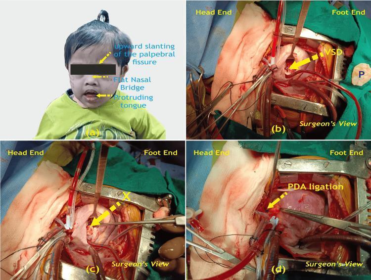
A multi-panel medical image illustrating the clinical presentation and surgical management of congenital heart disease in a pediatric patient with Trisomy 21 (Down Syndrome). Panel (a) is a clinical photograph of a child displaying characteristic dysmorphic facial features, including upward slanting palpebral fissures, a flat nasal bridge, and a protruding tongue. Panels (b), (c), and (d) provide intraoperative views during a median sternotomy. Panel (b) shows the exposed heart before surgical repair, identifying a ventricular septal defect (VSD) and a glutaraldehyde-treated pericardial patch (P) ready for use. Panel (c) demonstrates the intraoperative site after the VSD has been successfully closed with the patch (marked X). Panel (d) displays the ligation of a patent ductus arteriosus (PDA). The surgical images are labeled from the 'Surgeon's View' with 'Head End' and 'Foot End' orientations provided. This composite image serves to correlate the phenotypic features of Trisomy 21 with common associated cardiac anomalies (VSD and PDA) and their definitive surgical correction.
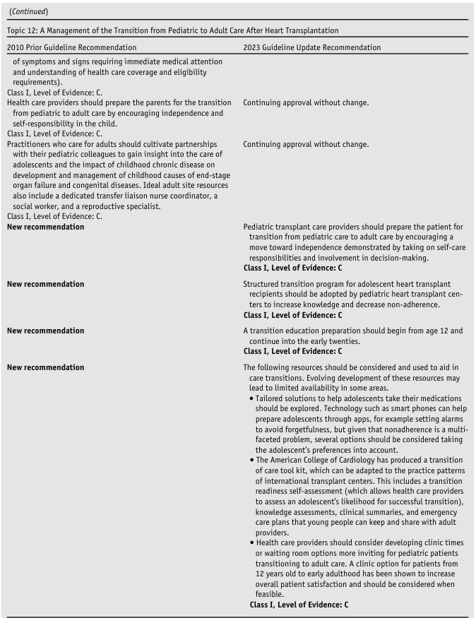
(Continued) <table><thead><tr><th colspan="2">Topic 12: A Management of the Transition from Pediatric to Adult Care After Heart Transplantation</th></tr><tr><th>2010 Prior Guideline Recommendation</th><th>2023 Guideline Update Recommendation</th></tr></thead><tbody><tr><td>of symptoms and signs requiring immediate medical attention and understanding of health care coverage and eligibility requirements).<br>Class I, Level of Evidence: C.</td><td></td></tr><tr><td>Health care providers should prepare the parents for the transition from pediatric to adult care by encouraging independence and self-responsibility in the child.<br>Class I, Level of Evidence: C.</td><td>Continuing approval without change.</td></tr><tr><td>Practitioners who care for adults should cultivate partnerships with their pediatric colleagues to gain insight into the care of adolescents and the impact of childhood chronic disease on development and management of childhood causes of end-stage organ failure and congenital diseases. Ideal adult site resources also include a dedicated transfer liaison nurse coordinator, a social worker, and a reproductive specialist.<br>Class I, Level of Evidence: C.</td><td>Continuing approval without change.</td></tr><tr><td>New recommendation</td><td>Pediatric transplant care providers should prepare the patient for transition from pediatric care to adult care by encouraging a move toward independence demonstrated by taking on self-care responsibilities and involvement in decision-making.<br>Class I, Level of Evidence: C</td></tr><tr><td>New recommendation</td><td>Structured transition program for adolescent heart transplant recipients should be adopted by pediatric heart transplant centers to increase knowledge and decrease non-adherence.<br>Class I, Level of Evidence: C</td></tr><tr><td>New recommendation</td><td>A transition education preparation should begin from age 12 and continue into the early twenties.<br>Class I, Level of Evidence: C</td></tr><tr><td>New recommendation</td><td>The following resources should be considered and used to aid in care transitions. Evolving development of these resources may lead to limited availability in some areas.<br>• Tailored solutions to help adolescents take their medications should be explored. Technology such as smart phones can help prepare adolescents through apps, for example setting alarms to avoid forgetfulness, but given that nonadherence is a multi-faceted problem, several options should be considered taking the adolescent's preferences into account.<br>• The American College of Cardiology has produced a transition of care tool kit, which can be adapted to the practice patterns of international transplant centers. This includes a transition readiness self-assessment (which allows health care providers to assess an adolescent's likelihood for successful transition), knowledge assessments, clinical summaries, and emergency care plans that young people can keep and share with adult providers.<br>• Health care providers should consider developing clinic times or waiting room options more inviting for pediatric patients transitioning to adult care. A clinic option for patients from 12 years old to early adulthood has been shown to increase overall patient satisfaction and should be considered when feasible.<br>Class I, Level of Evidence: C</td></tr></tbody></table>
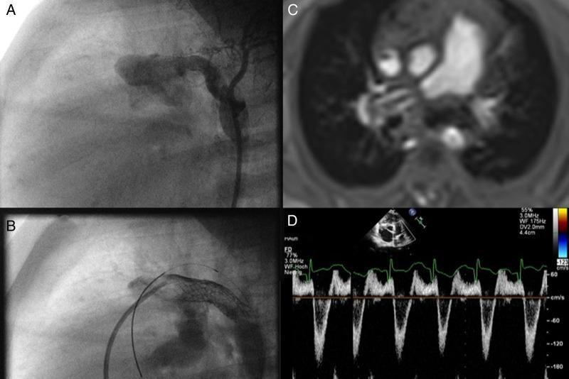
This composite figure illustrates advanced interventional and surgical management of pediatric pulmonary hypertension and heart failure through a four-panel multimodal display. Panels A and B are fluoroscopic angiograms showing a surgical reversed 'Potts' shunt (a connection between the descending aorta and the left pulmonary artery). Panel A demonstrates the initial 6mm PTFE shunt, while Panel B shows the same shunt dilated using an 8mm Formula stent, with a guidewire visible indicating the percutaneous procedure. Panel C is an MRI scan in the axial plane showing bilateral pulmonary artery banding, characterized by physical constrictions on both pulmonary arteries to balance circulation. Panel D presents a Doppler echocardiography trace with a spectral display demonstrating bidirectional flow dynamics: a systolic right-to-left shunt and a diastolic left-to-right shunt. The images collectively demonstrate the clinical challenge of maintaining a parallel fetal-like circulation and the physiological hemodynamics resulting from surgical and transcatheter interventions in complex congenital heart disease cases.
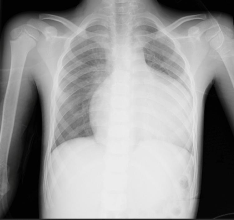
This diagnostic image is a posterior-anterior (PA) chest radiograph of a pediatric patient demonstrating severe pathology. The most prominent finding is massive cardiomegaly, with the cardiac silhouette occupying a significantly increased cardiothoracic ratio, nearly filling the lower half of the thoracic cavity. This global enlargement is consistent with advanced congenital heart disease, such as Ebstein's anomaly. Additionally, the bilateral hilar regions exhibit increased vascular density and indistinctness, characteristic of pulmonary hilar congestion and venous hypertension. The lung fields show prominent vascular markings extending from the hila, though the peripheral lung zones remain relatively clear. The bony structures, including the ribs and clavicles, appear developmentally appropriate for a child. This radiograph serves as a critical educational example of extreme cardiac remodeling and secondary pulmonary vascular changes resulting from chronic volume overload and right-sided heart failure.
pediatric congestive heart failure treatment guidelines drugs doses 2024
https://www.biomed.cas.cz/physiolres/pdf/2024/73_S597.pdf
https://www.mdpi.com/2227-9067/11/7/859
Congestive Heart Failure (CHF) in Pediatrics - Treatment
Key Differences from Adults
- The most common cause is congenital heart disease (CHD), not coronary artery disease
- Other causes include cardiomyopathy (dilated, hypertrophic), myocarditis, arrhythmias, and acquired conditions (Kawasaki disease, rheumatic heart disease)
- RCT evidence is very sparse - most drug dosing is extrapolated from adult trials
- Treatment must account for age-specific pharmacokinetics (neonates, infants, children, adolescents differ significantly)
- LVEF-based classification applies, but a broader functional assessment is needed
Causes by Age Group
| Age | Common Causes |
|---|---|
| Neonate (0-4 weeks) | Ductal-dependent lesions (HLHS, severe aortic/pulmonary stenosis, coarctation), hypoxic-ischemic cardiomyopathy, arrhythmias |
| Infant (1-12 months) | Large L-to-R shunts (VSD, PDA, AV canal), cardiomyopathy, myocarditis |
| Child/Adolescent | Cardiomyopathy, myocarditis, corrected/uncorrected CHD, rheumatic heart disease, drug toxicity (anthracyclines) |
A. Acute Decompensated Heart Failure (Emergency Management)
1. Oxygen & Ventilation
- Supplemental O₂ to maintain SpO₂ (note: in ductal-dependent lesions, avoid excess O₂ as it may close the PDA and worsen hypoperfusion)
- Non-invasive positive pressure ventilation (CPAP/BiPAP): Reduces work of breathing and improves gas exchange
- Intubation for refractory respiratory failure
2. Diuretics (First-Line)
| Drug | Dose | Notes |
|---|---|---|
| Furosemide (IV) | 0.5-2 mg/kg q6-12h, or 0.1-0.4 mg/kg/h continuous infusion | Mainstay; monitor K+, Mg²+, renal function |
| Bumetanide (IV) | 0.015-0.1 mg/kg/dose q6-24h | Alternative loop diuretic |
| Chlorothiazide | 4-10 mg/kg/day ÷ q12-24h (max 20 mg/kg/day) | Adjunct thiazide for diuretic resistance |
3. Vasoactive & Inotropic Support
| Drug | Dose | Indication |
|---|---|---|
| Milrinone | 0.25-1 mcg/kg/min | Low-output state, pulmonary hypertension; preferred as it works even on beta-blockade |
| Dopamine | 3-5 mcg/kg/min | Moderate hypoperfusion |
| Dobutamine | 2.5-10 mcg/kg/min | Low cardiac output with hypotension |
| Epinephrine | 0.01-0.1 mcg/kg/min | Cardiogenic shock, severe low output |
4. Vasodilators
- Nitroprusside: 0.3-4 mcg/kg/min (max 6 mcg/kg/min in neonates; 12 mcg/kg/min in children) - afterload reduction in warm, euvolemic patients
- Nitroglycerin (GTN): Can cause profound hypotension in children - NOT a first-line agent (unlike adults)
5. Special Neonatal Considerations
- Prostaglandin E1 (Alprostadil): 0.05-0.1 mcg/kg/min IV - maintains PDA open in ductal-dependent lesions (critical during stabilization pending surgery)
- Avoid oxygen in duct-dependent systemic circulation lesions (HLHS, severe coarctation)
B. Chronic Heart Failure - Pharmacological Treatment
1. ACE Inhibitors (First-Line RAAS Blockade)
| Drug | Starting Dose | Target Dose | Frequency |
|---|---|---|---|
| Captopril | 0.3 mg/kg/day | 0.5 mg/kg/day (neonates); 1-2 mg/kg/day (older children) | TID |
| Enalapril | 0.1 mg/kg/day | 0.2-0.4 mg/kg/day | BD |
| Lisinopril | 0.05 mg/kg/day | 0.2-0.4 mg/kg/day | OD |
| Ramipril | 0.05 mg/kg/day | 0.1-0.2 mg/kg/day | OD or BD |
Caution in neonates <4 months: Significant risk of renal dysfunction. Monitor creatinine - if it rises >50% above baseline, withhold ACEi. Expect a small BP drop after first dose; observe for 2 hours.
2. ARNI (Sacubitril/Valsartan - Entresto)
- FDA/EMA approved for children ≥1 year since 2019
- According to proposed 2024 ISHLT guidelines: Class IIa, Level B - reasonable alternative to ACEi/ARB in children >1 year with HFrEF
- The PANORAMA-HF trial did not meet its primary endpoint but showed improved natriuretic peptides
- Titrate slowly; do NOT combine with ACEi (36-hour washout required)
3. ARBs
- Losartan: 0.5-1.5 mg/kg/day - used if ACEi-intolerant (cough less common in children than adults)
- Avoid concurrent ACEi + ARB (hyperkalaemia/azotaemia risk)
4. Beta-Blockers
| Drug | Starting Dose | Target Dose | Notes |
|---|---|---|---|
| Carvedilol | <62.5 kg: 0.1 mg/kg/day ÷ BD; ≥62.5 kg: 6.25 mg BD | <62.5 kg: 0.8-1 mg/kg/day ÷ BD; ≥62.5 kg: 25 mg BD (25 mg TID if >75 kg) | Children <4 yrs: dose q8h (altered pharmacokinetics) |
| Metoprolol tartrate | <50 kg: 0.5 mg/kg/day ÷ q12h; ≥50 kg: 25 mg BD | <50 kg: max 4 mg/kg/day; ≥50 kg: max 200 mg q12h | Preferred when systolic BP is borderline low |
| Bisoprolol | >50 kg: 1.25 mg/day | Max 10 mg/day | Pediatric dosing extrapolated from adults |
| Propranolol | 1-6 mg/kg/day ÷ q6h | - | Use for arrhythmia control or HCM outflow obstruction; monitor for hypoglycaemia |
Never start beta-blockers during acute decompensation. Introduce only after stabilization.
5. Mineralocorticoid Receptor Antagonists (MRA)
- Spironolactone: Start 1 mg/kg/day; target max 2 mg/kg/day
- Monitor K+, creatinine before starting, shortly after, then every 3-6 months
- Monitor for gynecomastia in males
6. Diuretics (Chronic)
- Furosemide oral: 1-4 mg/kg/day ÷ OD-BD for maintenance - use lowest effective dose
- As RAAS/ARNI therapy is optimized, diuretic need often decreases - wean if euvolemic
7. Digoxin
- Used more frequently in children than adults (unlike adult guidelines which largely discourage it)
- However, some recent pediatric guidelines (Canadian CCS 2024) state it is not recommended for most children
- Still used in: SVT/AF rate control, cardiomyopathy with severe symptoms, post-surgical low output (especially in infants)
- Requires careful therapeutic drug monitoring (target level 0.5-1 ng/mL in HF)
C. Newer Therapies & Emerging Agents
| Drug Class | Status in Pediatrics |
|---|---|
| SGLT2 Inhibitors (dapagliflozin, empagliflozin) | Evidence emerging; 2024 ISHLT proposes Class IIb, Level C for HFpEF in children; no approved indication yet in most jurisdictions |
| Ivabradine | Class IIa in selected children with elevated HR in HFrEF on max-tolerated beta-blocker |
| Vericiguat | Being evaluated in pediatrics; no current recommendation |
D. Device Therapy in Children
| Device | Indication |
|---|---|
| ICD | HFrEF with LVEF ≤35%, documented VT/VF, high-risk channelopathies |
| CRT | Dyssynchrony with LBBB morphology, LVEF ≤35% - evidence extrapolated from adults; used selectively |
| VAD (Ventricular Assist Device) | End-stage HF as bridge to transplant; LVAD or biventricular |
| ECMO | Acute cardiogenic shock refractory to medical therapy (especially myocarditis, post-cardiotomy) |
E. Heart Transplantation
- Definitive treatment for end-stage pediatric HF
- Indications: Progressive HF despite GDMT, VAD dependency, failed surgical palliation
- Per the ISHLT 2024 Transplant Guidelines (PMID 39115488), structured transition from pediatric to adult care should begin at age 12 and continue into the early twenties for heart transplant recipients
F. Surgical / Catheter Interventions
- Definitive correction of the underlying CHD is the ultimate goal when feasible (e.g., VSD closure, valvuloplasty, arterial switch)
- Palliative procedures (e.g., pulmonary artery banding, Fontan palliation for single ventricle) reduce volume load pending repair
- Treat precipitants: arrhythmias, anaemia, infection, medication non-compliance
G. Non-Pharmacological / Supportive Measures
- Nutritional support: High-calorie feeds for infants with poor weight gain due to increased metabolic demand; nasogastric feeding if needed
- Fluid restriction: Individualized, especially with hyponatraemia
- Activity: Encourage developmentally appropriate activity; avoid strenuous exercise in severe HF
- Treat comorbidities: Anaemia, infections, arrhythmias
- Family education: Daily weight monitoring, symptom recognition, medication adherence
H. Pediatric CHF Management Algorithm (Summary)
Acute decompensation?
  YES → O₂/ventilation → IV furosemide → Vasodilators (nitroprusside)
        → Inotropes if low output (milrinone/dobutamine)
        → PGE1 if ductal-dependent lesion
        → ECMO if refractory shock

Stable/Chronic HF with reduced EF?
  Step 1: ACEi (Captopril in infants, Enalapril in older children)
  Step 2: Add Beta-blocker (Carvedilol or Metoprolol) - after stabilization
  Step 3: Add Spironolactone (MRA)
  Step 4: Consider ARNI (>1 year) or SGLT2i (investigational)
  Step 5: Add Digoxin if persistent symptoms
  Step 6: Device therapy / VAD / Transplant for refractory HF