
Bilirubin formation in detail from basica
I'll start by reading the medical library skill, then search for detailed bilirubin formation content.
Reading File
Now let me search the library for bilirubin formation content in parallel:
Finding Sources
Finding Sources
Excellent results. Let me now read the most relevant and detailed sources in parallel - Lippincott Biochemistry, Harrison's, and Goldman-Cecil:
Reading File
Reading File
Reading File
Excellent content. Now let me fetch the key diagrams and read further content about intestinal fate and the complete pathway:
Loading Image
Loading Image
Reading File
Now let me fetch the full catabolism diagram and get the Guyton physiology content for completeness:
Loading Image
Reading File
I now have all the detail needed to write a comprehensive answer. Let me compile it:
Bilirubin Formation - From Basics
Bilirubin is the major end product of heme catabolism. Its metabolism is a tightly organized, multi-organ process spanning the mononuclear phagocyte system (MPS), blood, liver, and intestine.
1. Sources of Heme (What Gets Degraded)
About 80-85% of daily bilirubin (~4 mg/kg/day) comes from the breakdown of hemoglobin in senescent red blood cells (RBC lifespan ~120 days). The remaining 15-20% comes from:
- Premature destruction of erythroid precursors in the bone marrow (ineffective erythropoiesis)
- Turnover of non-hemoglobin hemoproteins: myoglobin, cytochrome P-450, catalase, peroxidase
These sources are collectively called the "early-labeled" bilirubin fraction.
Harrison's Principles of Internal Medicine 22E, p. 364
2. Step 1 - RBC Destruction and Heme Liberation
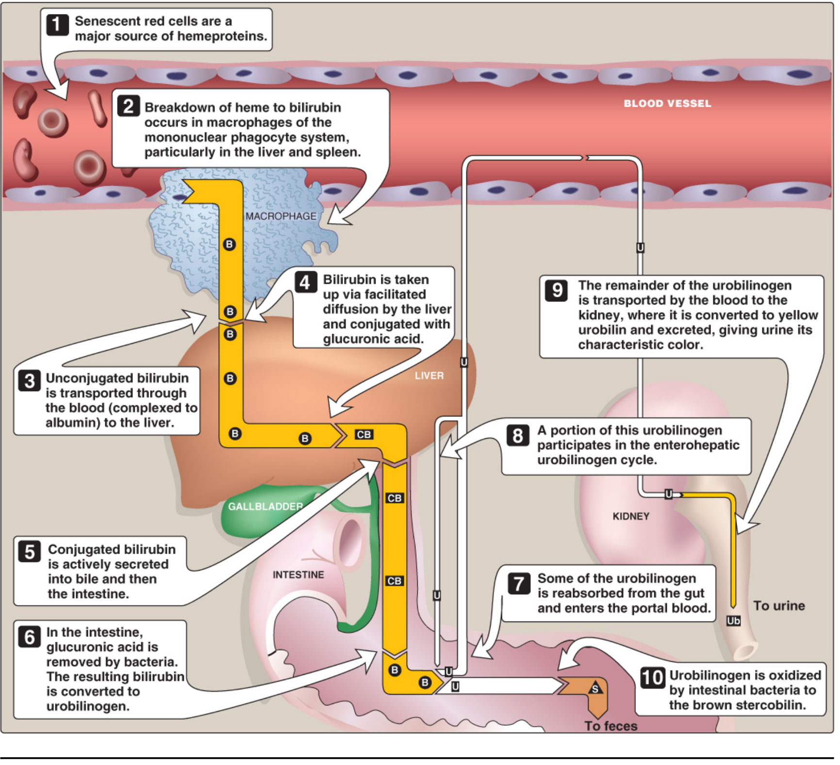
Senescent RBCs are engulfed by macrophages of the MPS - primarily in the spleen, liver (Kupffer cells), and bone marrow. Inside the macrophage:
- Hemoglobin is split into globin (recycled as amino acids) and heme (ferroprotoporphyrin IX)
- Heme = an iron-containing, cyclic tetrapyrrole (porphyrin ring with Fe²⁺ at the center)
3. Step 2 - Heme → Biliverdin (via Heme Oxygenase)
This is the rate-limiting step in bilirubin formation.
Enzyme: Microsomal heme oxygenase (located in the ER of macrophages)
Reaction: Three successive oxygenations using NADPH + O₂:
- The α-methene bridge carbon of the porphyrin ring is oxidatively cleaved
- The cyclic heme ring is opened to form a linear tetrapyrrole
- Products released: Biliverdin (green pigment) + CO (carbon monoxide) + Fe²⁺
The equation per molecule of heme:
Heme + 3O₂ + 3NADPH → Biliverdin + CO + Fe²⁺ + 3NADP⁺
By-products have functions:
- CO acts as a signaling molecule and anti-inflammatory mediator
- Fe²⁺ is recycled - bound to transferrin and transported back to bone marrow for re-use in new hemoglobin
In birds, reptiles, and amphibians, biliverdin is the final excretory product. In mammals, it is reduced further to bilirubin - this is evolutionarily important because bilirubin (unlike biliverdin) can cross the placenta, allowing fetal bilirubin to be excreted via the maternal liver.
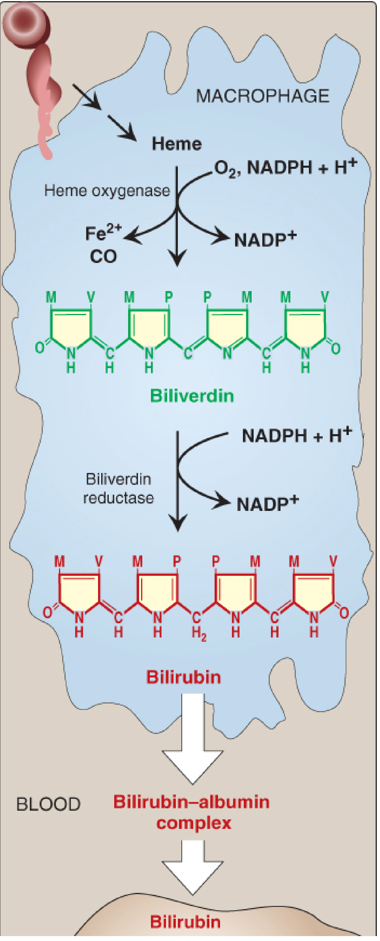
4. Step 3 - Biliverdin → Bilirubin (via Biliverdin Reductase)
Enzyme: Cytosolic biliverdin reductase
Reaction: Reduces the central methylene bridge of biliverdin using NADPH:
Biliverdin + NADPH + H⁺ → Bilirubin + NADP⁺
This converts the green biliverdin to the characteristic red-orange bilirubin.
Why bilirubin is insoluble: Bilirubin has tight internal hydrogen bonds between its propionic acid carboxyl groups and the imino/lactam groups of the opposite dipyrrolic half. This "ridge-tile" configuration:
- Places hydrophobic residues outward
- Buries polar residues inward
- Makes bilirubin virtually insoluble in water
Note: Bilirubin in mammals also has antioxidant function at low concentrations. It is oxidized back to biliverdin, which is then reduced again by biliverdin reductase - creating a redox cycle.
Lippincott's Illustrated Reviews: Biochemistry 8e, p. 795
5. Step 4 - Blood Transport (Unconjugated Bilirubin)
Because bilirubin is water-insoluble, it cannot travel freely in plasma. It is:
- Bound noncovalently to albumin (2 binding sites per albumin molecule)
- This form is called unconjugated (indirect) bilirubin or free bilirubin
- It is NOT filtered by the kidney in normal conditions (too large as albumin complex)
Clinical note: Certain drugs (salicylates, sulfonamides, furosemide, radiographic contrast agents) competitively displace bilirubin from albumin. In neonates, this raises free bilirubin which can cross the blood-brain barrier and cause kernicterus.
Goldman-Cecil Medicine, Chapter 133
6. Step 5 - Hepatic Uptake
When albumin-bilirubin complex reaches the liver sinusoids:
- Bilirubin dissociates from albumin (albumin stays in blood)
- Unconjugated bilirubin enters hepatocytes by facilitated diffusion (specific transporters, including OATP1B1/OATP1B3)
- Inside the hepatocyte cytosol, bilirubin binds to ligandin (glutathione-S-transferase Y) and protein Z - these reduce back-diffusion and shuttle bilirubin to the ER
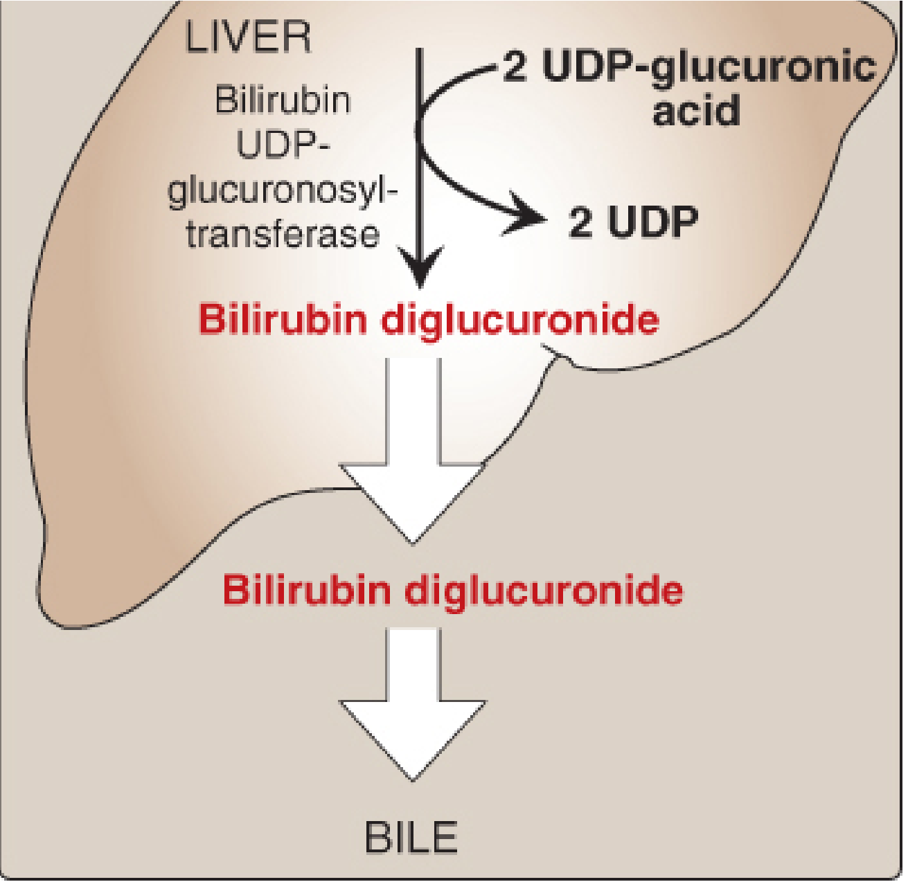
7. Step 6 - Conjugation in the Liver (Making Bilirubin Water-Soluble)
Location: Endoplasmic reticulum of hepatocytes
Enzyme: Bilirubin UDP-glucuronosyltransferase (UGT1A1), encoded by the UGT1 gene complex
Reaction: Sequential addition of two glucuronic acid molecules from UDP-glucuronic acid:
- Bilirubin + UDP-glucuronic acid → Bilirubin monoglucuronide (BMG)
- BMG + UDP-glucuronic acid → Bilirubin diglucuronide (BDG) ← predominant form
The conjugation disrupts the internal hydrogen bonds, making bilirubin water-soluble (conjugated/direct bilirubin). It can now be excreted.
According to Guyton and Hall: ~80% is conjugated as bilirubin glucuronide, ~10% as bilirubin sulfate, and ~10% with other substances.
Clinical relevance of UGT1A1:
- Gilbert syndrome: Mild UGT1A1 reduction → mild unconjugated hyperbilirubinemia
- Crigler-Najjar syndrome type I: Complete absence of UGT1A1 → severe, life-threatening unconjugated hyperbilirubinemia
- Crigler-Najjar syndrome type II (Arias): Markedly reduced UGT1A1
8. Step 7 - Biliary Secretion
Conjugated bilirubin is actively transported across the canalicular membrane into bile by:
- MRP2 (Multidrug Resistance-associated Protein 2), also called cMOAT/ABCC2 - an ATP-dependent transporter
- This is the rate-limiting step for bilirubin excretion (not conjugation)
- MRP3 transports some conjugated bilirubin back into sinusoidal blood when MRP2 is overloaded (regurgitation pathway)
Clinical note: Deficiency of MRP2 (canalicular transport) = Dubin-Johnson syndrome (conjugated hyperbilirubinemia, benign).
Conjugated bilirubin flows into bile, is stored in the gallbladder, and enters the duodenum.
9. Step 8 - Intestinal Fate and Enterohepatic Circulation
In the small intestine, conjugated bilirubin is NOT absorbed (too hydrophilic and large). In the terminal ileum and colon, intestinal bacteria:
- Hydrolyze the glucuronide conjugate (beta-glucuronidase) → free unconjugated bilirubin
- Reduce bilirubin → urobilinogen (colorless compound), a series of compounds (mesobilirubinogen, stercobilinogen)
Fate of urobilinogen:
- Most (~80%) is further oxidized by bacteria to stercobilin (brown pigment) → excreted in feces (gives stool its brown color)
- ~20% is reabsorbed from the gut into portal blood:
- Most is re-extracted by the liver and re-secreted into bile = enterohepatic circulation of urobilinogen
- A small fraction escapes into systemic blood → kidneys → oxidized to urobilin (yellow) → excreted in urine (gives urine its yellow color)
Summary Table
| Stage | Location | Key Enzyme/Protein | Product |
|---|---|---|---|
| RBC destruction | Spleen/liver/BM macrophages | - | Heme liberated |
| Heme → Biliverdin | Macrophage ER | Heme oxygenase | Biliverdin + CO + Fe²⁺ |
| Biliverdin → Bilirubin | Macrophage cytosol | Biliverdin reductase | Unconjugated bilirubin |
| Blood transport | Plasma | Albumin | Bilirubin-albumin complex |
| Hepatic uptake | Hepatocyte cytosol | Ligandin/OATP | Unconjugated bilirubin (intrahepatic) |
| Conjugation | Hepatocyte ER | UGT1A1 | Bilirubin diglucuronide (conjugated) |
| Biliary secretion | Bile canaliculus | MRP2 (ATP-dependent) | Conjugated bilirubin in bile |
| Intestinal reduction | Colon bacteria | Bacterial reductases | Urobilinogen |
| Fecal excretion | Intestine | Bacterial oxidases | Stercobilin (brown) |
| Urinary excretion | Kidney | - | Urobilin (yellow) |
Normal Reference Values
- Total serum bilirubin: ≤1 mg/dL
- Conjugated (direct): ≤0.3 mg/dL
- Jaundice clinically visible: ≥2-3 mg/dL
Sources: Lippincott's Illustrated Reviews: Biochemistry 8e (pp. 794-799) | Harrison's Principles of Internal Medicine 22E (p. 364) | Goldman-Cecil Medicine, Chapter 133 | Guyton and Hall Medical Physiology 14e, Chapter 71
This is a shared conversation. Sign in to Orris to start your own chat.