
Stoppa approach ms ortho 20 marks answer
I'll read the medical library skill first, then research the Stoppa approach comprehensively.
Searching sources...
Reading File
Now let me search the library in parallel for all relevant content on the Stoppa approach.
Finding Sources
Finding Sources
Excellent! Found the primary sources. Now let me read all relevant sections in parallel.
Reading File
Reading File
Reading File
Let me now read more detailed content from the Campbell's and Rockwood & Green's sections on the Stoppa approach.
Reading File
Reading File
Reading File
Finding Sources
Reading File
Reading File
Reading File
Reading File
Reading File
I now have comprehensive information from three authoritative textbooks. Let me fetch the key diagram image to include in the answer.
Loading Image
Loading Image
I now have all the information needed for a comprehensive 20-mark MS orthopaedics answer. Here is the full structured answer:
Stoppa Approach (Anterior Intrapelvic / Modified Stoppa Approach)
Sources: Campbell's Operative Orthopaedics 15th Ed 2026 (Technique 61.1), Rockwood & Green's Fractures in Adults 10th Ed 2025, Miller's Review of Orthopaedics 9th Ed
1. Introduction and Historical Background
The Stoppa approach, formally termed the Anterior Intrapelvic (AIP) approach or Modified Stoppa approach, is an anterior extraperitoneal approach to the pelvis and acetabulum. It was originally described by Stoppa for hernia repair and subsequently modified and popularized by Cole and Bolhofner (1994) for acetabular fracture fixation. It has largely replaced the traditional medial window of the ilioinguinal approach for many fracture patterns, offering superior access to the quadrilateral surface with less vascular risk.
2. Relevant Surgical Anatomy
Understanding the anatomy in this region is mandatory before performing the approach:
- Space of Retzius (retropubic space): A preperitoneal extraperitoneal space between the pubic symphysis anteriorly and the bladder posteriorly - the key working space of this approach
- Rectus abdominis: Paired muscles attached to the pubic symphysis; elevated off the pubis to develop exposure
- Linea alba: Midline decussation of fascial fibers - the key fascial interval used
- Quadrilateral surface: The internal surface of the acetabulum, formed by the body of the ilium above the obturator foramen - the main target of this approach
- Corona mortis ("Crown of Death"): An anastomotic connection between the external iliac (or inferior epigastric) artery/vein and the obturator vessels. Present in 10-30% of patients, located approximately 5 cm medial to the pubic symphysis. Inadvertent transection can retract into the pelvis causing uncontrollable hemorrhage - must be identified and ligated
- Obturator neurovascular bundle: Traverses the operative field just below the pelvic brim; injury causes hypesthesia of the inner thigh and adductor weakness. Reported injury rate up to 26% (most transient, resolving within 12 months)
- Lumbosacral trunk (L4-L5): Crosses the pelvic brim and sacral ala; at risk during posterior dissection
- External iliac vessels: Run superior and lateral along the pelvic brim; protected by Deaver retractor
- Urinary bladder: Lies immediately posterior to the symphysis; protected with a malleable retractor and damp sponge
3. Indications
The Modified Stoppa approach is used for:
- Anterior column fractures - especially those with quadrilateral surface involvement
- Anterior column with posterior hemitransverse (ACPHT) fractures
- Both-column (associated, ABC) fractures - most commonly used indication
- T-type fractures with anterior displacement or quadrilateral comminution
- Transverse fractures - those associated with disruption of the symphysis pubis
- Anterior wall fractures
- Fractures previously treated through the ilioinguinal approach (medial window) - the AIP offers equivalent or superior access without middle window dissection
- Combined with the lateral window of ilioinguinal for comminuted patterns requiring iliac wing exposure
- Revision fixation and correction of malunion of acetabular fractures
The approach provides the best access to the quadrilateral surface and is preferred when medial wall comminution is the primary problem.
4. Contraindications / Limitations
- Pre-existing midline laparotomy scar (relative - adhesions to bladder/peritoneum)
- Obesity (limits proximal extension and visualization)
- Cannot visualize the articular surface of the acetabulum directly
- Limited access to the posterior column; posterior column fractures require indirect reduction or combined approaches
- Does not provide access to the external surface of the iliac wing
5. Patient Positioning and Setup
- Position: Supine on a radiolucent flat-top table
- A triangle under the hip relaxes the iliopsoas and improves visualization through the lateral window when combined
- Affected limb is prepped into the field (free-draped) to allow intraoperative manipulation and skeletal traction
- A Foley catheter is mandatory to decompress the bladder and monitor for intraoperative bladder injury
- Surgeon stands on the side opposite the fracture to improve visualization of the true intrapelvic cavity
- A headlamp or fiberoptic light retractor is strongly recommended
- C-arm fluoroscopy available
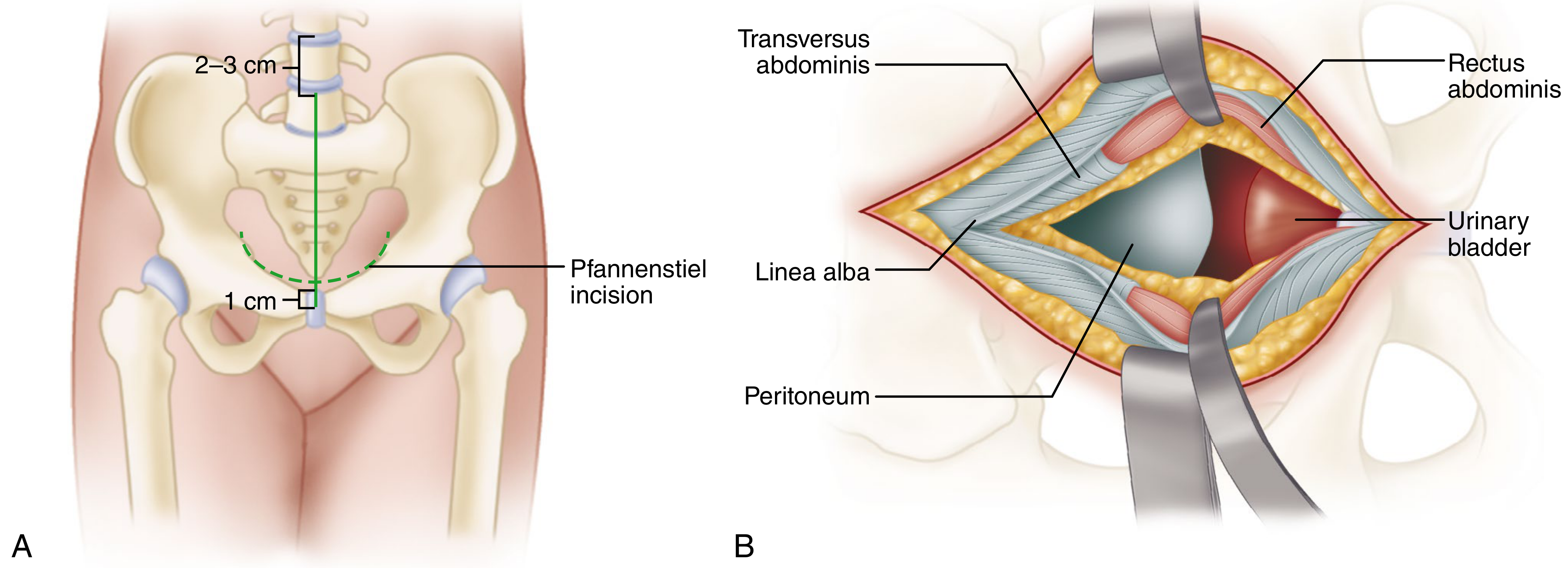
6. Surgical Technique (Step-by-Step)
Skin Incision
Step 1. Make a Pfannenstiel incision 2 cm above the pubic symphysis, extending approximately from one external inguinal ring to the other (approximately 8-10 cm). Alternatively, a vertical midline skin incision starting 1 cm inferior to the symphysis and ending 2-3 cm below the umbilicus may be used (especially if prior laparotomy incision exists).
Fascial Dissection
Step 2. Divide subcutaneous tissue in line with the incision to expose the fascia overlying both rectus muscles. Identify the linea alba (midline decussation of fascial fibers).
Step 3. Make a small 0.5-1 cm transverse nick in the fascia near the midline to identify the interval between the two rectus muscle bellies. Extend the incision vertically along the linea alba, retracting both rectus bellies laterally.
Retroperitoneal Dissection
Step 4. The entire approach is maintained in the preperitoneal (extraperitoneal) space - avoid entering the peritoneum, especially in the proximal portion. Extending the muscular interval proximally increases muscular excursion and visualization.
Step 5. Bluntly dissect into the Space of Retzius (retropubic space). Pack a damp laparotomy sponge loosely into this space and place a malleable retractor to protect the bladder (Fig. C).
Pubic Bone Exposure
Step 6. Release the insertion of the rectus abdominis over the anterior aspect of the pubic tubercle bilaterally - increased release improves later visualization.
Step 7. Sharply dissect the thick periosteum from the superior surface of the pubic bone to allow deeper blunt dissection. Place a sharp Hohmann retractor lateral to the pubic tubercle over the superior ramus on the injured side (Fig. D).
Corona Mortis
Step 8. Identify the upper border of the superior pubic ramus (pectin pubis) and carry the dissection laterally along the pelvic brim. Place a Deaver retractor laterally to protect the iliac vessels.
Step 9. (CRITICAL) Dissecting carefully along the medial surface of the superior ramus, identify the corona mortis - anastomotic vessels between external iliac/inferior epigastric and obturator vessels. Ligate all such vessels to prevent retraction and uncontrollable hemorrhage (Fig. E).
Deeper Pelvic Exposure
Step 10. Continue subperiosteal dissection laterally, following the upper border of the superior pubic bone to the pelvic brim, exposing the iliopectineal eminence.
Step 11. Dissect the beginning of the iliopectineal arch from the bone to allow elevation of the femoral vessels and nerve (Fig. F). Continue over the anterior wall of the acetabulum.
Step 12. Place a sharp Hohmann or custom retractor over the acetabular rim near the iliopubic eminence. Continue subperiosteal lateral dissection along the pelvic brim. The entire internal surface of the superior pubic ramus is now exposed for plate fixation.
Quadrilateral Surface Exposure
Step 13. As the quadrilateral surface is reached, identify the obturator neurovascular bundle, which traverses the field just below the pelvic brim. Mobilize the bundle as necessary and protect it using a custom pelvic floor retractor or malleable retractor placed into the lesser sciatic notch medial to the bundle (Fig. G).
Step 14. Elevate the periosteum and obturator internus with a Cobb elevator to expose the quadrilateral surface.
Step 15. A sharp Hohmann retractor may be impacted on the posterior top of the acetabulum into the ilium, slightly lateral to the SI joint. Take great care to avoid injuring the external iliac vein near the elevators (Fig. H).
Step 16. Dissection can be carried around the pelvic brim all the way to the sacroiliac joint if needed. A blunt retractor placed into the greater sciatic notch must be placed medial to the obturator nerve to avoid nerve tension.
7. Structures Exposed
Through the Modified Stoppa approach, the following structures are directly visible:
| Structure | Notes |
|---|---|
| Internal surface of superior pubic ramus | From symphysis to iliopectineal eminence |
| Pelvic brim (arcuate line) | Full length to SI joint |
| Quadrilateral surface | Best access of any anterior approach |
| Medial wall of acetabulum | Key for fracture reduction |
| Anterior column (inner surface) | Subperiosteal |
| Anterior sacroiliac joint (with extension) | Posterolateral dissection |
The articular surface of the acetabulum is NOT directly visualized.
8. Advantages over Ilioinguinal Approach
| Feature | Modified Stoppa | Ilioinguinal (Medial Window) |
|---|---|---|
| Femoral vessel dissection | Not required | Required (middle window) |
| Quadrilateral surface access | Superior | Inferior |
| Vascular risk | Lower | Higher (femoral vessels) |
| Learning curve | Shorter | Longer |
| Lymphatic morbidity | None | Risk of lymphocele |
| Combination with lateral window | Easy | Standard |
9. Extensions and Combinations
- Lateral window of ilioinguinal combined with AIP: avoids middle window (no femoral vessel dissection) while gaining access to the iliac wing. Recommended by Campbell's for comminuted anterior fracture patterns.
- ASIS osteotomy addition: significantly improves exposure of the anterior wall or psoas gutter, otherwise accessible only through the middle window.
- Combined anterior + posterior (Kocher-Langenbeck): for T-type, both-column with posterior wall, or ACPHT fractures where indirect posterior reduction is inadequate.
10. Closure
- Irrigate the space of Retzius
- Inspect the Foley bag for hematuria to exclude iatrogenic bladder injury
- Place a drain if desired
- Close the rectus fascia in interrupted figure-of-eight fashion to prevent abdominal hernia
- Routine subcutaneous and skin closure
11. Complications
| Complication | Notes |
|---|---|
| Obturator nerve injury | Most common nerve injury; up to 26%; mostly transient (resolves within 12 months); weakness of adductors, hypesthesia inner thigh |
| Corona mortis hemorrhage | If unrecognized and divided; vessel retracts into pelvis |
| Bladder injury | Avoided by Foley catheter decompression and careful retraction |
| External iliac vein injury | During posterior dissection near SI joint |
| Lumbosacral trunk injury | Stretching during sciatic notch retraction |
| Sciatic nerve injury | Retractors in sciatic notch; worse with hip and knee in flexion |
| Incisional hernia | Inadequate fascial closure of linea alba |
| Peritoneal breach | If proximal dissection is not carefully maintained extraperitoneal |
| DVT / thromboembolism | Standard pelvic fracture risk |
| Heterotopic ossification | Less than posterior approaches |
12. Comparison with Other Anterior Approaches
| Feature | Modified Stoppa | Ilioinguinal | Extended Iliofemoral |
|---|---|---|---|
| Skin incision | Pfannenstiel | Iliac crest-based | Extensive lateral |
| Posterior column access | Indirect | Indirect | Direct |
| Abductor muscle stripping | No | No | Yes |
| HO risk | Low | Low | High |
| Morbidity | Low-moderate | Moderate | High |
Summary Points for Exam
- The Stoppa approach = Anterior Intrapelvic (AIP) approach; works through the Space of Retzius in a preperitoneal plane
- Incision: Pfannenstiel, 2 cm above symphysis pubis
- Deep interval: along the linea alba, between two rectus abdominis bellies
- Corona mortis: must always be identified and ligated (present in 10-30%)
- Best access to quadrilateral surface of any anterior approach
- Obturator nerve most commonly injured (26%; mostly transient)
- Surgeon stands contralateral to fracture
- Does NOT expose articular surface; does NOT require femoral vessel dissection
- Combine with lateral ilioinguinal window to avoid middle window dissection
- Close linea alba in figure-of-eight to prevent hernia
- Check Foley bag for hematuria before closing to exclude bladder injury
References: Campbell's Operative Orthopaedics 15th Ed 2026, Technique 61.1 (p. 3507-3509); Rockwood & Green's Fractures in Adults 10th Ed 2025 (p. 2511-2512); Miller's Review of Orthopaedics 9th Ed
This is a shared conversation. Sign in to Orris to start your own chat.